




Medical education
Snapshot
Volume 219 - Issue 3
A unique pattern of urothelial calcifications secondary to disseminated tuberculosis
Authors: Samuel Lau, Bertrand Ang and Sandra Tan
Med J Aust 2023; 219 (3): 103-103. || doi: 10.5694/mja2.52006
Published online: 7 August 2023
Published online: 7 August 2023
A 35-year-old woman presented with haematuria, unintentional weight loss, and night sweats for two months
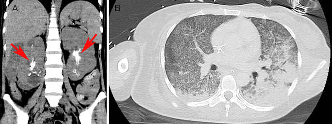
A 35‐year‐old woman presented with haematuria, unintentional weight loss, and night sweats for two months. On examination, she was febrile (38.8°C), hypotensive (71/56mmHg) with low oxygen saturations (89% on room air), and right flank tenderness.
A computed tomography scan showed extensive urothelial calcifications involving bilateral renal pelvis and calyces and a right perinephric collection (Figure, A, arrows). Diffuse miliary nodules with consolidation were seen in the lungs (Figure, B). Disseminated Mycobacterium tuberculosis was suspected as the patient was from a country where tuberculosis is endemic. Bacterial pneumonia with urinary tract infection and concomitant hyperparathyroidism were differentials, but urine and sputum cultures had negative bacterial growth and serum calcium was normal.
Fluid from the collection was positive for M. tuberculosis on polymerase chain reaction (PCR) test and cultures (sensitive to rifampicin). Nephrocalcinosis is a manifestation of renal tuberculosis,1,2 and recognition of radiological features with concordant clinical presentation can lead to earlier diagnosis and treatment.
Competing interests
No relevant disclosures.
References
- Jung YY, Kim JK, Cho KS. Genitourinary tuberculosis: comprehensive cross‐sectional imaging. AJR Am J Roentgenol 2005; 184: 143‐150.
- Wong A, Dhingra S, Surabhi VR. AIRP best cases in radiologic‐pathologic correlation: Genitourinary tuberculosis. Radiographics 2012; 32: 839‐844.
Costs of Delivering the Hep B PAST Model of Care in Remote Australia: A Retrospective Pre–Post Study
Hoa Nguyen, Anh Le Tuan Nguyen, Paula Binks, Melita McKinnon, Emily Vintour-Cesar, Karen Wills, Nicola Stephens, Benjamin Cowie, Joshua S. Davis, Yuejen Zhao, Peter Nihill, Julie A. Campbell, Andrew J. Palmer, Jane Davies, Barbara de Graaff
Buruli Ulcer Acquired From Ringtail Possum Rescue
Daniel P. O'Brien, Naomi Clarke, Sara Bonnici, Mohammad A. Hussain
Q Fever Visceral Abscesses After Contact With Native Australian Marsupials
Samantha J. Laws, Enzo Binotto, James Stewart, Jennifer Ho, Trent Yarwood, Josh Hanson, Simon Smith
Hospital-Admitted Injection-Related Infections Among Incarcerated People Who Inject Drugs in Australia: A Retrospective Cohort Study
Andrew Palmer, Matthew Carter, Jeremy Yeo, Cecilia Shim, Jason Connor, Jeremy Hayllar, Gerald Holtmann, Naomi Moy, Elliott G. Playford, Naomi Runnegar, Paul J. Clark
The Live Attenuated Influenza Vaccine in Australia: An Additional Tool for Influenza Prevention
Cyra Patel, Alexis Pillsbury, Tran Nguyen, Xia Wang, Helen E. Quinn, Clayton K. Chiu, Allen C. Cheng, Katie L. Flanagan, Zhicheng Wang
Reflex Testing for Hepatitis D Infection: A Unique Opportunity to Reduce Hepatitis D-Related Chronic Liver Disease Deaths in Australia
Jessica Howell, Lauren Andersson, Miriam T. Levy, James O'Beirne, Leon Adams, Katharine Irvine, Avik Majumdar, Golo Ahlenstiel, Kathy Jackson, Krispin Hajkowicz, Joseph Doyle, Jane Davies, Sarah Cherian, Wayne Dimech, Alexander J. Thompson