




Medical education
Lessons from practice
Volume 219 - Issue 10
Human pulmonary dirofilariasis: an unexpected differential diagnosis for a solitary lung lesion
Authors: Sigrid G Theodore, Hannah J Sawkins, Manish Mathew, Sumit Yadav and Robert Norton
Med J Aust 2023; 219 (10): 455-456. || doi: 10.5694/mja2.52115
Published online: 20 November 2023
Published online: 20 November 2023
A 56-year-old woman of European ancestry from a suburban area of Far North Queensland was referred to the local cardiothoracic unit for a right lower lobe lung lesion
Clinical record
A 56‐year‐old woman of European ancestry from a suburban area of Far North Queensland was referred to the local cardiothoracic unit for a right lower lobe lung lesion. Her past medical history included a 43 pack‐year smoking history, chronic obstructive pulmonary disease, hypertension, dyslipidaemia, diabetes mellitus and obesity. She had resided in Malaysia for almost three years as a child and had previously travelled to Papua New Guinea several years before presentation. A lung lesion was incidentally detected while the patient was undergoing imaging to investigate hepatic steatosis. She had no clinical features of infection or malignancy.
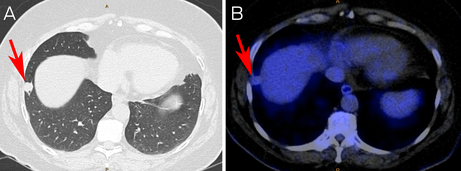
Chest computed tomography (CT) scan characterised a solitary 13mm anterolateral lesion in the lower lobe of the right lung abutting the pleura without associated lymphadenopathy (Box 1, A). This lesion was not present on previous imaging completed nine months prior. A positron emission tomography (PET) scan revealed a minimally 18F‐fluorodeoxyglucose (18F‐FDG) avid lesion concerning for primary lung malignancy (Box 1, B). The maximum standardised uptake value (SUV) was 2.7. Tissue biopsy performed under CT guidance was non‐diagnostic. Blood tests revealed normal white cell and eosinophil counts.
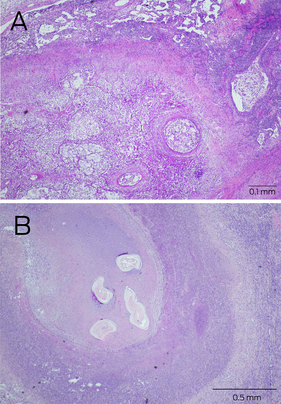
The patient was referred for surgery following discussion of the suspicious lung lesion at the Lung Multidisciplinary Meeting. A right lower lobe wedge resection was performed, with a frozen section revealing necrotising granulomatous inflammation (Box 2, A). Histopathology confirmed helminthiasis (Box 2, B). Tissue and mycobacterial cultures were negative. Expert opinion concluded a diagnosis of pulmonary Dirofilaria infection based on histological characteristics. A subsequent reactive Strongyloides serology result was considered secondary to cross‐reactivity. Filaria serology was equivocal. There were no post‐operative complications.
Discussion
Human pulmonary dirofilariasis is a rare, non‐lethal infection caused by the nematode Dirofilaria immitis.1 This parasite is responsible for canine heartworm disease.1 Mosquitos are vectors for D. immitis, hence its prevalence in tropical and subtropical climates.2 Definitive hosts include various mammals, frequently domestic and wild dogs.1,3 Humans are considered incidental hosts following inoculation of larvae via an infected mosquito.3 The larvae enter the pulmonary vasculature and embolise into smaller peripheral arterial branches.2 Here, an inflammatory reaction leads to destruction of the immature worm.2 This process can result in a pulmonary nodule.2
There remains a paucity of literature regarding the incidence of human pulmonary dirofilariasis in Australia over the past two decades.2 Typically, human pulmonary dirofilariasis presents as an incidentally detected solitary pulmonary nodule, though reports of multiple nodules have occurred previously.2 The pulmonary nodule is typically described as a “coin lesion” frequently located within the right lower lobe.1,4 Patients are commonly asymptomatic, but haemoptysis, chest pain, fever and eosinophilia can occur.1,3 Varying 18F‐FDG avidity has been reported in cases of human pulmonary dirofilariasis, ranging from non‐avid to an SUV of 7.5.4,5,6 Malignancy is frequently suspected, although fine needle biopsy is typically non‐diagnostic.7 Diagnosis is generally made on histological specimen following surgical resection.1,3 Serological testing is not validated in isolation but may be complementary to diagnostic workup.1,3 As the worm cannot mature in human pulmonary dirofilariasis, the disease is self‐limiting. Hence, no antihelminthic therapy is required.1 Where surgery has occurred, resection is considered definitive.1,3
The incidence of D. immitis is increasing in dog populations around the world, attributed in part due to climate change, prevalence of vector mosquitos, and affordability and accessibility of chemoprophylaxis.2,4,7,8 In Australia, the routine use of macrocyclic lactone prophylaxis for dogs was introduced in the 1990s.8 Despite an overall reduction of heartworm disease in Australia, a recent study of D. immitis in Queensland dog populations revealed disproportionately high rates of canine infection in north Queensland compared with southeast Queensland.8 This geographical difference is multifactorial, reflecting north Queensland's tropical climate, vector prevalence, wild dog populations, and possibly reduced dog owner education and compliance with preventive treatments.8 This highlights the ongoing challenges of D. immitis infection control in tropical regions of Australia and the possible implications for humans as dead‐end hosts. As such, primary prevention should remain a priority, in addition to improved diagnostic strategies of human pulmonary dirofilariasis to avoid invasive and costly interventions for this self‐limiting disease.
Lessons from practice
- Dirofilaria immitis is the parasite responsible for canine heartworm disease.
- Human pulmonary dirofilariasis is a rare cause of a solitary lung nodule.
- Nodules may be avid on positron emission tomography. Fine needle biopsy is often inconclusive. Diagnosis is typically made on histological specimen obtained following surgery for suspected malignancy.
- This parasitic infection is self‐limiting in humans. Management is centred around symptom management. Surgery is considered curative if operative resection has occurred.
Box 1 – (A) Computed tomography scan demonstrated a focal right lower lobe lung nodule. (B) Positron emission tomography scan revealed a mildly avid lung lesion concerning for primary pulmonary malignancy
Box 2 – (A) Frozen section assessment at the time of surgery revealed necrotising granulomatous inflammation (20×magnification). (B) Microscopic paraffin sections demonstrated multiple degenerate helminths surrounded by a zone of karyorrhectic debris with a rim of epithelioid macrophages, multinucleate giant cells, lymphocytes and plasma cells (10×magnification)
Competing interests
No relevant disclosures.
References
- Saha BK, Bonnier A, Chong WH, et al. Human pulmonary dirofilariasis: a review for the clinicians. Am J Med Sci 2022; 363: 11‐17.
- Simón F, Siles‐Lucas M, Morchón R, et al. Human and animal dirofilariasis: the emergence of a zoonotic mosaic. Clin Microbiol Rev 2012; 25: 507‐544.
- Maxwell G, Sood J, Allpress S. An unexpected zoonosis: pulmonary dirofilaria infection mimicking pulmonary neoplasm. Respirol Case Rep 2019; 7: e00484.
- Stone M, Dalal I, Stone C, Dalal B. 18‐FDG uptake in pulmonary dirofilariasis. J Radiol Case Rep 2015; 9: 28‐33.
- Moore W, Franceschi D. PET findings in pulmonary dirofilariasis. J Thorac Imaging 2005; 20: 305‐306.
- Higashi K, Ueda Y, Sakuma T, et al. Comparison of [18F]FDG PET and 201Tl SPECT in evaluation of pulmonary nodules. J Nucl Med 2001; 42:1489‐1496.
- Flieder DB, Moran CA. Pulmonary dirofilariasis: a clinicopathologic study of 41 lesions in 39 patients. Hum Pathol 1999; 30: 251‐256.
- Constantinoiu C, Croton C, Paterson MB, et al. Prevalence of canine heartworm infection in Queensland, Australia: comparison of diagnostic methods and investigation of factors associated with reduction in antigen detection. Parasit Vectors 2023; 16: 63.
Provenance: Not commissioned; externally peer reviewed.
Barriers and Facilitators to Timely Detection and Optimal Management of Chronic Wet Cough in Aboriginal Children Across Australia From the Perspectives of Carers of Aboriginal Children: A Qualitative Study
Gloria T. Y. Lau, Pam Laird, Robyn Aitken, Melanie Barwick, Liam Bedford, Emily R. Bowden, Jamie Everingham, Angela Fuery, Reece Griffin, Neha Jain, Mina Kinghorn, Renae Knapp, Gabrielle B. McCallum, Peter Morris, Richard Norman, Peter C. Richmond, Aarti Saiganesh, Slade Sibosado, Cameron Taylor, Maree Toombs, Roz Walker, Joan Wilson, Anne B. Chang, André Schultz
Barriers and Facilitators to Timely Detection and Optimal Management of Chronic Wet Cough in Aboriginal Children Across Australia From the Perspective of Health Care Providers: A Qualitative Study
Gloria T. Y. Lau, Pamela Laird, Robyn Aitken, Melanie Barwick, Liam Bedford, Emily R. Bowden, Corey Dalton, Jamie Everingham, Angela Fuery, Reece Griffin, Neha Jain, Mina Kinghorn, Renae Knapp, Gabrielle B. McCallum, Peter Morris, Richard Norman, Peter C. Richmond, Aarti Saiganesh, Slade Sibosado, Maree Toombs, Roz Walker, Joan Wilson, Anne B. Chang, André Schultz
Respiratory Syncytial Virus Disease Burden and Evidence Gaps in Australia Before Immunisation Introduction: A Retrospective Population-Based Study Using 2016–2019 Hospitalisation and 2023 Laboratory Surveillance Data
Chisato Imai-Boulter, Jean Li-Kim-Moy, Clayton Chiu, Sanjay Jayasinghe, Kristine Macartney
Not Gone but Often Forgotten: Action Needed to Address Tuberculosis in Aboriginal and Torres Strait Islander Communities
Nikkita Joshua, Emma L. Smith, Eunice (Cheeky) Love, Christopher P. Lowbridge, Nicole Tukana, Amy Bowden, Megan A. Campbell, Dawn Casey, James Ward
Uneven Ground: Survival Differences Among Victorian Lung Cancer Patients by Location of Residence (2011–2023): A Retrospective Cohort Study
Evangeline Samuel, Eldho Paul, Mike Lloyd, Sanuki Tissera, Craig Underhill, Sagun Parakh, Phillip Parente, Inger Olesen, Javier Torres, Katharine See, Gavin M. Wright, David Langton, Thomas John, Matthew Conron, James Bartlett, Nicola Atkin, Nikolajs Zeps, Susan V. Harden, Wasek Faisal, John R. Zalcberg, Rob G. Stirling
Treatment and Survival Outcomes for Indigenous and Non-Indigenous Australians Within the Victorian Lung Cancer Registry: A Retrospective Cross-Sectional Cohort Study
Melanie Wong, Mike Lloyd, Jessie Zeng, Sanuki Tissera, Kalinda E. Griffiths, Justine Clark, Jonathan Gillies, Lisa Briggs, Jacqueline Lesage, Tom Wood, Craig Underhill, Sagun Parakh, Louis B. Irving, Wasek Faisal, Rob Blum, Gary E. Richardson, Phillip Parente, Michelle Caldecott, Inger Olesen, Javier Torres, Evangeline Samuel, Christopher Lyne, Katharine See, David Langton, Thomas John, Gavin Wright, Matthew Conron, James Bartlett, Golsa Adabi, Maggie Moore, Susan Harden, Zoe K. McQuilten, John R. Zalcberg, Rob Stirling