




Medical education
Lessons from practice
Volume 218 - Issue 2
Keratinocyte cancer in chronic smokers: is this arsenic exposure?
Authors: Lachlan DW Lau, Anneliese Willems and Laura Scardamaglia
Med J Aust 2023; 218 (2): 68-69. || doi: 10.5694/mja2.51798
Published online: 6 February 2023
Published online: 6 February 2023
An 83-year-old woman of European ancestry had an initial diagnostic consideration of arsenical keratosis
Clinical record
An 83‐year‐old woman of European ancestry had an initial diagnostic consideration of arsenical keratosis in 2014 on the presumed background of childhood arsenic exposure reportedly from a cough mixture containing arsenic flakes. She disclosed smoking an average of ten cigarettes per day since she was 14years old and reported a history of chronic obstructive pulmonary disease and dyslipidaemia, for which she received inhaled corticosteroids and statin therapy. She attended routine interval skin checks during an 8‐year period, when a surplus of 15 total basal cell carcinomas and squamous cell carcinomas were excised and confirmed with subsequent histopathological diagnosis. Both acitretin and nicotinamide were unsuccessfully trialled, alongside extensive photoprotection measures.
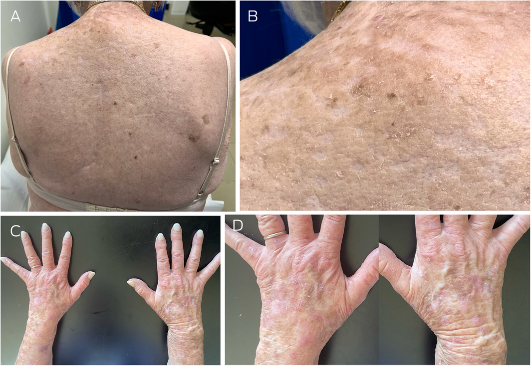
Her case was re‐evaluated, and detailed clinical examination at this time revealed yellowish, hyperkeratotic plaques diffusely distributed across the upper back, in addition to extensively photodamaged skin with multiple actinic keratoses (Box 1; A–D).
She underwent arsenical blood and urine testing, which found a serum arsenic level of 0.182μmol/L (reference interval [RI], <0.160μmol/L) and urine arsenic 3564nmol/24h (RI, <470nmol/24h). Toxicologist consultation was sought and the opinion was that her levels of serum and urine arsenic were inappropriately high after suspected childhood exposure and that her levels should have been corrected over the years. History was revisited and she denied evidence to indicate other sources of arsenic such as occupational exposure or significant ingestion of possible contaminated seafood, groundwater or well water, or assorted herbal medications. Further history revealed that in her 35‐pack‐year smoking history, she was predominantly partaking in self‐rolled cigarettes with overseas’ sourced tobacco.
A trial of smoking cessation for a week and a repeat arsenical urine test documented near‐complete resolution of her arsenic levels. The relationship between the history of current chronic cigarette use and the correction of arsenic laboratory tests after the period of smoking cessation supported the diagnosis of cigarette‐induced arsenical keratosis.
Discussion
Arsenical keratosis is a pre‐cancerous dermatosis that manifests due to chronic arsenic toxicity, which is most commonly attributed to environmental, medicinal and occupational exposure.1 Arsenic ingestion, in higher levels and repetitive doses, accumulates within the bodily tissues. It is a well documented human carcinogen disrupting the cellular pathway activity of DNA synthesis and repair.2 Cutaneous manifestations of arsenic exposure include keratinocyte cancers, skin pigmentation changes such as raindrop hypopigmentation, hyperkeratosis, transverse band‐like deposits along the nail bed (Mees lines), and deposits within the hair.2,3 Thus, a prudent and thorough history is imperative to identify the source of arsenic exposure (Box 2), and immediate cessation is necessary to prevent the development of dermatological malignancies. In this case, we report a patient with proven arsenic exposure, induced by chronic smoking, resulting in arsenical keratosis and keratinocyte cancer.
The health risks of smoking are well established, particularly lung, bladder and kidney carcinomas, as well as cardiovascular diseases and stroke.5 However, arsenic exposure secondary to smoking may be an overlooked association, as it is challenging to identify on history. This case highlights arsenic exposure as a source of morbidity from smoking. In particular, arsenic exposure is associated with the development of recurrent dermatological malignancies as a result of toxicity from gradual arsenic deposition in bodily tissues. Arsenic is a known carcinogenic element in tobacco, and this case highlights the link between skin malignancies and smoking, emphasising the role of the clinician in counselling for smoking cessation.2
Since 2016, Australia has ceased the manufacturing of all tobacco products, and cigarette supply has been sought from overseas, particularly from the United States and the United Kingdom.6 The average factory‐made cigarette has a mean concentration of 0.3μg of arsenic per gram of tobacco.7 In addition, roll‐your‐own cigarettes, despite being an attractive alternative due to its cheaper purchase price, are at least as harmful as manufactured cigarettes, with often higher amounts of toxic metal additives.8,9 As such, overall tobacco consumption poses a significant arsenic risk, but roll‐your‐own cigarettes pose an increased risk factor for arsenic exposure because the tobacco content can be increased per cigarette as per individual preference.
In summary, chronic heavy smoking may be a significant risk factor for skin cancer from arsenic exposure. Therefore, clinicians should be vigilant for possible arsenic exposure through tobacco products in patients with multiple keratinocyte cancers without associated risk factors. In these cases, it is prudent to consider urinary and serological arsenic testing to exclude arsenic exposure. Smoking cessation in these patients can reduce disease load and aid in the prevention of further keratinocyte cancer development.
Lessons from practice
- A detailed smoking history, and patient education to encourage smoking cessation, plays a key role in the assessment and management of keratinocyte cancers.
- Clinical concern for arsenic exposure should be raised in patients with multiple keratinocyte cancers without associated risk factors.
- Patients with suspicion of arsenic exposure should undergo baseline serum and urine arsenic levels and require regular full skin examinations.
Box 1 – (A) Arsenical keratosis to upper back, as demonstrated by yellowish, hyperkeratotic plaques. (B) Magnified image of panel A displaying arsenical hyperkeratosis. (C) Multiple actinic keratoses to dorsal aspect of hands and forearms. (D) Magnified image of panel C to visualise multiple actinic keratoses
Box 2 – Common culprits of arsenic exposure1,4
|
Categorical source of exposure |
Common examples |
||||||||||||||
|
|
|||||||||||||||
|
Dietary ingestion |
|
||||||||||||||
|
Occupational |
|
||||||||||||||
|
Medicinal |
|
||||||||||||||
|
|
|||||||||||||||
|
|
|||||||||||||||
Competing interests
No relevant disclosures.
References
- Austin Health. Austin Clinical Toxicology Service Guideline: arsenic, 2021. https://www.austin.org.au/Assets/Files/Arsenic_Sept2021%20FINAL.pdf (viewed Apr 2022).
- Ratnaike R. Acute and chronic arsenic toxicity. Postgrad Med J 2003; 79: 391‐396.
- Ngan V. Chronic arsenic poisoning [website]. DermNet, 2021. https://dermnetnz.org/topics/chronic‐arsenic‐poisoning (viewed Aug 2022).
- World Health Organization. Arsenic [fact sheet]. https://www.who.int/news‐room/fact‐sheets/detail/arsenic (viewed Apr 2022).
- Australian Government, Department of Health and Aged Care. What are the effects of smoking and tobacco [website]. https://www.health.gov.au/health‐topics/smoking‐and‐tobacco/about‐smoking‐and‐tobacco/what‐are‐the‐effects‐of‐smoking‐and‐tobacco (viewed Aug 2022).
- Tobacco in Australia. The manufacturing and wholesaling industry in Australia — major international companies [website]. https://www.tobaccoinaustralia.org.au/chapter‐10‐tobacco‐industry/10‐3‐the‐manufacturing‐and‐wholesaling‐industry‐in‐australia (viewed Aug 2022).
- Fresquez MR, Pappas RS, Watson CH. Establishment of toxic metal reference range in tobacco from US cigarettes. J Anal Toxicol 2013; 37: 298‐304.
- Edwards R. Roll your own cigarettes are less natural and at least as harmful as factory rolled tobacco. BMJ 2014; 348: f7616.
- Koszowski B, Rosenberry ZR, Viray LC, et al. Make your own cigarettes: toxicant exposure, smoking topography, and subjective effects. Cancer Epidemiol Biomarkers Prev 2014; 23: 1793‐1803.
Provenance: Not commissioned; externally peer reviewed.
Designing Housing to Reduce Overcrowding-Related Harms: Rheumatic Heart Disease as the Canary in the Coal Mine
Simon Quilty, Veronica Matthews, Angus Baumann, James Marangou, Bo Remenyi, Gavin Wheaton, Serena Morton Nabanunga, Norman Frank Jupurrurla, Simon Robinson, Steve Mintern, Cary Duffield, Joshua R. Francis, Paul C. Memmott
Australian Climate Leadership in 2026: COP-Out or Step-Up for Health?
Catherine G. A. Pendrey, Angie Bone, Aletha Ward, Francis Nona, Michelle Isles, Paul M. Kelly, Nicholas J. Talley
The impact of the Breast Screen NSW transition from film to digital mammography, 2002–2016: a linked population health data analysis
Rachel Farber, Nehmat Houssami, Katy J L Bell
Shifting focus to adolescent wellbeing and inclusive participation in the digital age
Allyson R Todd, Elena Wang, Stephanie R Partridge
The CURE Asthma roadmap
Gary P Anderson, Anthony Flynn, Phil G Bardin, John D Blakey, Shyamali C Dharmage, Paul Foster, Peter G Gibson, Adam Jaffe, Alan James, Christine R Jenkins, Sundram Sivamalai, Peter D Sly, Guy B Marks, Vanessa M McDonald, Judy Wetttenhall
Protecting public interest journalism as a public health good
Bronwyn Fredericks, Neha Lalchandani, Melissa A Sweet, Alex Cramb, Carmel Williams