




Medical education
Lessons from practice
Volume 217 - Issue 4
Microangiopathic haemolytic anaemia: a rare first presentation of lung cancer
Authors: Nicholas M Stacey and Martin Feddersen
Med J Aust 2022; 217 (4): 183-185. || doi: 10.5694/mja2.51659
Published online: 15 August 2022
Published online: 15 August 2022
A 76-year-old woman with no significant medical history presented to hospital after 3 days of vomiting
Clinical record
A 76‐year‐old woman with no significant medical history presented to hospital after 3days of vomiting. There were no concerning features to her vomiting, no diarrhoea, no abdominal pain or distension, no fevers, and no recent weight loss. She was an ex‐smoker, took no regular medications, and was the full‐time carer for her husband. Her physical examination was unremarkable.
Initial blood tests showed a creatinine concentration of 693μmol/L (reference interval [RI], 63–97μmol/L), a platelet count of 46×109/L (RI, 150–400×109/L) and a haemoglobin level of 104g/L (RI, 115–165g/L). An urgent diagnostic workup was commenced to determine the cause of her renal failure, thrombocytopenia and anaemia (Box 1).
A coagulopathy screen showed a fibrinogen level of 4.5g/L (RI, 2–4g/L), a D‐dimer level>20mg/L (RI, <0.5mg/L), and a normal international normalised ratio. This ruled out a coagulopathic cause but was suspicious for venous thromboembolism. Her haemolytic screen was consistent with haemolysis, showing a lactate dehydrogenase level of 2407U/L (RI, 129–250U/L), a haptoglobin level<0.1g/L (range, 0.5–2.2), a reticulocyte count of 71×109/L (RI, 10–100×109/L), and a peripheral blood film with schistocytes and spherocytes (Box 2). Her direct antiglobulin test result was negative, ruling out immune haemolysis and confirming microangiopathic haemolytic anaemia (MAHA).
Key differentials included thrombotic thrombocytopenic purpura (TTP), haemolytic uraemic syndrome (HUS) and complement‐mediated (atypical) HUS. She was admitted to the intensive care unit and treated provisionally for TTP with plasmapheresis, steroids and haemodialysis. Her ADAMTS‐13 level was 59% (RI, 60–130%), ruling out TTP. Her stool sample was negative for Shiga toxin and she had normal complement C3/C4 levels.
A computed tomography pulmonary angiogram was ordered to investigate her raised D‐dimer level and the possibility of underlying malignancy. It showed multiple pulmonary emboli, which were treated with a heparin infusion owing to her renal failure, and a right upper lobe mass suspicious for malignancy (Box 3, A). A follow‐up positron emission tomography scan confirmed fluorodeoxyglucose‐avid lesions in the right lung, bilateral iliac bones, and rectum (Box 3, B). Tissue biopsy samples of her lung and iliac lesions were taken. The microscopic appearance, molecular testing and immunohistochemistry results in both samples were consistent with a diagnosis of primary lung adenocarcinoma (Box 4). A colonoscopy showed rectal polyps with benign appearance (biopsy was not performed because of her bleeding risk).
Acutely, she responded well to plasmapheresis and haemodialysis. Once stabilised, she began rapid treatment with combination chemotherapy and immunotherapy of carboplatin, pemetrexed and pembrolizumab, to which she responded extremely well. She recently completed palliative radiotherapy and at the time of writing was functioning fully independently at home while receiving maintenance palliative chemotherapy.
Discussion
Thrombotic microangiopathies (TMAs) are a group of conditions that cause endothelial damage and microthrombi deposition in arterioles and capillaries. This causes physical shearing of red blood cells and increases platelet activation, resulting in a MAHA characterised by schistocytes on peripheral blood film, consumptive thrombocytopenia, and end‐organ damage such as renal failure or neurological dysfunction.1,2 The underlying cause of the TMA needs to be urgently differentiated to guide treatment, with primary causes first being considered:- ▪ TTP: a deficiency in ADAMTS‐13 leads to abnormal von Willebrand factor clearance, ultimately leading to aggregation of platelet von Willebrand factor complexes in microvasculature.2
- ▪ HUS: infection with Shiga toxin‐producing bacteria causes endothelial damage and platelet aggregation in microvasculature.2
- ▪ Complement‐mediated TMA: uncontrolled complement activation in microvasculature causes platelet aggregation and endothelial damage.2
However, as this case demonstrates, TMAs can also be secondary to systemic conditions such as malignant hypertension, infection or malignancy, and these must also be excluded.3,4 Malignancy‐associated TMA is well described in the literature, almost always in patients with a known active or past malignancy.3,4 It is extremely rare for a patient with no history of malignancy to present with a malignancy‐associated TMA, with only a handful of documented cases of lung adenocarcinoma presenting in this way.5,6
All TMAs require supportive care, often including haemodialysis. In addition, TTP requires emergency plasmapheresis and immunosuppression to correct abnormal von Willebrand factor and ADAMTS‐13 profiles, while complement‐mediated TMA also requires specific anti‐complement agents.2 While it is reasonable to start plasmapheresis on suspicion of TTP (particularly with severe end‐organ injury present), investigation for an underlying malignancy should be a priority. Malignancy‐associated TMA has a poor prognosis, but rapid identification does provide the opportunity for specific antineoplastic treatment which may reverse advanced neoplastic disease if started quickly.3,6
TMAs are life‐threatening and have a dismal prognosis if emergency therapy is not instituted rapidly. Alongside emergency supportive management, determining the underlying cause is critically important to guide correct treatment. While malignancy presenting as MAHA is rare, it should be part of the initial diagnostic workup to enable potentially life‐saving chemotherapy to commence as quickly as possible.
Lessons from practice
- • In any patient with suspected thrombotic microangiopathy, supportive care (including haemodialysis if required) must be started as soon as possible.
- • Urgent differentiation of the cause is a priority, as it guides further emergency treatment. Primary causes, including thrombotic thrombocytopenic purpura, haemolytic uraemic syndrome and complement‐mediated thrombotic microangiopathy, must be ruled out.
- • Malignancy can present with microangiopathic haemolytic anaemia, which requires radically different treatment, and must be considered in the differentials and investigated with imaging if suspected, to enable potentially life‐saving chemotherapy to commence.
Box 1 – Flow chart of diagnostic workup and differential diagnoses
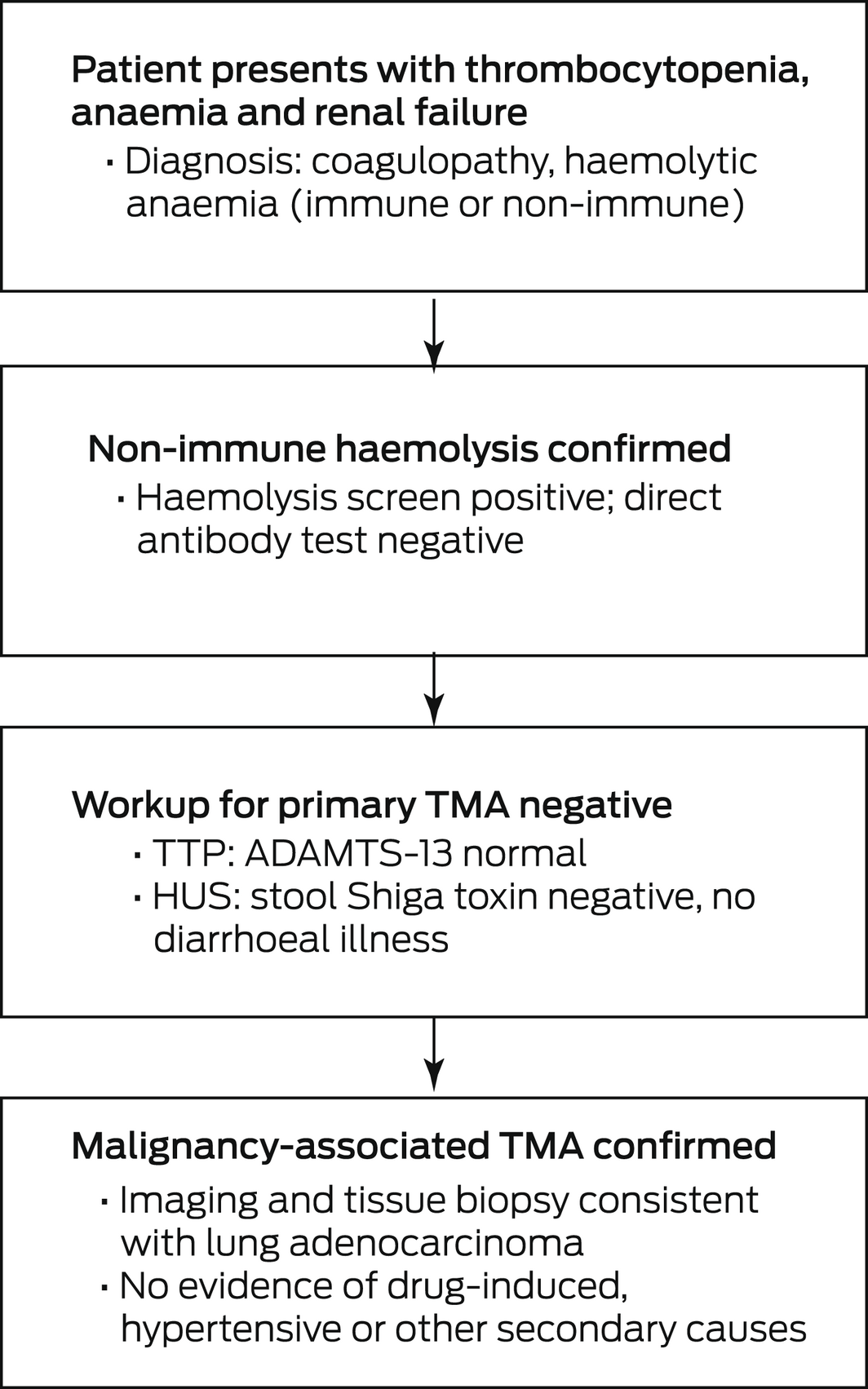
HUS = haemolytic uraemic syndrome; TMA = thrombotic microangiopathy; TTP = thrombotic thrombocytopenic purpura.
Box 2 – Blood film from initial presentation showing schistocytes (blue arrow) and spherocytes (red arrow)
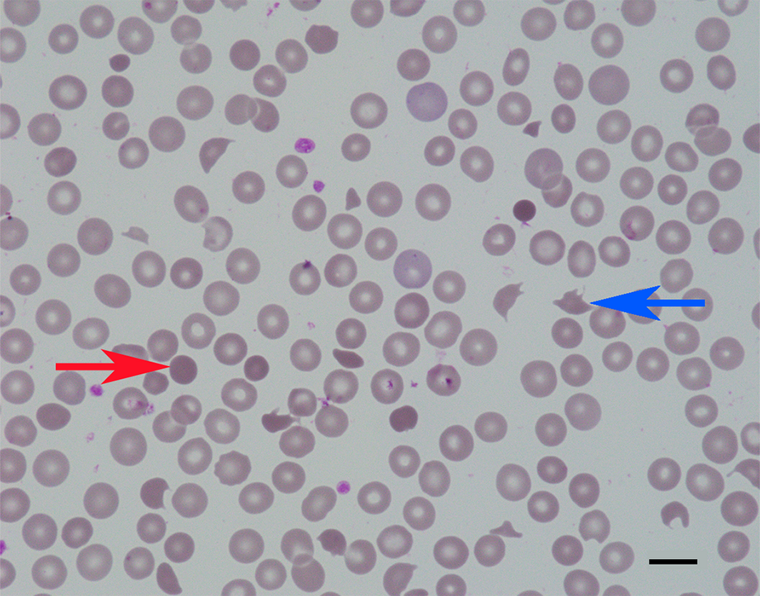
Magnification 1000×, scale bar 10μm.
Box 3 – CT pulmonary angiogram and full body PET scan
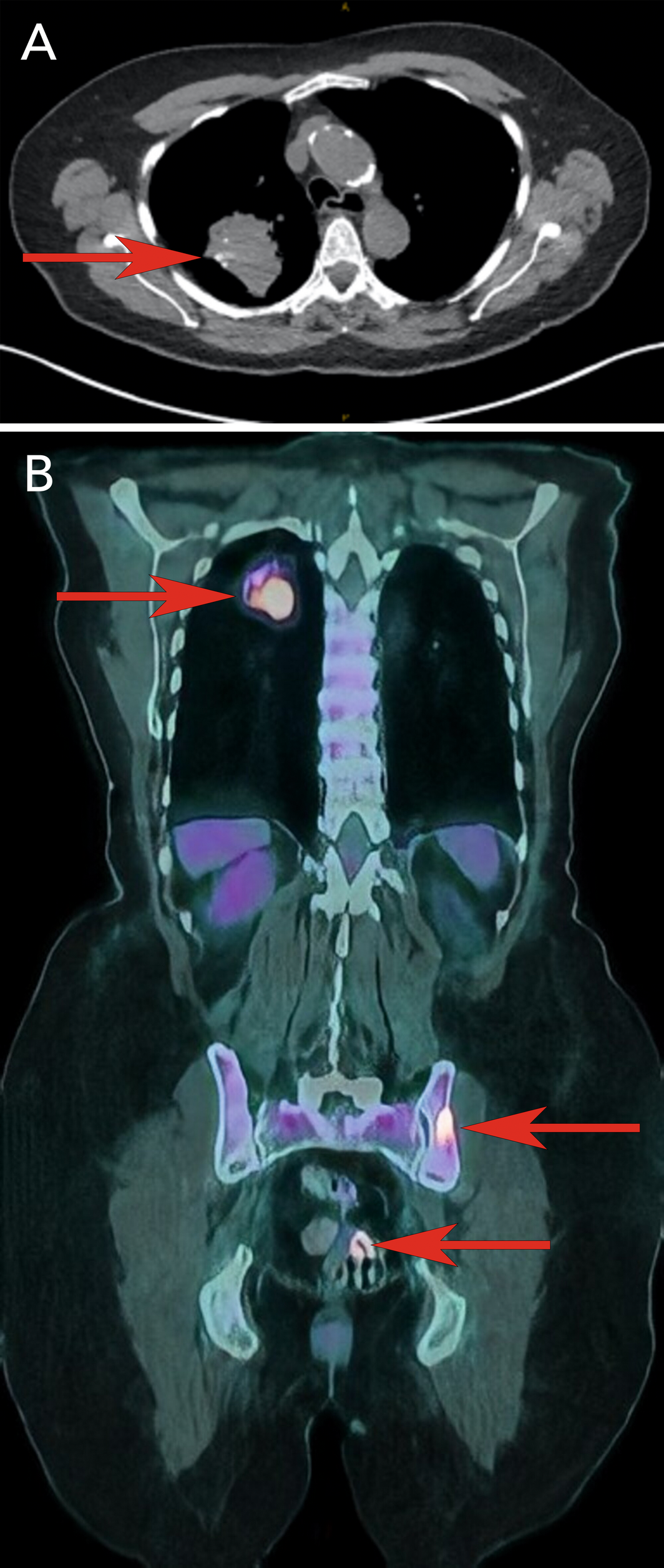
CT = computed tomography; PET = positron emission tomography.
A: CT pulmonary angiogram showing a large right upper lobe lung mass (arrows); it also identified multiple non‐occlusive pulmonary emboli and a second nodule in the right lower lobe (not visible in this slice). B: Full body PET scan showing fluorodeoxyglucose‐avid lesions suspicious for malignancy in the right lung, iliac bones and upper rectum (arrows).
Box 4 – Biopsy samples of lung and iliac bone
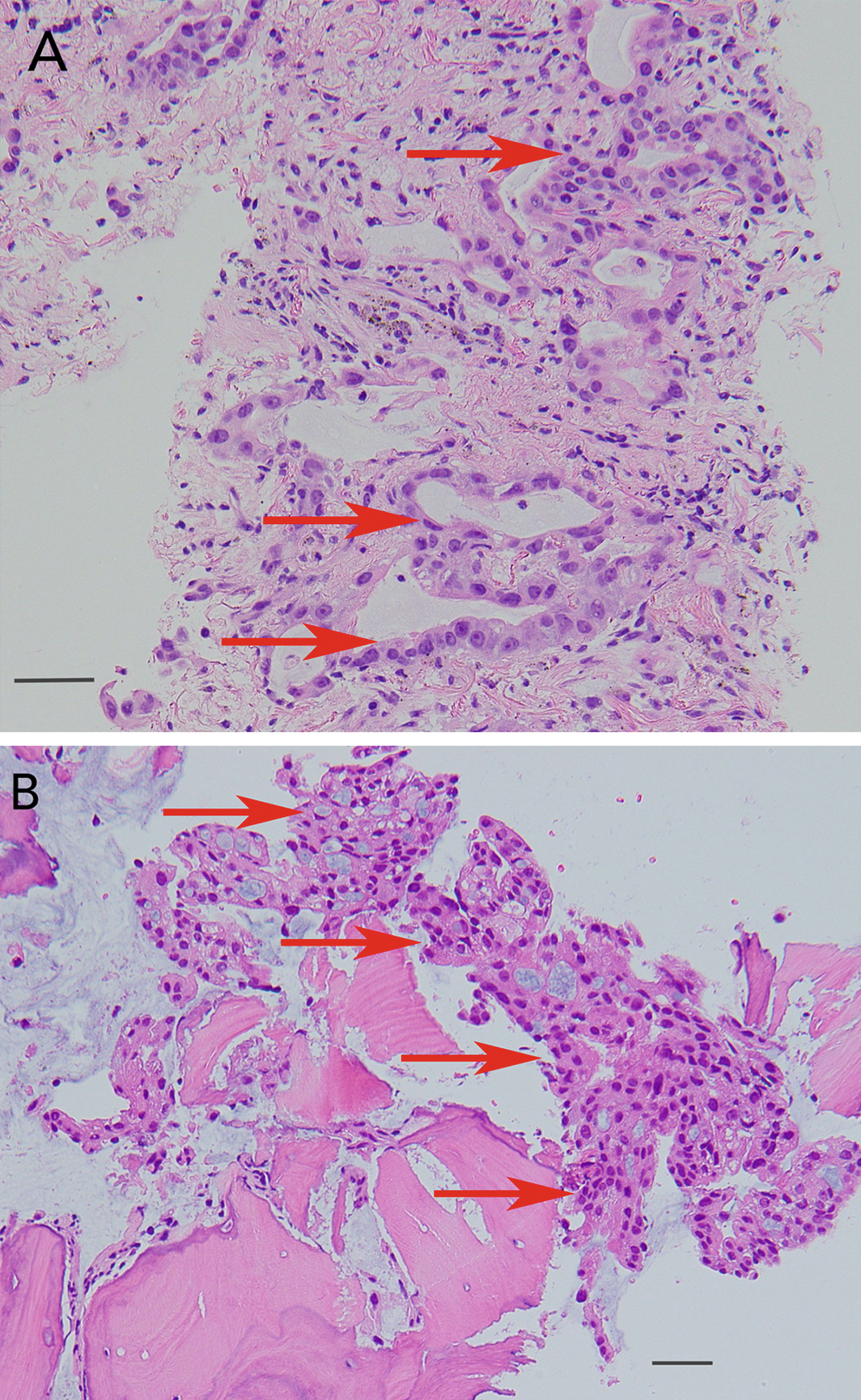
A: Lung mass biopsy showing infiltrating neoplastic tissue consisting of large cells arranged in glands and cords (arrows) (magnification 200×, scale bar 50μm). B: Iliac bone biopsy showing heavy infiltration by metastatic adenocarcinoma, which has formed complex glands (arrows) (magnification 200×, scale bar 50μm).
Competing interests
No relevant disclosures.
Acknowledgements
References
- Winters JL. Plasma exchange in thrombotic microangiopathies (TMAs) other than thrombotic thrombocytopenic purpura (TTP). Hematology Am Soc Hematol Educ Program 2017; (1): 632‐638.
- George JN, Nester CM. Syndromes of thrombotic microangiopathy. N Engl J Med 2014; 371: 654‐666.
- Elliott MA, Letendre L, Gastineau DA, et al. Cancer‐associated microangiopathic hemolytic anemia with thrombocytopenia: An important diagnostic consideration. Eur J Haematol 2010; 85: 43‐50.
- Bayer G, Von Tokarski F, Thoreau B, et al. Etiology and outcomes of thrombotic microangiopathies. Clin J Am Soc Nephrol 2019; 14: 557‐566.
- Dirweesh A, Siddiqui W, Khan M, et al. Adenocarcinoma of the lung presenting as thrombotic thrombocytopenic purpura. Respir Med Case Reports 2017; 21: 82‐83.
- Francis KK, Kalyanam N, Terrell DR, et al. Disseminated malignancy misdiagnosed as thrombotic thrombocytopenic purpura: a report of 10 patients and a systematic review of published cases. Oncologist 2007; 12: 11‐19.
Provenance: Not commissioned; externally peer reviewed.
Splenic injury in severe cases of the zoonoses Q fever and rickettsial infection: diagnostic challenges
Ashleigh Drury, Philippa Harrison, Aiveen Bannan
Health and economic benefits of improving pre‐hospital identification of stroke in Australian women: a modelling study
Thomas Gadsden, Lei Si, Emily R Atkins, Cheryl Carcel, Xia Wang, Stephen Jan, Mark Woodward, Laura E Downey
Updating the diagnosis and management of elevated serum ferritin levels in the era of routine ferritin testing of blood donors by Australian Red Cross Lifeblood
Gary D Zhang, James Chen, Daniel M Johnstone, Martin B Delatycki, Katie Allen, John K Olynyk
Early cardiovascular collapse after envenoming by snakes in Australia, 2005–2020: an observational study (ASP‐31)
Geoffrey K Isbister, Katherine Z Isoardi, Angela L Chiew, Shane Jenkins, Nicholas A Buckley
Updated recommendations for warfarin reversal in the setting of four‐factor prothrombin complex concentrate
Danielle Robinson, James McFadyen, Eileen Merriman, Chee Wee Tan, Ross Baker, Huyen Tran
Updating the diagnosis and management of iron deficiency in the era of routine ferritin testing of blood donors by Australian Red Cross Lifeblood
Gary D Zhang, Daniel Johnstone, Michael F Leahy, John K Olynyk