




Medical education
Lessons from practice
Volume 216 - Issue 7
Lung cancer in non‐smokers: a diagnosis of increasing importance
Authors: Samantha Dean, Rachel Lennox, Clare Senko and Sagun Parakh
Med J Aust 2022; 216 (7): 342-343. || doi: 10.5694/mja2.51471
Published online: 18 April 2022
Published online: 18 April 2022
A previously well 32-year-old Caucasian man with no history of smoking presented with a 6-week history of progressive exertional dyspnoea, a productive cough, and night sweats
Clinical record
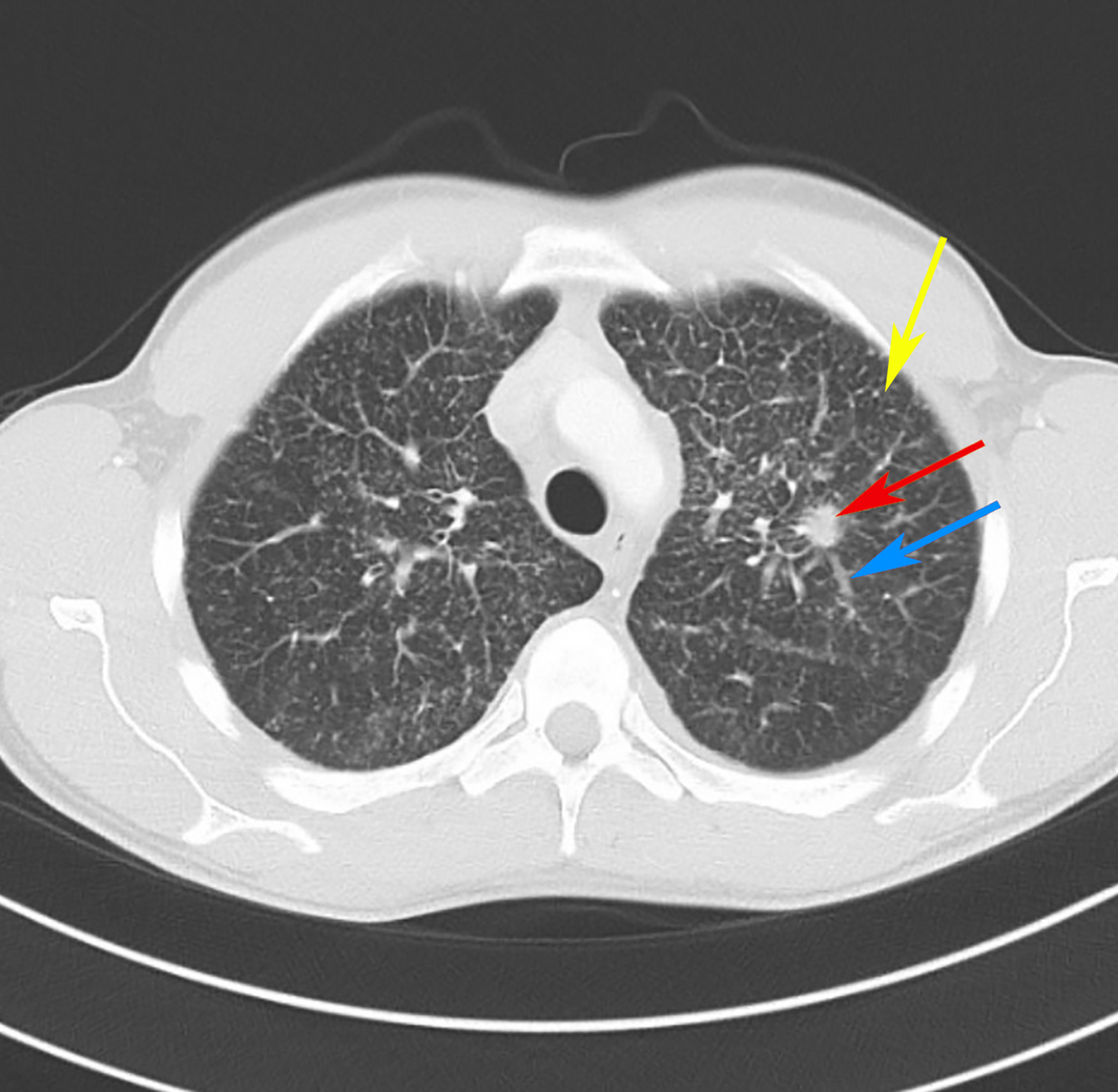
A previously well 32‐year‐old Caucasian man with no history of smoking presented to his general practitioner with a 6‐week history of progressive exertional dyspnoea, a productive cough, and night sweats. There was no prior history of dust or small particle exposure and no family history of cancer or lung disease. A chest x‐ray showed diffuse severe reticulation in both lungs. Despite a course of oral antibiotics, the patient’s symptoms worsened, prompting a computed tomography (CT) scan of the chest. The CT scan demonstrated extensive interlobular septal thickening, centrilobular nodules bilaterally, and a 15 mm spiculated density in the apex of the left upper lobe, most consistent with interstitial lung disease (Box). A bronchoscopy revealed a biopsy‐confirmed primary lung adenocarcinoma. Molecular testing of epidermal growth factor receptor (EGFR) mutations by Idylla EGFR Mutation Test (Biocartis), and programmed death‐ligand 1 (PD‐L1), ALK and ROS1 rearrangement by immunohistochemistry were negative. Further molecular testing by next generation sequencing could not be performed due to insufficient tissue. While awaiting his oncology appointment, the patient deteriorated and presented to the emergency department with type 1 respiratory failure. Laboratory tests revealed mild thrombocytopenia (94 × 109/L; reference interval, 150–300 × 109/L), a normal autoimmune screen, and negative human immunodeficiency virus (HIV) serology. A concurrent infective process was unlikely given that the recent bronchoscopy did not culture any organisms and that the acid‐fast bacilli smear, extended respiratory viral culture and coronavirus disease 2019 (COVID‐19) nucleic acid tests were negative. A CT pulmonary angiogram did not identify any pulmonary emboli; however, it demonstrated worsening ground‐glass changes and progressive interlobular thickening. The patient was transferred to the intensive care unit and given the severity of his symptoms was commenced on intravenous antibiotics to cover for potential underlying infection and hydrocortisone. A transthoracic echocardiogram demonstrated severe pre‐capillary pulmonary hypertension, with an estimated pulmonary artery systolic pressure of 61 mmHg and a dilated right ventricle with severe systolic dysfunction prompting commencement of sildenafil 25 mg orally every 8 hours. A brain CT scan and a whole body fluorodeoxyglucose (FDG)‐positron emission tomography (PET) scan demonstrated a left pre‐central brain metastasis with diffuse FDG uptake throughout both lungs in keeping with pulmonary metastases with no discrete FDG‐avid primary pulmonary lesion and extensive bony metastatic disease. A repeat biopsy to complete further molecular testing was not performed due to the severity of the patient’s respiratory failure. On day 5 of his admission, the patient was commenced on chemotherapy with carboplatin and pemetrexed. The patient’s condition continued to decline with increasing oxygen requirements, acute anaemia, worsening thrombocytopenia and acute kidney injury. Blood film and coagulation studies showed findings consistent with acute disseminated intravascular coagulation and microangiopathic haemolytic anaemia. The patient rapidly deteriorated and died 7 days after his hospital admission.
Discussion
Approximately 12% of patients with lung cancer in the United States are never‐smokers, with the incidence being highest in this population among those aged 20–49 years.1 While the overall incidence for lung cancer has continued to decline over the past decade, a notable increase has been observed in adults with no history of smoking. These patients are often diagnosed with advanced and aggressive cases of non‐small cell lung cancer (NSCLC), with distinct clinicopathological and molecular features and a higher incidence of targetable mutations compared with lung cancer seen in smokers.2 NSCLC can have diverse clinical and radiological presentations, including masquerading as interstitial lung disease,3 as seen in our patient. Lung cancer screening with low dose chest CT scan has shown to significantly reduce lung cancer mortality in patients aged 55–74 years with a smoking history of at least 30 pack‐years, who are current smokers or have quit smoking in the past 15 years, based on the National Lung Screening Trial.4 There are currently no guidelines to recommend lung cancer screening in non‐smokers, as the feasibility and effectiveness of these programs are still uncertain. The challenge lies in identifying never‐smokers with risk factors for lung cancer who would benefit from screening.5 It is important for general practitioners and general physicians to be aware that NSCLC may present in a diverse manner and, importantly, to consider a differential of NSCLC in young, non‐smoking patients who present with acute and/or atypical respiratory symptoms. Chest CT imaging and diagnostic testing should be sought early to avoid delays in diagnosis and to initiate appropriate management. With the availability of effective targeted therapies, patients with lung cancer with a light‐ or never‐smoking history should have comprehensive molecular tissue testing performed on their tumour biopsy to identify potential driver mutations.
Lessons from practice
- Despite the incidence for lung cancer declining over the past decade, a notable increase has been observed in adults with no history of smoking.
- There are currently no guidelines to recommend lung cancer screening in non‐smokers and in younger patients in particular.
- It is important to be aware that non‐small cell lung cancer may present in a diverse manner and to consider it as a differential in non‐smoking patients who present with acute and/or atypical respiratory symptoms.
- With the availability of effective targeted therapies, patients with lung cancer with a light‐ or never‐smoking history should have comprehensive molecular tissue testing to identify potential driver mutations.
Competing interests
No relevant disclosures.
References
- Siegel DA, Fedewa SA, Henley SJ, et al. Proportion of never smokers among men and women with lung cancer in 7 US states. JAMA Oncol 2021; 7: 302–304.
- Dubin S, Griffin D. Lung cancer in non‐smokers. Mo Med 2020; 117: 375–379.
- Pathak V, Rendon ISH. Pulmonary adenocarcinoma masquerading as diffuse inflammatory interstitial lung disease. Respir Med CME 2011; 4: 67–69.
- National Lung Screening Trial Research Team; Aberle DR, Adams AM, Berg CD, et al. Reduced lung‐cancer mortality with low‐dose computed tomographic screening. N Engl J Med 2011; 365: 395–409.
- Kerpel‐Fronius A, Tammemägi M, Cavic M, et al. Screening for lung cancer in individuals who never smoked: an International Association for the Study of Lung Cancer Early Detection and Screening Committee report. J Thorac Oncol 2021; 17: 56–66.
Linked content
-
MJA Perspective: Lung cancer: progress with prognosis and the changing state of play
-
MJA Podcast: Professor Fraser Brims
-
InSight+: Lung cancer: pay attention to never smokers
Provenance: Not commissioned; externally peer reviewed.
Sovereignty and Survival: Understanding Lung Cancer Outcomes for Aboriginal and Torres Strait Islander People in Australia, a Case–Control Study
Alice R. T. Bergin, Luc te Marvelde, Roger L. Milne, Benjamin J. Solomon, Kris Ivanova, Peter Savas, Stephen J. Luen, Jay Hamann, Andrea Casey, Nicole Watt, Sue M. Evans, Gail Garvey, Sherene Loi
The Changing Indications for Adult Liver Transplantation in Australia: A Narrative Review
Fadak Mohammadi, Alan J. Wigg, Ken Liu, Simone I. Strasser
Uneven Ground: Survival Differences Among Victorian Lung Cancer Patients by Location of Residence (2011–2023): A Retrospective Cohort Study
Evangeline Samuel, Eldho Paul, Mike Lloyd, Sanuki Tissera, Craig Underhill, Sagun Parakh, Phillip Parente, Inger Olesen, Javier Torres, Katharine See, Gavin M. Wright, David Langton, Thomas John, Matthew Conron, James Bartlett, Nicola Atkin, Nikolajs Zeps, Susan V. Harden, Wasek Faisal, John R. Zalcberg, Rob G. Stirling
Early-Onset Colorectal Cancer With Liver-Only Metastases: A Retrospective Cohort Study Integrating Prospectively Collected Real-World Clinical and Molecular Data From an Australian National Database (2009–2024) to Guide Treatment Planning
Savio G. Barreto, Christos S. Karapetis, Shahid Ullah, Matthew Burge, Susan Caird, Angus Campbell, Azim Jalali, Ross Jennens, Muhammad A. Khattak, Belinda Lee, Stephanie H. Lim, Shehara Mendis, Louise Nott, Timothy J. Price, Jeremy D. Shapiro, Jeanne Tie, Javier Torres, Colin Williams, Rachel Wong, Vanessa Wong, Peter Gibbs
Australian Pathways for Specialist Pain Management and Early Palliative Care for People With Pancreatic Cancer: Developed Using a Community Consensus Approach
Jennifer Philip, Melanie R. Lovell, Kylee Bellingham, Gail Garvey, Gregory B. Crawford, Nicole M. Rankin, Kara Burns, Isabel Young, Vivienne Milch, Dorothy Keefe, Katrina Anderson, James Lawson, Meinir Krishnasamy
Program Guidelines for the National Lung Cancer Screening Program: Targeted Lung Cancer Screening in High-Risk Individuals in Australia
Nicole M. Rankin, Rebecca Zosel, Lisa J. Whop, Raglan Maddox, Annette McWilliams, Miranda Siemienowicz, Jon Emery, Maria A. R. Lantin, Georgia Bartlett, Mikayla Wolfe, Abbey Diaz, Katrina Anderson, Lillian Liu, Cindy Toms, Sarah McDermott, Peter Bligh, Jeremy Chalke, Stephen Melsom, Claire E. Nightingale, Alison Brown, Sam Pope, Julia Brotherton, Anne Fidler, Michel Itel, Mark Brooke, Diane M. Pascoe, Fraser Brims, Tracy L. Leong, Emily Stone, Dorothy Keefe, Vivienne Milch