




Medical education
Lessons from practice
Volume 216 - Issue 7
Abdominal discomfort leading to the incidental finding of complete heart block
Authors: Angus AW Baumann, Nicholas J Montarello and Glenn D Young
Med J Aust 2022; 216 (7): 340-342. || doi: 10.5694/mja2.51469
Published online: 18 April 2022
Published online: 18 April 2022
A 62-year-old man presented complaining of 3 days of abdominal discomfort on a background of a month-long history of fatigue and fevers
Case record
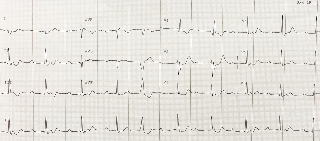
A 62‐year‐old man presented to hospital complaining of 3 days of abdominal discomfort on a background of a month‐long history of fatigue and fevers. He was a past smoker of 12 years and did not use illicit drugs. His medical history was otherwise unremarkable. His abdominal discomfort was epigastric, constant, non‐radiating and unaffected by food. There was no associated change in bowel habit. In the emergency department, he was afebrile (37.2°C) but looked unwell. His heart rate was 30 beats per minute, blood pressure was 105/65 mmHg, heart sounds were normal without audible murmurs, and peripheral examination was unremarkable. Jugular venous pressure was normal. He was tender in the epigastrium without guarding or rebound tenderness. Murphy sign was negative. The liver edge was palpable but not tender or pulsatile. Electrocardiography showed complete heart block (Box 1).
A complete blood picture revealed an elevated white cell count 15.81 × 10⁹/L (reference interval [RI], 4.5–11 × 109/L). Serum biochemistry demonstrated abnormal liver function, with alkaline phosphatase 185 U/L (RI, 30–110 U/L) and γ‐glutamyl transferase 141 U/L (RI, < 60 U/L), consistent with cholestasis. Bilirubin was mildly raised at 30 μmol/L (RI, < 24 μmol/L). Arterial blood gas showed a normal pH of 7.40, bicarbonate 15 mmol/L (RI, 22–31 mmol/L) and lactate 5.7 mmol/L (RI, 0.2–2.0 mmol/L). Blood cultures were obtained.
Chest x‐ray was normal. A computed tomography (CT) scan of the abdomen revealed pericholecystic fat stranding, consistent with cholecystitis, and intravenous ceftriaxone and metronidazole were administered.
Due to active systemic infection and haemodynamic stability, permanent pacemaker implantation was deferred until his infection was controlled.
Twelve hours after presentation he developed cardiogenic shock, with transient hypotension (72/50 mmHg) and anuria despite intravenous fluid resuscitation.
Transthoracic echocardiography was performed (Box 2). Parasternal images demonstrated diffuse thickening of aortic valve leaflets with restriction, asymmetric leaflet closure, mild regurgitation and bicuspid aortic valve morphology. The aortic annulus was thickened, suggesting perivalvular abscess formation. Apical five‐chamber and subcostal views revealed colour Doppler flow from the left ventricle into the right atrium consistent with Gerbode defect.
The patient was diagnosed with infective endocarditis complicated by perivalvular abscess formation. He proceeded to emergency cardiac surgery with abscess drainage, bioprosthetic aortic valve insertion, and Gerbode defect repair using bovine pericardium. Tissue culture identified Streptococcus oralis, a Streptococcus viridans species, as the causative organism.
He received a 6‐week course of intravenous vancomycin followed by permanent pacemaker implantation and recovered fully.
Discussion
This patient had no formal risk factors for infective endocarditis elicited on history. However, he had an undiagnosed congenitally bicuspid aortic valve, a common congenital heart condition with a prevalence of 0.5–2.0%,1 conferring a significantly greater risk of infective endocarditis, with an increased relative risk of 23.1 times compared with patients with a trileaflet aortic valve.2 Patients with bicuspid aortic valve and infective endocarditis are often younger, have fewer comorbid conditions, and are more likely to require valve replacement than other patients with infective endocarditis.1,2 In cases of native valve infective endocarditis, patients with a bicuspid aortic valve are more likely to have aortic valve involvement (93% v 34%; P < 0.0001) and annular abscess formation at diagnosis (27% v 4%; P < 0.0001) than those with trileaflet aortic valve.2 Infection involving bicuspid aortic valve has lower in‐hospital mortality1 and lower mortality at one and 5 years compared with unadjusted cohorts of patients with trileaflet aortic valve infective endocarditis,2 perhaps reflecting that bicuspid aortic valve infective endocarditis is more likely due to S. viridans than Staphylococcus aureus infection.1,2
Perivalvular abscess formation can cause conduction abnormalities, including PR prolongation, bundle branch block, and complete heart block,3 and is associated with poorer prognosis. A Gerbode defect is a rare complication of perivalvular abscess resulting in communication between the left ventricle and the right atrium,4 exposing the right‐sided chambers to left ventricular systolic pressure and increasing right atrial pressure. In this case, resultant hepatic venous congestion manifested as abdominal discomfort and masqueraded as cholecystitis on liver function tests and CT imaging. Cardiogenic shock may have developed due to a progressive left to right shunt, falling cardiac output in the setting of elevated right atrial pressure, progressive sepsis, or a combination of all three. Although elevated jugular venous pressure or peripheral oedema was not noted in this case, thorough physical examination for signs of right ventricular failure as well as valvular murmurs is essential and may have prompted an earlier diagnosis.
This case demonstrates the insidious nature of subacute bacterial infective endocarditis and the significant and often unheralded risk posed to patients with bicuspid aortic valve, and highlights the need to exclude infective endocarditis in any patient presenting with complete heart block and non‐specific constitutional or infective symptoms.
Lessons from practice
- Complete heart block in patients presenting with non‐specific constitutional or infective symptoms may be due to the presence of an aortic perivalvular abscess. The diagnosis of infective endocarditis should always be considered.
- A bicuspid aortic valve is a common congenital heart abnormality that is often undiagnosed until later in life and associated with an increased risk of infective endocarditis.
- Infective endocarditis in patients with a bicuspid aortic valve is more likely to be due to Streptococcus viridans species and be complicated by a perivalvular abscess.
- A Gerbode defect is a rare complication of infective endocarditis or aortic valve surgery. It is a communication between the left ventricle and right atrium and can lead to signs and symptoms of decompensated right ventricular failure.
Box 1 – Electrocardiography showing complete heart block with a normal axis and QRS width and no ischaemic changes (a single premature ventricular complex is evident)
Box 2 – Transthoracic echocardiography showing evidence of infective endocarditis
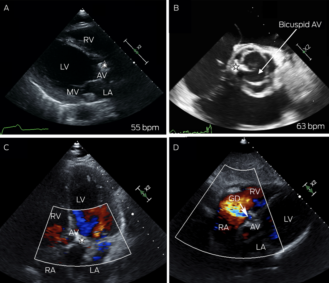
AV = aortic valve; bpm = beats per minute; GD = Gerbode defect; LA = left atrium; LV = left ventricle; MV = mitral valve; RA = right atrium; RV = right ventricle; ★ = vegetation. (A) Parasternal long axis image demonstrating thickened aortic valve leaflets (★) and an asymmetric coaptation point. (B) Parasternal short axis image showing bicuspid morphology and leaflet and annular thickening (★). (C) Apical five‐chamber image showing the aortic valve vegetation (★) and evidence of colour Doppler flow moving into the right atrium. (D) Subcostal image showing colour Doppler flow from the left ventricular outflow tract into the right atrium.
Competing interests
No relevant disclosures.
References
- Zegri‐Reiriz I, de Alarcón A, Muñoz P, et al. Infective endocarditis in patients with bicuspid aortic valve or mitral valve prolapse. J Am Coll Cardiol 2018; 71: 2731‐2740.
- Kiyota Y, Della Corte A, Montiero Vieira V, et al. Risk and outcomes of aortic valve endocarditis among patients with bicuspid and tricuspid aortic valves. Open Heart 2017; 4: e000545.
- Weisse AB, Khan MY. The relationship between new cardiac conduction defects and extension of valve infection in native valve endocarditis. Clin Cardiol 1990; 13: 337‐345.
- Saker E, Bahri GN, Montalbano MJ, et al. Gerbode defect: A comprehensive review of its history, anatomy, embryology, pathophysiology, diagnosis, and treatment. J Saudi Heart Assoc 2017; 29: 283‐292.
Provenance: Not commissioned; externally peer reviewed.
Fatty Liver Disease in Australia: A Narrative Review on the Epidemiology, Natural History, Prognostication and Management in People With Metabolic Dysfunction
Karl Vaz, Daniel Clayton-Chubb, William W. Kemp, Stuart K. Roberts, Ammar Majeed
Sex Differences in Management and Outcomes of People With ST-Elevation Myocardial Infarction, New South Wales, 2011–2020: A Retrospective Cohort Study
Adam C. Livori, Jedidiah I. Morton, Dianna J. Magliano, Jonathan E. Shaw
Rethinking Rheumatic Fever Diagnosis: Progress, Pitfalls and the Path Towards a Definitive Test
Anna P. Ralph, Rachel Webb, Tom Parks, Nicole J. Moreland, Asha C. Bowen, Benjamin Jones, Emma Ndagire, Andrea Z. Beaton
Sex Differences in Management and Outcomes of People With ST-Elevation Myocardial Infarction, New South Wales, 2011–2020: A Retrospective Cohort Study
Samia Kazi, Simone Marschner, Clara K. Chow
Distribution of Cardiovascular Disease Risk Based on the Updated 2023 Guideline-Recommended Australian Cardiovascular Disease Risk Algorithm and Comparison With the 2012 Algorithm: An Observational Study
Nina Lazarevic, Meghana Bhat, Grace Joshy, Danielle C. Butler, Mark Woodward, Anushka Patel, Rod T. Jackson, Garry Jennings, Rosemary Wyber, Ellie Paige, Emily Banks
They Brought the Hospital to Me
Hugh N. Burrill