




Medical education
Lessons from practice
Volume 216 - Issue 6
The urgency of phlegmasia cerulea dolens: management for physicians and surgeons
Authors: Mitra Rahmatzadeh, Jonathan Clarke, Joseph Jaya, Limi Lee, Sam Farah, Roger Bell and Ming Yii
Med J Aust 2022; 216 (6): 285-286. || doi: 10.5694/mja2.51458
Published online: 4 April 2022
Published online: 4 April 2022
A 45-year-old Sudanese man who was an ex-smoker with no past medical or family history presented with a painful, swollen left lower limb of uncertain duration
Clinical record
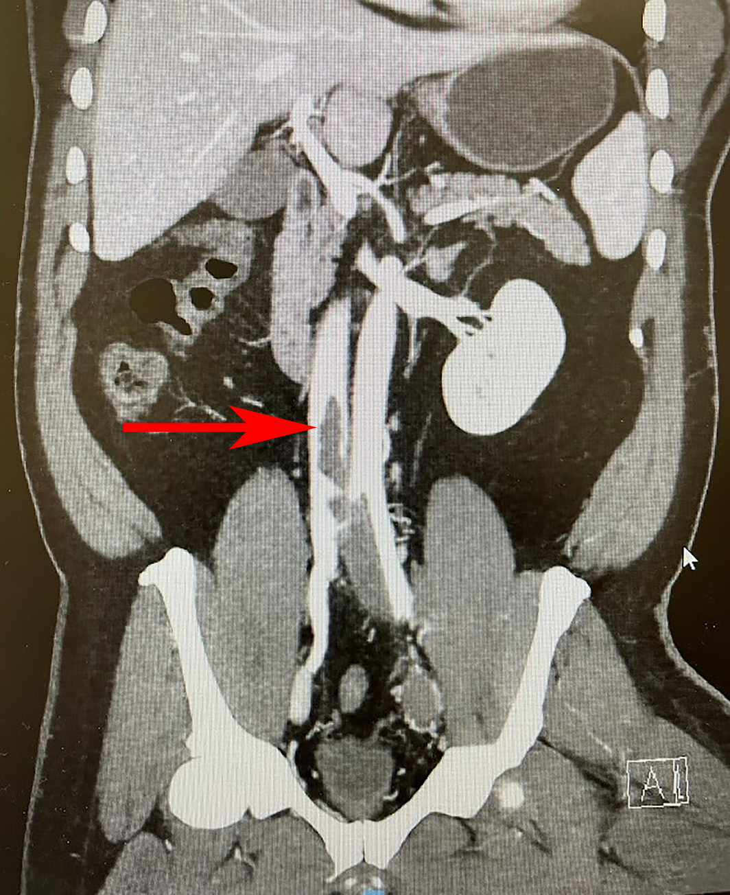
A 45‐year‐old Sudanese man who was an ex‐smoker with no past medical or family history presented to our emergency department with a painful, swollen left lower limb of uncertain duration. Due to the patient’s dark complexion, skin discolouration could not be appreciated. The patient was unable to move the lower limb and while only a femoral pulse was palpable due to severe swelling, the lower limb appeared well perfused. Venous duplex ultrasound showed thrombosis of the superficial and deep veins of the left lower limb. Computed tomography (CT) venogram confirmed proximal extension of thrombosis to the infrarenal inferior vena cava (Box 1). The patient was commenced on heparin infusion and taken to angiography, where an inferior vena cava filter was inserted. Catheter‐directed thrombolysis with alteplase was initiated via the left popliteal vein access, using a 9Fr sheath, and continued overnight in an intensive care unit. The next morning, the patient underwent suction thrombectomy using an 8Fr Penumbra device. The right common femoral vein was accessed via a 5Fr sheath and the thrombus was cleared from the left common iliac and external iliac veins. Subsequently, angioplasty and stenting of the proximal left common iliac vein to the distal external iliac vein was undertaken using three SinusRepo (Optimed) self‐expanding stents. Alteplase infusion was continued overnight, with surveillance venography the following day revealing patent stents and inferior vena cava. Fasciotomies of the left lower limb were undertaken, which at this point was swollen and tense with blister formation, concerning for acute compartment syndrome. Intraoperative appraisal of fasciotomy wounds 16 days after the initial fasciotomies was suggestive of largely non‐viable muscle in the anterior and posterior compartments without evidence of venous gangrene. A through knee amputation was proposed to the patient once the lower limb swelling had improved. Surveillance CT venogram to investigate ongoing lower limb swelling despite a period of elevation and compression revealed stenosis of the proximal left common iliac vein, which was addressed with repeat angiogram, requiring high pressure balloon angioplasty and a self‐expanding stent. Following further elevation and compression of the lower limb, a through knee amputation was performed. The amputation stump became complicated by wound dehiscence 6 days later, ultimately healing with a combination of vacuum and basic dressings. Development of heparin‐induced thrombocytopenia 6 days after left lower limb fasciotomies required transition from heparin to bivalirudin infusion, switching to an oral anticoagulant, rivaroxaban, one month later. Acute kidney injury on admission, likely secondary to rhabdomyolysis, required one month of dialysis via a Permcath catheter (Medtronic). The patient was discharged to a rehabilitation centre 8 weeks after admission.
Discussion
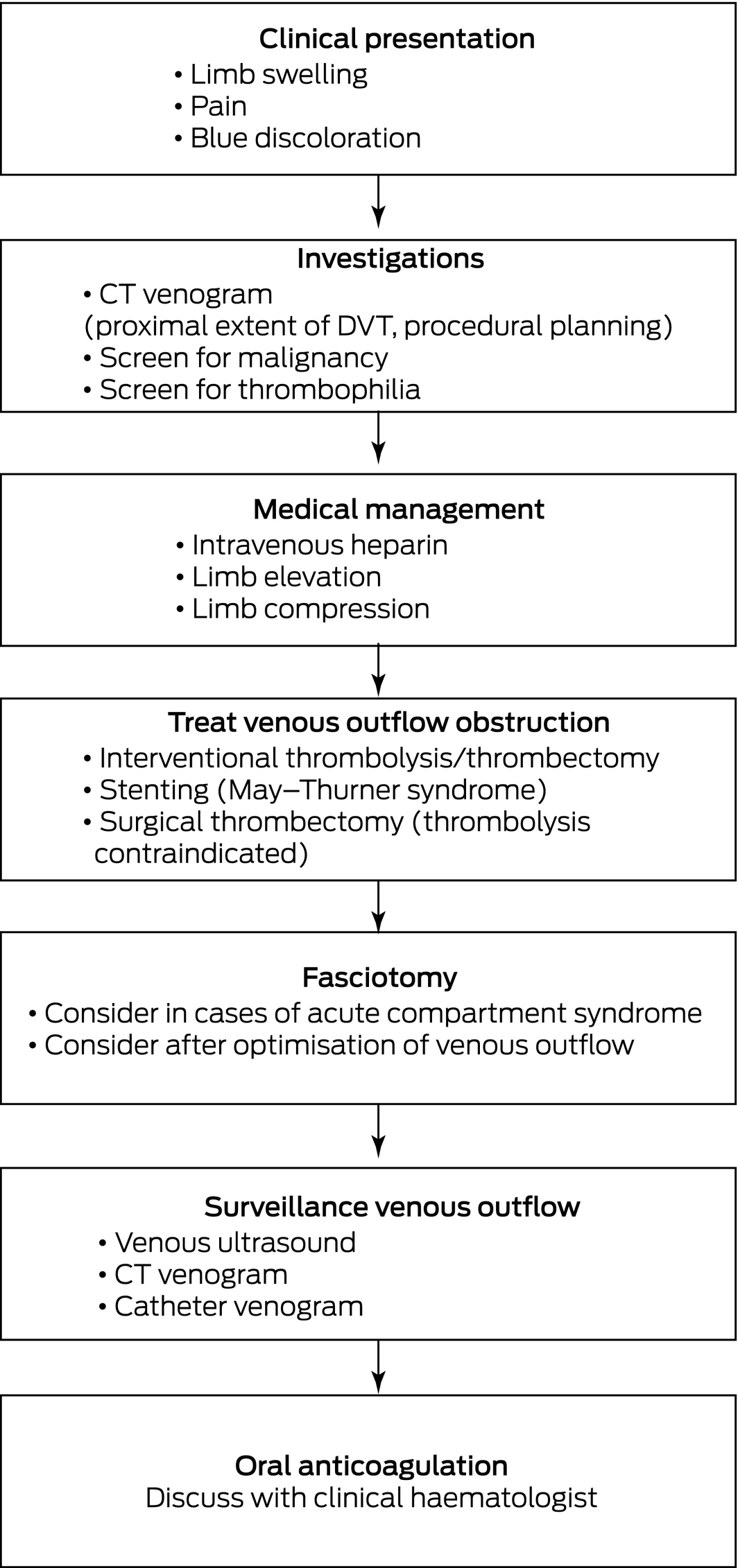
Phlegmasia cerulea dolens is an emergency.1 It is a limb‐ and life‐threatening form of thrombosis involving the deep and superficial veins of the lower limb.2 The patient presents with a painful and swollen limb that is blue discoloured; hence, the term “cerulea”, meaning blue.2 Extreme venous hypertension causes an increase in interstitial pressure in the lower limb, resulting in arrest of blood flow and tissue ischaemia.1 This venous gangrene, which is irreversible, can occur in 50% of cases, leading to limb loss.3 Fluid extravasation and loss of intravascular fluid can result in severe hypotension and shock, with a mortality rate of up to 40%.3 Clinical diagnosis of phlegmasia cerulea dolens by the physician requires prompt referral to a vascular surgery centre where logistics to treat venous outflow obstruction are available. Delay in management or presentation, potentially in the case of this patient, can lead to a poor prognosis. On presentation, an urgent CT venogram must be organised to determine the proximal extent of the deep vein thrombosis and facilitate procedural planning. Venous duplex ultrasound often provides inadequate imaging and leads to unnecessary delay in interventional planning. Bed rest, lower limb elevation and compression to reduce swelling must be instigated (Box 2). Heparin infusion must be commenced to prevent progression of the existing thrombus. In the management of phlegmasia cerulea dolens, “time is tissue”. A trial of treatment with intravenous anticoagulation alone or systemic thrombolysis has been shown to be inadequate in the treatment of the phlegmasia cerulea dolens, delaying the definitive treatment that is required.2,4 Interventional thrombolysis and thrombectomy techniques, which aim to resolve the venous outflow obstruction, may require intensive care unit admission for ongoing thrombolysis. Patients diagnosed with May–Thurner syndrome — the anatomic compression of the left common iliac vein by the right common iliac artery — must be considered for angioplasty and/or stenting of the affected segment. Surveillance scans to ensure resolution of venous outflow obstruction are important in this context. Choice of oral anticoagulation peri‐discharge should be discussed with a clinical haematologist. The timing of fasciotomy in cases of acute compartment syndrome remains ill‐defined in the context of phlegmasia cerulea dolens, but any delay in urgent treatment of venous outflow obstruction can lead to onset of venous gangrene requiring an amputation. Screening for thrombophilia and malignancy, the most common cause of phlegmasia cerulea dolens, will be crucial.
Lessons from practice
- • Phlegmasia cerulea dolens is an emergency that must be promptly recognised by both the physician and the surgeon.
- • Immediate commencement of intravenous anticoagulation on patient presentation is crucial in the prevention of thrombus propagation.
- • “Time is tissue”, and definitive intervention to resolve venous outflow obstruction is urgent.
- • Although the timing of fasciotomy for acute compartment syndrome remains ill‐defined in the context of phlegmasia cerulea dolens, delay in treatment of venous outflow obstruction can lead to onset of venous gangrene requiring amputation.
Competing interests
No relevant disclosures.
References
- Khou CTJ, Batzlaff C, Bezzant ML, Sjulin T. Phlegmasia cerulea dolens: a life‐threatening manifestation of deep vein thrombosis. Cureus 2020; 12: e8587.
- Elsaid A, Alqattan A, Elashaal E, et al. The ugly face of deep vein thrombosis: phlegmasia cerulea dolens — case report. Int J Case Rep 2019; 59: 107–110.
- Chaochankit W, Akaraborworn O. Phlegmasia cerulea dolens with compartment syndrome. Ann Vasc Dis 2018; 11: 355–357.
- Singh AD, Makkar N, Ray A, Sood R. Phlegmasia cerulea dolens presenting with acute compartment syndrome and pulmonary embolism. BMJ Case Rep 2018; 2018: bcr2018224879.
Provenance: Not commissioned; externally peer reviewed.
Splenic injury in severe cases of the zoonoses Q fever and rickettsial infection: diagnostic challenges
Ashleigh Drury, Philippa Harrison, Aiveen Bannan
Health and economic benefits of improving pre‐hospital identification of stroke in Australian women: a modelling study
Thomas Gadsden, Lei Si, Emily R Atkins, Cheryl Carcel, Xia Wang, Stephen Jan, Mark Woodward, Laura E Downey
Updating the diagnosis and management of elevated serum ferritin levels in the era of routine ferritin testing of blood donors by Australian Red Cross Lifeblood
Gary D Zhang, James Chen, Daniel M Johnstone, Martin B Delatycki, Katie Allen, John K Olynyk
Early cardiovascular collapse after envenoming by snakes in Australia, 2005–2020: an observational study (ASP‐31)
Geoffrey K Isbister, Katherine Z Isoardi, Angela L Chiew, Shane Jenkins, Nicholas A Buckley
Updated recommendations for warfarin reversal in the setting of four‐factor prothrombin complex concentrate
Danielle Robinson, James McFadyen, Eileen Merriman, Chee Wee Tan, Ross Baker, Huyen Tran
Updating the diagnosis and management of iron deficiency in the era of routine ferritin testing of blood donors by Australian Red Cross Lifeblood
Gary D Zhang, Daniel Johnstone, Michael F Leahy, John K Olynyk