




Volume 216 - Issue 2
Vision loss and methamphetamine use
Authors: Yi Fan Tang and Elaine Chong
Med J Aust 2022; 216 (2): 72-72. || doi: 10.5694/mja2.51377
Published online: 7 February 2022
Published online: 7 February 2022
A 57-year-old man presented with a 2-day history of painless vision loss in his left eye
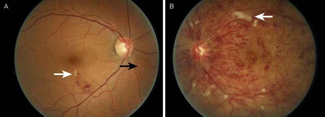
A 57‐year‐old man presented to the emergency department with a 2‐day history of painless vision loss in his left eye. He had a history of smoking tobacco and methamphetamine, with increased methamphetamine use over the previous week. He had no history of hypertension, dyslipidaemia or diabetes. Visual acuity was 6/6 in the right eye and count fingers in the left eye. He had a left relative afferent pupillary defect, and on fundoscopy there were widespread flame haemorrhages and cotton wool spots (Figure, B, arrow), suggestive of an ischaemic central retinal vein occlusion. In the right eye, he also had some flame haemorrhages and a cotton wool spot (Figure, A, white arrow) inferior to the macula, suggestive of a small area of branch retinal vein occlusion. A sclerosed branch retinal vein extending from the inferonasal part of the right optic nerve (Figure, A, black arrow) was also visible, suggestive of a previous (presumably long‐standing) branch retinal vein occlusion. Methamphetamine‐induced ocular complications include severe corneal ulceration and scarring, keratitis, episcleritis, scleritis, crystalline retinopathy, central retinal artery occlusion, ischaemic retinopathy, and retinal vasculitis.1,2 The sympathomimetic effects of methamphetamine, which might induce extensive vasoconstriction or vascular spasm,2 are believed to be the cause of ischaemic central retinal vein occlusion.
Competing interests
No relevant disclosures.
References
- Hazin R, Cadet JL, Kahook MY, Saed D. Ocular manifestations of crystal methamphetamine use. Neurotox Res 2009; 15: 187–191.
- Guo J, Tang W, Liu W, et al. Bilateral methamphetamine‐induced ischemic retinopathy. Am J Ophthalmol Case Rep 2019; 15: 100473.
A Diagnostic Headache
Thomas Glynn, Michelle T. Leech, Stacy K. Goergen, Emily Lin, Nadeem Toodayan, Ralph Junckerstorff
Neisseria meningitidis: an uncommon cause of conjunctivitis
Chloe Story, Tina Marinelli, Simeon Crawford, Amrita Ronnachit
Dismantling barriers to research and clinical care for individuals with a vision impairment
Eden G Robertson, Kate Hetherington, Meredith Prain, Julia Hall, Leighton Boyd AM, Rosemary Boyd OAM, Emily Shepard, Hollie Feller, Sally Karandrews, Fleur O'Hare, Kanae Yamamoto, Matthew P Simunovic, Robyn V Jamieson, Alan Ma, Lauren Ayton AM, Anai Gonzalez‐Cordero
The New South Wales Pharmacy Trial for herpes zoster: on the nose?
Christian P Pappas, Timothy R Holmes, Minas T Coroneo
Skin fragility disorder misdiagnosed as child abuse: a cautionary tale
Heather G Mack, Fred K Chen, John R Grigg
Health economic aspects of inherited retinal diseases: looking for cost‐effective treatments
Benjamin Kamien, Rachael Heath Jeffery, Fred K Chen