




Volume 216 - Issue 10
Neglected tropical diseases in Australia: a narrative review
Authors: Johanna Kurcheid, Catherine A Gordon, Naomi E Clarke, Kinley Wangdi, Matthew Kelly, Aparna Lal, Polydor N Mutombo, Dongxu Wang, Mary L Mationg, Archie CA Clements, Stephen Muhi, Richard S Bradbury, Beverley‐Ann Biggs, Wendy Page, Gail Williams, Donald P McManus and Darren Gray
Med J Aust 2022; 216 (10): 532-538. || doi: 10.5694/mja2.51533
Published online: 6 June 2022
Published online: 6 June 2022
Neglected tropical diseases represent a threat to the health, wellbeing and economic prosperity of billions of people worldwide, often causing serious disease or death
Summary
- • Neglected tropical diseases (NTDs) represent a threat to the health, wellbeing and economic prosperity of billions of people worldwide, often causing serious disease or death.
- • Commonly considered diseases of low and middle‐income nations, the presence of NTDs in high income countries such as Australia is often overlooked.
- • Seven of the 20 recognised NTDs are endemic in Australia: scabies, soil‐transmitted helminths and strongyloidiasis, echinococcosis, Buruli ulcer, leprosy, trachoma, and snakebite envenoming.
- • Dengue, while not currently endemic, poses a risk of establishment in Australia. There are occasional outbreaks of dengue fever, with local transmission, due to introductions in travellers from endemic regions.
- • Similarly, the risk of introduction of other NTDs from neighbouring countries is a concern. Many NTDs are only seen in Australia in individuals travelling from endemic areas, but they need to be recognised in health settings as the potential consequences of infection can be severe.
- • In this review, we consider the status of NTDs in Australia, explore the risk of introducing and contracting these infections, and emphasise the negative impact they have on the health of Australians, especially Aboriginal and Torres Strait Islander peoples.
Neglected tropical diseases (NTDs) refer to a diverse group of primarily infectious diseases prevalent in low and middle‐income countries in tropical regions where sanitation is poor and access to health services is limited.1,2 This group of diseases generally receives less attention and research funding compared with other major infectious diseases, hence the term “neglected”. Less than 1% of official development funding worldwide is directed towards addressing NTDs;3 yet they represent the most common infections of the world’s most vulnerable people.
NTDs are associated with high morbidity and, in some cases, mortality, particularly in individuals harbouring co‐infections. NTDs can be highly disfiguring, leading to social stigma. Consequently, NTDs can have significant adverse social and economic effects, trapping people in an ongoing cycle of poverty. Disability‐adjusted life‐years (DALYs) measure overall disease burden, expressed as number of healthy years lost due to ill health. In the 2019 Global Burden of Disease Study, it was estimated that NTDs account for about 11 million DALYs.4
The World Health Organization NTDs portfolio includes 20 diseases, encompassing bacterial, viral, fungal, protozoal, arthropod and helminthic origin, and snakebite envenoming.2 NTDs are diverse biologically and in their transmission characteristics, adding to the complexity and challenge to their control, particularly when resources are limited and already stretched. The WHO and its partners set a roadmap of targets and milestones for NTD elimination for 2012–2020,5 which focused on implementation of five key strategies: preventive chemotherapy, intensified disease management, vector and intermediate host control, an integrated human and animal health approach, and provision of safe water, sanitation and hygiene. NTDs were also officially included in the health targets of the Sustainable Development Goals in 2015.6 A new WHO roadmap for NTDs was released in 2020 for the period 2021–2030, acknowledging that not all targets had been met in the 2012–2020 roadmap and listing diagnostics as a priority area.1 NTD control has had recent setbacks due to the coronavirus disease 2019 (COVID‐19) pandemic, which led the WHO to issue guidance for halting NTD control programs to limit COVID‐19 spread.7 This will likely cause increased prevalence and morbidity for a number of NTDs.
Prevalence and distribution of NTDs in other regions has been previously reviewed;8 however, there is no comprehensive nor recent review of the prevalence and distribution of NTDs in Australia. This narrative review thus provides an overview of the status of NTDs currently found in Australia (Box 1 and Supporting Information, section 1). An overview of clinically relevant NTD diagnostics can be found in the Supporting Information, table 1.9 We conducted a literature search on PubMed, Google, Google Scholar and Web of Science using “disease name OR commonly used terms” OR “causative agent” AND “Australia” (specific search terms are provided in the Supporting Information, table 2).
Australia has a population of 25.69 million people (as of 2020), with a diverse climate ranging from equatorial and tropical zones in the north, subtropical areas along the coasts, desert in the centre, and more temperate climates in the south. NTDs are generally found in tropical and subtropical areas of the world, but can be found in temperate zones, and several NTDs are endemic outside tropical and subtropical areas in Australia (Box 2).
The WHO list of neglected tropical diseases
Of the 20 NTDs listed by the WHO,1 Australia is either free from the majority or they occur only as rare imported cases in travellers or immigrants. Of those, onchocerciasis, mycetoma, schistosomiasis, dracunculiasis, and human African trypanosomiasis are unlikely to become established. As such, they are considered low risk and will not be discussed further in this review. Lymphatic filariasis was previously endemic to Australia, and even though competent mosquito vector species are still present, it is unlikely to re‐emerge and will also not be discussed.10
In this review, we concentrate on NTDs currently endemic in Australia. The information on NTDs that are either not present or sporadic in humans but which infect animals is provided in the Supporting Information, section 1.
Endemic neglected tropical diseases
Scabies
Scabies is a parasitic infestation of the skin caused by the mite Sarcoptes scabiei var. hominis. Transmission occurs by direct skin‐to‐skin contact, although indirect transmission (via clothing, bedding and other fomites) has been proposed.11
Scabies and skin infections are endemic in many resource‐poor tropical settings where there is overcrowding and poverty and limited access to treatment. Scabies is endemic in many remote Aboriginal communities in central and northern Australia. The prevalence of scabies in Australia can reach as high as 30% in endemic areas, with higher a prevalence often seen in children of up to 35%, this represents some of the highest rates of scabies in the world.12,13 A large number of individuals with crusted scabies, the most severe form, require hospitalisation.14 Secondary Staphylococcus aureus and Streptococcus pyogenes bacterial infections, introduced through scratching‐induced skin abrasions, are an added complication of scabies infestation.15 Scabies outbreaks also occur in residential and nursing care homes where they cause significant morbidity and distress.15
Crusted (also called Norwegian) scabies is highly contagious, debilitating and disfiguring, and results from a failure of the host immune response to control the proliferation of scabies mites in the skin leading to high mite burden (up to 4700 mites per gram of skin).14 In some communities, individuals with crusted scabies are considered core transmitters. Community‐based mass treatment with oral ivermectin and topical creams has resulted in a decline in scabies prevalence in some, but not all, endemic areas.16,17 Sustainable control has proven difficult to achieve, particularly where there is high mobility between communities and households.18 Topical medications are effective for treatment of individual cases but less so as population‐level interventions because they require a long duration of application and cause skin irritation, resulting in decreased treatment compliance.17,19,20
Soil‐transmitted helminths
Soil‐transmitted helminths (phylum Nematoda) are parasitic helminths comprising Ascaris lumbricoides, Trichuris trichiura and the hookworms Ancylostoma spp. and Necator americanus. They are the most prevalent NTDs, with 900 million people infected worldwide (www.thelancet.com/gbd/summaries).21 Adult helminths reside in the human gastrointestinal tract. Heavy infections lead to iron‐deficiency anaemia and impaired absorption of micronutrients causing impairments to child growth and cognitive development.22,23
In Australia, soil‐transmitted helminths are commonly found among migrants from endemic countries.24,25 T. trichiura and Ancylostoma duodenale are endemic to parts of Australia, although epidemiological data are limited. As. lumbricoides has not been reported since the 1980s.26,27,28 but human cases of the zoonotic Ascaris suum, normally found in pigs, have been reported.29 Hookworms were once endemic across parts of New South Wales, Queensland, Western Australia and the Northern Territory,30,31 but endemic hookworm infections in Australia are now rare.26,30 The zoonotic hookworm Ancylostoma ceylanicum has been recently identified in dogs in various parts of Australia,32 and in humans from WA.33,34
A control program known as the Aboriginal Community Children’s Deworming Program was implemented in the NT commencing in 1995, and involved regular treatment of children aged 6 months to 16years with albendazole.35 Recent data regarding soil‐transmitted helminths in northern Australia are scarce but indicate that hookworm and T. trichiura are in decline. Hookworm and T. trichiura infections were found to be predominant among Aboriginal people in recent studies, with T. trichiura mainly infecting children and hookworm mainly found in adults.35,36 As recent data are limited to individual communities or audits of stool samples collected for other purposes, the true extent of soil‐transmitted helminth infections in northern Australia remains unclear.
Strongyloidiasis
The cause of strongyloidiasis, Strongyloides stercoralis (phylum Nematoda; class Chromadorea; order Rhabditida), is a soil‐transmitted helminth but is distinct from other soil‐transmitted helminth species due to differing life cycle, clinical features and management. S. stercoralis can persist for decades, even lifelong, following initial infection,37 and is often asymptomatic. In immunocompromised or immunosuppressed hosts, however, infection can be fatal, and screening and treatment of patients before immunosuppressive treatment is recommended.38 Ivermectin is the recommended treatment but there are no specific public health strategies advocated for strongyloidiasis control.39,40
Strongyloidiasis is endemic in parts of Australia, predominantly occurring in remote Aboriginal and Torres Strait Islander communities in tropical areas of WA, the NT, Queensland, northern South Australia, and northern NSW (Box 2).27,41,42,43,44,45,46,47,48 A limited number of epidemiological surveys, both retrospective and prospective, have been undertaken in Australia, reporting highly variable prevalence (0–60%).42,48,49,50 The true prevalence and burden of strongyloidiasis in Australia is unclear, due to the large number of asymptomatic cases, underdiagnosis, and limited surveillance data.51 There have been recent calls for strongyloidiasis to be made a nationally notifiable disease in order to improve surveillance and inform the planning, implementation, and evaluation of control measures.48,51
The human T‐lymphotropic virus type 1 (HTLV‐1) is also endemic in remote communities, primarily in Northern Australia and is associated with bacteraemia and disseminated strongyloidiasis.41,49 Other risk factors for S. stercoralis infection in Australian Aboriginal people include age (highest risk in school‐aged children)42,52 and malnutrition.53 Outside remote communities, other groups at risk of infection in Australia include immigrants from endemic countries,24,54 returned travellers,55,56 and those with occupational exposure in endemic locations.57
Echinococcosis
Echinococcosis is caused by tapeworms of the genus Echinococcus (phylum Platyhelminthes; class Cestoda). In humans, there are two major forms of the disease: cystic echinococcosis, caused by Echinococcus granulosus, and alveolar echinococcosis, due to Echinococcus multilocularis. E. multilocularis does not occur in Australia. The life cycle of E. granulosus involves animals as both intermediate (primarily ungulates in the domestic life cycles and macropods in the Australian sylvatic life cycle) and definitive (canine) hosts (Box 2).58 Echinococcosis is transmitted to humans through ingestion of tapeworm eggs, mainly through food, water or soil contaminated by the faeces of the definitive host, or following direct handling of an infected definitive host.59 Both alveolar echinococcosis and cystic echinococcosis are characterised by long asymptomatic periods, with clinical illness developing after many years.60 Cystic echinococcosis leads to formation of large, fluid‐filled cysts, most commonly in the liver and lungs.58 Alveolar echinococcosis causes a slow growing, invasive lesion, usually in the liver, that can metastasise to distant sites and has a high fatality rate.60 Both are managed with prolonged courses of anthelmintic drugs and/or surgical interventions.
In Australia, dogs, dingoes and foxes act as definitive hosts of E. granulosus, and kangaroos, wallabies, wombats, feral pigs, sheep and cattle are intermediate hosts.61 There are an estimated 80–100 cases of human echinococcosis diagnosed each year in Australia, including locally acquired and imported cases.58 Cystic echinococcosis is prevalent nationwide in animals, and human cases have been reported in all states and territories; there has been no evidence of transmission to humans in Tasmania since 1996, when the state declared eradication of hydatid disease.57,61,62,63,64,65 A review of human cases between 1996 and 2012 found a number of human cases of hydatid, but these were all linked back to exposure in the 1960s and 1970s.62 Data from WA show residents of rural areas and Aboriginal Australians have higher rates of infection.58
Control of cystic echinococcosis in Australia is complicated by the sylvatic life cycle, but overall, there is a relatively low incidence of human disease.61,65
Buruli ulcer
Buruli ulcer is a rapidly emerging NTD. Buruli ulcer is endemic in at least 30 countries worldwide, mainly those with tropical and subtropical climates. The disease is caused by infection with the toxin‐producing environmental pathogen Mycobacterium ulcerans and primarily affects the skin and subcutaneous tissue, often progressing without pain or systemic symptoms. Symptoms may begin with a nodule, plaque or oedema and often progress to large skin ulcerations and sometimes osteomyelitis.66 The incubation period for the disease is about 4.5 months.67
Australia is the only high income nation globally that experiences local transmission of Buruli ulcer, and has the highest reported global incidence outside Africa.68 The disease was first identified and described by Australian scientists in 1948, and it was classified as a notifiable condition in 2004 as a subset of non‐tuberculosis mycobacteria in Queensland, the NT and SA, and specifically as M. ulcerans in Victoria; it is not notifiable in NSW or WA.69 Although Buruli ulcer is usually curable with the use of antibiotics, the treatment is expensive and is associated with side effects.70,71 New infections are possible after treatment, with the majority of new cases resulting from reinfection rather than recurrence of a previous infection.72
While Buruli ulcer is also present in north and central Queensland, referred to as Daintree ulcer,73 the main focus of infection is in the southern coastal areas of Victoria, where more than 80% of cases occur (Box 2). As early cases were reported from the Bairnsdale district, it is referred to locally as the Bairnsdale ulcer.74 In Victoria, the disease is of major concern due to the increasing infection incidence and expanding geographic distribution. The areas where Buruli ulcer has been identified for the longest period are in Gippsland, Phillip Island, Frankston, and the Mornington and Bellarine peninsulas.69 Geographic patterns have changed over the past 70years since the disease was first identified. In general, endemic areas for Buruli ulcer have been moving steadily westwards and Buruli ulcer is now present in inner Melbourne.75
There is a lack of understanding of the environmental reservoirs for the disease and the mode of transmission. Human‐to‐human transmission appears to be very rare,76 suggesting an animal reservoir or environmental source is likely.66,77,78 Mosquitos may act as a transmission vector, and dogs, cats, koalas, possums and bandicoots have been found to carry M. ulcerans.79,80
A distinctive characteristic of Buruli ulcer is its confinement to sharply defined geographic areas, with endemic and non‐endemic areas only a few kilometres apart (Box 2).81 M. ulcerans is most common in wetland areas, with one hypothesis being that current outbreaks in Victoria may be caused by environmental disturbances in wetland areas through building work, flooding, and using recycled water for gardening.70
Leprosy (Hansen disease)
Leprosy is caused by the bacterium Mycobacterium leprae. Leprosy rates in Australia are low (less than one case per million population) with few cases reported annually, primarily in the north of the country, although there have been cases in Queensland,82 Victoria,83,84 Adelaide85 and Sydney86 — most of these cases were imported rather than locally acquired (Box 2).
More recently, leprosy has been reported sporadically in the NT, Queensland and WA,83,87 with Aboriginal Australians suffering disproportionately from the disease. Between 1986 and 2002, 28 new cases of leprosy, all but one in Aboriginal patients, were notified to the Kimberley Public Health Unit in WA.88
Delay in diagnosis of rare and unusual infections such as leprosy is common. This may be partly due to a lengthy latency period and lack of familiarity with clinical manifestations by clinicians.87 Diverse presentations of leprosy include lesions or thickened peripheral nerves. Although leprosy is curable with antibiotics, a delay in treatment can result in physical deformity and long term disability.89
Trachoma
Chronic infection with the bacterium Chlamydia trachomatis (serotypes A, B and C) is the leading cause of infectious blindness worldwide. The disease is commonly found in impoverished communities with poor housing, inadequate sanitation and limited sewage disposal systems.90 Trachoma was unknown in Australia before British colonisation but eventually became so common that certain places were named in association with trachoma or “sandy blight”, as it was commonly known.90
Trachoma is reported to be hyperendemic in parts of Australia, and is still considered an important public health problem among certain groups of Aboriginal people (Box 2).91,92 Australia’s first national population‐based random cluster sample survey of eye health among Aboriginal children and older adults, conducted in 2008, found that the overall rate of follicular trachomatous inflammation among children was 3.8%, ranging from 0.6% in major cities to 7.3% in very remote areas.93 Trachomatous scarring occurred in 15.7%, corneal opacity among 0.3%, and trachomatous trichiasis among 1.4% of infected adults.93
A finer grading scheme detailing the pathogenesis, distribution and natural history of trachoma for the more precise identification of clinical status than the current WHO disease classification has been suggested.94
Snakebite envenoming
There are over 5 million incidences of snakebites globally, resulting in 2.7 million envenomings (poisoning from snake bites) and between 81000 and 138000 deaths each year.95 In 2017, the WHO listed snakebite envenoming as a highest priority NTD to help raise public awareness of this issue, particularly in developing nations where most cases occur.
Australia is well known for having a number of different venomous snakes, including some considered among the deadliest in the world due to the lethality of their venom. However, cases of snakebite envenoming, as distinguished from snakebites (where envenoming does not occur), are rare in Australia.96 A study examining snakebite envenoming in Australia from 2005 to 2015 enrolled 1548 patients with suspected snakebites, of which 835 had been envenomed; this represented a median of 87 envenomations per year and a median of two deaths annually.96 The high level of awareness of snakebites and immobilisation methods in Australia, as well as the remoteness of many venomous snake locations are the likely reason for the low number of bites and envenomations.
Conclusion
The overall countrywide prevalence of NTDs in Australia is low, although the lack of robust prevalence studies, case finding, and notifiability97 of many of these diseases means that the true infection rates and distribution are uncertain and difficult to ascertain (Box 1). Hyperendemicity of some NTDs occurs, particularly in remote communities in the northern parts of Australia, where many NTDs have an overlapping distribution (Box 2). A lack of health infrastructure and distance from health services and testing facilities contribute to the underdiagnosis of NTDs in these communities, with residents often having to travel considerable distances to access the nearest health care facility.
Chikungunya, dengue, rabies, bat lyssavirus, and leprosy are notifiable NTDs in Australia. Dengue, in particular, has a high risk of becoming endemic due to the widespread distribution of competent mosquito vectors and regular introductions of the virus from returning travellers and immigrants, which can lead to outbreaks.98 Most tend to occur in north Queensland, around Cairns and Townsville, although there have been recent cases of locally acquired infection in Rockhampton, in central Queensland.99 Climate change will almost certainly extend the range of Aedes aegypti, a vector of dengue and chikungunya, which is present in Queensland and has previously been identified in WA, the NT and NSW.100,101 Aedes albopictus, a very aggressive invasive species, is currently in the Torres Strait, and there are real fears of it spreading to mainland Australia particularly as, in contrast to Ae. aegypti, it is not dependent on a tropical climate. Wolbachia, a bacterial endosymbiont of certain insects, has been introduced to mosquitoes in north Queensland as a dengue blocking agent.102 Since the initial release of Wolbachia‐infected mosquitoes, the transmission rate of dengue has been reduced and the intervention is now being trialled internationally.
Of the NTDs present in Australia that are currently not notifiable, trachoma, scabies and strongyloidiasis have the widest distribution, the highest prevalence and the greatest overall disease burden (Box 1 and Box 2). Making these NTDs nationally notifiable would provide improved data on their true prevalence and distribution in Australia and would inform public health and treatment strategies. For example, Strongyloides infection can cause major complications if an infected individual is co‐infected with HTLV‐1 or otherwise immunosuppressed, and can lead to hyperinfection and disseminated strongyloidiasis, often with disastrous clinical consequences.40,49 Obtaining quality prevalence data is a priority to addressing NTDs in Australia. Making NTDs notifiable would help gather this information. Laboratory notification could be done electronically to a central database, something that is now more feasible due to advances in technology. Resources would need to be allocated by the government for this task, but would provide valuable data for measuring the disease burden and combating NTDs in Australia.
The potential for NTDs to enter Australia through infected immigrants and returned travellers is considerable and already occurs with dengue and Chagas disease — a parasitic protozoan infection by Trypanosoma cruzi. Indeed, immigration from areas endemic with Chagas has resulted in a global increase of the disease outside Latin America.103 Recent recommendations have been documented for appropriate and comprehensive screening of new arrivals into Australia from refugee‐like backgrounds.104 However, only two NTDs (strongyloidiasis and schistosomiasis) are covered in depth; other NTDs are covered in the chapter on skin lesions, and soil‐transmitted helminths and Taenia are briefly covered under intestinal parasites. The document provides a good overview of treatment options but needs to be combined with a more comprehensive understanding of the epidemiology, aetiology and treatment options for NTDs, including those that can be imported and may lead to local transmission. This information is imperative for the effective prevention, control and elimination of these diseases of poverty which should not be present in such an economically advanced country as Australia.
Box 1 – Public health risks of contracting neglected tropical diseases (NTDs) in Australia using a traffic light system*
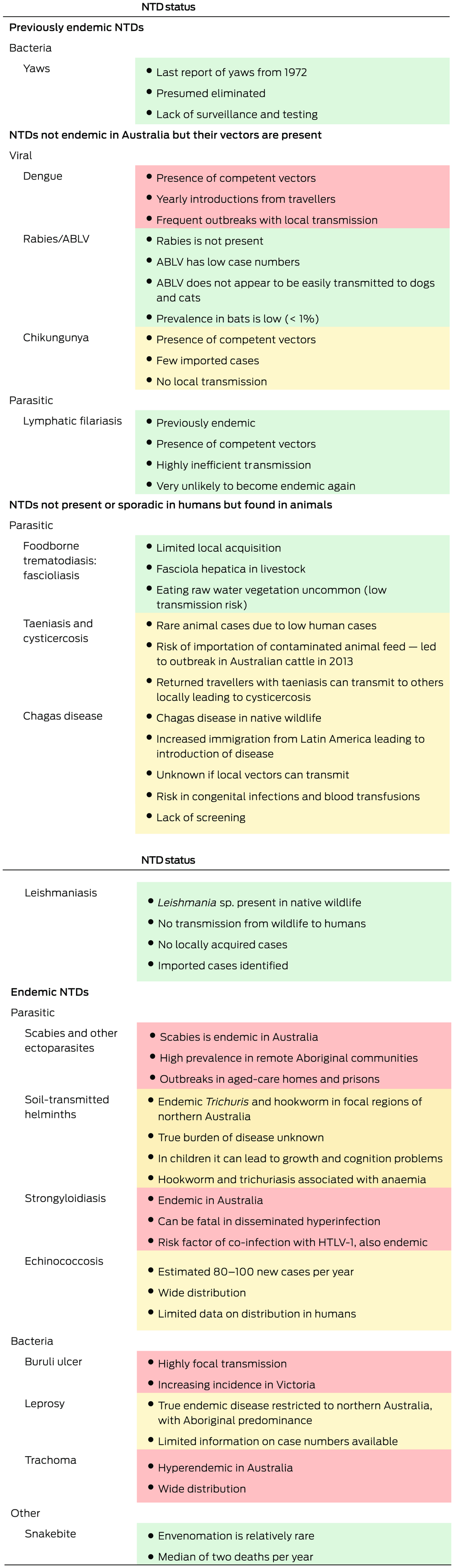
ABLV = Australian bat lyssavirus; HTLV‐1 = human T‐lymphotropic virus type 1. The following NTDs are either not present in Australia, or present only in imported cases (travellers or immigrants): lymphatic filariasis, onchocerciasis, mycetoma, schistosomiasis, dracunculiasis, taeniasis and neurocysticercosis, and human African trypanosomiasis. * Green = low risk; amber = intermediate risk; red = high risk.
Box 2 – Distribution of endemic neglected tropical diseases (NTDs) in Australia*
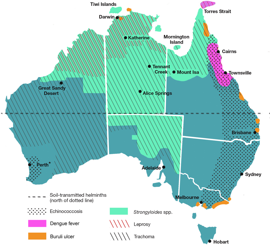
* Not shown are snakebite envenomation, which is rare in Australia, and scabies, which has a purported distribution across the whole of Australia. Distribution of echinococcosis is similarly widespread across mainland Australia; thus, only high transmission areas are shown. Soil‐transmitted helminths are present north of the dotted line. Dengue, although not endemic in Australia, exhibits frequent outbreaks that occur primarily in the areas marked on the map.
Competing interests
No relevant disclosures.
Acknowledgements
We thank Jennifer Shield for her help in edits and comments on the manuscript.
References
- World Health Organization. Ending the neglect to attain the Sustainable Development Goals: a road map for neglected tropical diseases 2021–2030. https://www.who.int/publications/i/item/9789240010352 (viewed Mar 2021).
- World Health Organization. Report of the tenth meeting of the WHO Strategic and Technical Advisory Group for Neglected Tropical Diseases. https://cdn.who.int/media/docs/default‐source/ntds/strategic‐and‐advisory‐group‐on‐neglected‐tropical‐diseases‐(stag‐ntds)/tenth‐ntd‐stag‐report‐2017.pdf?sfvrsn=9ec99065_2&download=true (viewed Apr 2022).
- Molyneux DH, Savioli L, Engels D. Neglected tropical diseases: progress towards addressing the chronic pandemic. Lancet 2017; 389: 312‐325.
- GBD cause and risk summaries. Lancet, 2020. https://www.thelancet.com/gbd/summaries (viewed Aug 2021).
- World Health Organization. Accelerating work to overcome the global impact of neglected tropical diseases: a roadmap for implementation. https://apps.who.int/iris/bitstream/handle/10665/70809/WHO_HTM_NTD_2012.1_eng.pdf?sequence=1&isAllowed=y (viewed Apr 2022).
- United Nations. Transforming our world: the 2030 agenda for Sustainable Development. https://sustainabledevelopment.un.org/post2015/transformingourworld/publication (viewed Apr 2022).
- World Health Organization. Considerations for implementing mass treatment, active case‐finding and population‐based surveys for neglected tropical diseases in the context of the COVID‐19 pandemic [interim guidance]. https://www.who.int/publications/i/item/WHO‐2019‐nCoV‐neglected‐tropical‐diseases‐2020‐1 (viewed Sept 2020).
- Hotez PJ. The rise or fall of neglected tropical diseases in East Asia Pacific. Acta Trop 2020; 202: 105182.
- Rogers MJ, McManus DP, Muhi S, Gordon CA. Membrane technology for rapid point‐of‐care diagnostics for parasitic neglected tropical diseases. Clin Microbiol Rev 2021; 34: e0032920.
- Gordon CA, Jones M, McManus DP. The history of bancroftian lymphatic filariasis in Australasia and Oceania: is there a threat of re‐occurrence in mainland Australia? Trop Med Infect Dis 2018; 3: 58.
- Mellanby K. Transmission of scabies. Br Med J 1941; 2: 405.
- Romani L, Steer AC, Whitfeld MJ, Kaldor JM. Prevalence of scabies and impetigo worldwide: a systematic review. Lancet Infect Dis 2015; 15: 960‐967.
- Engelman D, Cantey PT, Marks M, et al. The public health control of scabies: priorities for research and action. Lancet 2019; 394: 81‐92.
- Lokuge B, Kopczynski A, Woltmann A, et al. Crusted scabies in remote Australia, a new way forward: lessons and outcomes from the East Arnhem Scabies Control Program. Med J Aust 2014; 200: 644‐648. https://www.mja.com.au/journal/2014/200/11/crusted‐scabies‐remote‐australia‐new‐way‐forward‐lessons‐and‐outcomes‐east
- Pruksachatkunakorn C, Wongthanee A, Kasiwat V. Scabies in Thai orphanages. Pediatr Int 2003; 45: 719‐723.
- Carapetis JR, Connors C, Yarmirr D, et al. Success of a scabies control program in an Australian Aboriginal community. Pediatr Infect Dis J 1997; 16: 494‐499.
- Kearns TM, Speare R, Cheng AC, et al. Impact of an ivermectin mass drug administration on scabies prevalence in a remote Australian Aboriginal community. PLoS Negl Trop Dis 2015; 9: e0004151.
- Wong LC, Amega B, Barker R, et al. Factors supporting sustainability of a community‐based scabies control program. Australas J Dermatol 2002; 43: 274‐277.
- La Vincente S, Kearns T, Connors C, et al. Community management of endemic scabies in remote aboriginal communities of northern Australia: low treatment uptake and high ongoing acquisition. PLoS Negl Trop Dis 2009; 3: e444.
- Engelman D, Cantey PT, Marks M, et al. The public health control of scabies: priorities for research and action. Lancet 2019; 394: 81‐92.
- Global Health Metrics. Intestinal nematode infections — Level 3 cause. Lancet 2020; 396: S28‐S29.
- Bethony J, Brooker S, Albonico M, et al. Soil‐transmitted helminth infections: ascariasis, trichuriasis, and hookworm. Lancet 2006; 367: 1521‐1532.
- Mutombo PN, Man NWY, Nejsum P, et al. Diagnosis and drug resistance of human soil‐transmitted helminth infections: a public health perspective. Adv Parasitol 2019; 104: 247‐326.
- Caruana SR, Kelly HA, Ngeow JY, et al. Undiagnosed and potentially lethal parasite infections among immigrants and refugees in Australia. J Travel Med 2006; 13: 233‐239.
- Ryan N, Plackett M, Dwyer B. Parasitic infections of refugees. Med J Aust 1988; 148: 491‐494.
- Robertson G, Lade C, Norton R, Bradbury R. The burden of helminth disease in Queensland, 2003–2013 [poster]. Melbourne: Australian Society of Microbiology, 2014.
- Shield J, Aland K, Kearns T, et al. Intestinal parasites of children and adults in a remote Aboriginal community of the Northern Territory, Australia, 1994–1996. Western Pac Surveill Response J 2015; 6: 44‐51.
- Fryar D, Hagan S. Pilot screening program for intestinal parasites and anaemia in adults in a Top End Aboriginal community. Northern Territory Communicable Diseases Bulletin 1997; 4: 20‐21.
- Goldsmid JM. Human infection with “Ascaris suum” in Tasmania? Ann ACTM 2005; 6: 16.
- Prociv P, Luke RA. The changing epidemiology of human hookworm infection in Australia. Med J Aust 1995; 162: 150‐154.
- Sweet WC. Final report of the Australian Hookworm Campaign: 1st October 1919 to 30th September 1924. Brisbane: Australian Hookworm Campaign; 1924. https://nla.gov.au/nla.obj‐648199287/view?partId=nla.obj‐648199552 (viewed Apr 2022).
- Smout FA, Skerratt LF, Butler JR, et al. The hookworm Ancylostoma ceylanicum: an emerging public health risk in Australian tropical rainforests and Indigenous communities. One Health 2017; 3: 66‐69.
- Speare R, Bradbury RS, Croese J. A case of Ancylostoma ceylanicum infection occurring in an Australian soldier returned from Solomon Islands. Korean J Para 2016; 54: 533.
- Koehler AV, Bradbury RS, Stevens MA, et al. Genetic characterization of selected parasites from people with histories of gastrointestinal disorders using a mutation scanning‐coupled approach. Electrophoresis 2013; 34: 1720‐1728.
- Davies J, Majumdar SS, Forbes R, et al. Hookworm in the Northern Territory: down but not out. Med J Aust 2013; 198: 278‐281. https://www.mja.com.au/journal/2013/198/5/hookworm‐northern‐territory‐down‐not‐out
- Crowe AL, Smith P, Ward L, et al. Decreasing prevalence of Trichuris trichiura (whipworm) in the Northern Territory from 2002 to 2012. Med J Aust 2014; 200: 286‐289. https://www.mja.com.au/journal/2014/200/5/decreasing‐prevalence‐trichuris‐trichiura‐whipworm‐northern‐territory‐2002‐2012
- Page W, Judd JA, Bradbury RS. The unique life cycle of Strongyloides stercoralis and implications for public health action. Trop Med Infect Dis 2018; 3: 53.
- Marcos LA, Terashima A, DuPont HL, Gotuzzo E. Strongyloides hyperinfection syndrome: an emerging global infectious disease. Trans R Soc Trop Med Hyg 2008; 102: 314‐318.
- World Health Organization. Control of neglected tropical diseases: strongyloidiasis. https://www.who.int/teams/control‐of‐neglected‐tropical‐diseases/soil‐transmitted‐helminthiases/strongyloidiasis (viewed Apr 2022).
- Page W, Speare R. Chronic strongyloidiasis — don’t look and you won’t find. Aust Fam Physician 2016; 45: 40‐44.
- Einsiedel L, Fernandes L. Strongyloides stercoralis: a cause of morbidity and mortality for Indigenous people in Central Australia. Intern Med J 2008; 38: 697‐703.
- Johnston FH, Morris PS, Speare R, et al. Strongyloidiasis: a review of the evidence for Australian practitioners. Aust J of Rural Health 2005; 13: 247‐254.
- Prociv P, Luke R. Observations on strongyloidiasis in Queensland Aboriginal communities. Med J Aust 1993; 158: 160‐163.
- Hays R, Esterman A, McDermott R. Control of chronic Strongyloides stercoralis infection in an endemic community may be possible by pharmacological means alone: results of a three‐year cohort study. PLoS Negl Trop Dis 2017; 11: e0005825.
- Page WA, Dempsey K, McCarthy JS. Utility of serological follow‐up of chronic strongyloidiasis after anthelminthic chemotherapy. Trans R Soc Trop Med Hyg 2006; 100: 1056‐1062.
- Miller A, Young EL, Tye V, et al. A community‐directed integrated Strongyloides control program in Queensland, Australia. Trop Med and Infect Dis 2018; 3: 48.
- Page WA, Judd JA, MacLaren DJ, Buettner P. Integrating testing for chronic strongyloidiasis within the Indigenous adult preventive health assessment system in endemic communities in the Northern Territory, Australia: an intervention study. PLoS Negl Trop Dis 2020; 14: e0008232.
- Shield J, Braat S, Watts M, et al. Seropositivity and geographical distribution of Strongyloides stercoralis in Australia: a study of pathology laboratory data from 2012–2016. PLoS Negl Trop Dis 2021; 15: e0009160.
- Gordon CA, Shield JM, Bradbury RS, et al. HTLV‐1 and Strongyloides in Australia: the worm lurking beneath. Adv Parasitol 2021; 111: 119‐201.
- Hanieh S, Ryan N, Biggs B. Assessing enteric helminths in refugees, asylum seekers and new migrants. Microbiology Australia 2016; March: 15‐9.
- Beknazarova M, Whiley H, Judd J, et al. Argument for inclusion of strongyloidiasis in the Australian national notifiable disease list. Trop Med and Infect Dis 2018; 3: 61.
- Kearns TM, Currie BJ, Cheng AC, et al. Strongyloides seroprevalence before and after an ivermectin mass drug administration in a remote Australian Aboriginal community. PLoS Negl Trop Dis 2017; 11: e0005607.
- Kukuruzovic R, Robins‐Browne RM, Anstey NM, Brewster DR. Enteric pathogens, intestinal permeability and nitric oxide production in acute gastroenteritis. Pediatr Infect Dis J 2002; 21: 730‐739.
- De Silva S, Saykao P, Kelly H, et al. Chronic Strongyloides stercoralis infection in Laotian immigrants and refugees 7–20years after resettlement in Australia. Epidemiol Infect 2002; 128: 439‐444.
- Looke DF, Robson J. 9: Infections in the returned traveller. Med J Aust 2002; 177: 212‐219.
- Swaminathan A, Torresi J, Schlagenhauf P, et al. A global study of pathogens and host risk factors associated with infectious gastrointestinal disease in returned international travellers. J Infect 2009; 59: 19‐27.
- Soulsby HM, Hewagama S, Brady S. Case series of four patients with Strongyloides after occupational exposure. Med J Aust 2012; 196: 444. https://www.mja.com.au/journal/2012/196/7/case‐series‐four‐patients‐strongyloides‐after‐occupational‐exposure
- Jenkins DJ. Cystic echinococcosis in Australia: the current situation. Southeast Asian J Trop Med Public Health; 2004; 35 (Suppl): 183‐188.
- World Health Organization. Factsheet: echinococcosis, 2021. https://www.who.int/news‐room/fact‐sheets/detail/echinococcosis (viewed Apr 2022).
- McManus DP, Zhang W, Li J, Bartley PB. Echinococcosis. Lancet 2003; 362: 1295‐1304.
- Jenkins DJ. Echinococcus granulosus in Australia, widespread and doing well! Parasitol Int 2006; 55 (Suppl: S203‐S206).
- O’Hern JA, Cooley L. A description of human hydatid disease in Tasmania in the post‐eradication era. Med J Aust 2013; 199: 117‐120. https://www.mja.com.au/journal/2013/199/2/description‐human‐hydatid‐disease‐tasmania‐post‐eradication‐era
- Lin M, Roche P, Spencer J, et al. Australia’s notifiable diseases status, 2000. Commun Dis Intell 2002; 26: 118‐203.
- Owen R, Roche PW, Hope K, et al. Australia’s notifiable diseases status, 2005: annual report of the National Notifiable Diseases Surveillance System. Commun Dis Intell Q Rep 2007; 31: 1‐70.
- Jenkins DJ. Hydatid control in Australia: where it began, what we have achieved and where to from here. Int J Parasitol 2005; 35: 733‐740.
- van Ravensway J, Benbow ME, Tsonis AA, et al. Climate and landscape factors associated with Buruli ulcer incidence in Victoria, Australia. PLoS One 2012; 7: e51074.
- Loftus MJ, Trubiano JA, Tay EL, et al. The incubation period of Buruli ulcer (Mycobacterium ulcerans infection) in Victoria, Australia remains similar despite changing geographic distribution of disease. PLoS Negl Trop Dis 2018; 12: e0006323.
- Omansen TF, Erbowor‐Becksen A, Yotsu R, et al. Global epidemiology of Buruli ulcer, 2010–2017, and analysis of 2014 WHO programmatic targets. Emerg Infect Dis 2019; 25: 2183‐2190.
- Buultjens AH, Vandelannoote K, Meehan CJ, et al. Comparative genomics shows that Mycobacterium ulcerans migration and expansion preceded the rise of Buruli ulcer in southeastern Australia. Appl Environ Microbiol 2018; 84: 2612‐2617.
- O’Brien D, Athan E, Blasdell K, De Barro P. Tackling the worsening epidemic of Buruli ulcer in Australia in an information void: time for an urgent scientific response. Med J Aust 2018; 208: 287‐289. https://www.mja.com.au/journal/2018/208/7/tackling‐worsening‐epidemic‐buruli‐ulcer‐australia‐information‐void‐time‐urgent
- O’Brien DP, Friedman D, Hughes A, et al. Antibiotic complications during the treatment of Mycobacterium ulcerans disease in Australian patients. Intern Med J 2017; 47: 1011‐1019.
- Wynne JW, Stinear TP, Athan E, et al. Low incidence of recurrent Buruli ulcers in treated Australian patients living in an endemic region. PLoS Negl Trop Dis 2018; 12: e0006724.
- Steffen CM, Smith M, McBride WJ. Mycobacterium ulcerans infection in North Queensland: the “Daintree ulcer”. ANZ J Surg 2010; 80: 732‐736.
- Hayman J. Clinical features of Mycobacterium ulcerans infection. Australas J Dermatol 1985; 26: 67‐73.
- Lewin E. Melbourne GPs told to consider Buruli ulcers when assessing lesions. News GP 2021; 25 Feb. https://www1.racgp.org.au/newsgp/clinical/buruli‐ulcers‐discovered‐in‐non‐coastal‐areas‐in‐v (viewed Apr 2021).
- Muleta A J, Lappan R, Stinear TO, Greening C. Understanding the transmission of Mycobacterium ulcerans: a step towards controlling Buruli ulcer. PLOS Negl Trop Dis 2021; 15: e0009678.
- Carson C, Lavender CJ, Handasyde KA, et al. Potential wildlife sentinels for monitoring the endemic spread of human Buruli ulcer in south‐east Australia. PLoS Negl Trop Dis 2014; 8: e2668.
- Fyfe JAM, Lavender CJ, Handasyde KA, et al. A major role for mammals in the ecology of Mycobacterium ulcerans. PLoS Negl Trop Dis 2010; 4: e791.
- Lavender CJ, Fyfe JAM, Azuolas J, et al. Risk of Buruli ulcer and detection of Mycobacterium ulcerans in mosquitoes in southeastern Australia. PLoS Negl Trop Dis 2011; 5: e1305.
- Röltgen K, Pluschke G, Johnson PDR, Fyfe J. Mycobacterium ulcerans DNA in bandicoot excreta in Buruli ulcer‐endemic area, Northern Queensland, Australia. Emerg Infect Dis 2017; 23: 2042‐2045.
- Yerramilli A, Tay EL, Stewardson AJ, et al. The location of Australian Buruli ulcer lesions — implications for unravelling disease transmission. PLoS Negl Trop Dis 2017; 11: e0005800.
- Genever G. Queensland’s Black Leper Colony. Queensland Review 2016; 15: 59‐68.
- Australian Government Department of Health. National notifiable diseases surveillance system 2020. http://www9.health.gov.au/cda/source/cda‐index.cfm (viewed Dec 2020).
- Australian Government Department of Health. National notifiable diseases surveillance system 2021. http://www9.health.gov.au/cda/source/rpt_3.cfm (viewed Feb 2021).
- Bourne AJ, Dymock RB, Parry WD, Turner TW. Leprosy in Indo‐Chinese refugees. Med J Aust 1980; 1: 275‐276.
- van Hal SJ, Hudson BJ. Leprosy: an uncommon infection with varied presentations. Med J Aust 2006; 184: 473‐474.
- Barkla S, Modi S. Lepromatous leprosy: a rare presentation in Australia. Australas Med J 2013; 6: 175‐177.
- Mak DB, Platt EM, Heath CH. Leprosy transmission in the Kimberley, Western Australia: still a reality in 21st‐century Australia. Med J Aust 2003; 179: 452. https://www.mja.com.au/journal/2003/179/8/leprosy‐transmission‐kimberley‐western‐australia‐still‐reality‐21st‐century
- Hempenstall A, Smith S, Hanson J. Leprosy in Far North Queensland: almost gone, but not to be forgotten. Med J Aust 2019; 211: 182‐183. https://www.mja.com.au/journal/2019/211/4/leprosy‐far‐north‐queensland‐almost‐gone‐not‐be‐forgotten
- Taylor HR, Anjou MD. Trachoma in Australia: an update. Clin Exp Ophthalmol 2013; 41: 508‐512.
- Australian trachoma surveillance report 2017. Sydney: Kirby Institute, UNSW Sydney; 2018.
- Mak DB, O’Neill LM, Herceg A, McFarlane H. Prevalence and control of trachoma in Australia, 1997–2004. Commun Dis Intell Q Rep 2006; 30: 236‐247.
- Taylor HR, Fox SS, Xie J, et al. The prevalence of trachoma in Australia: the National Indigenous Eye Health Survey. Med J Aust 2010; 192: 248‐253. https://www.mja.com.au/journal/2010/192/5/prevalence‐trachoma‐australia‐national‐indigenous‐eye‐health‐survey
- Michel CE, Roper KG, Divena MA, et al. Correlation of clinical trachoma and infection in Aboriginal communities. PLoS Negl Trop Dis 2011; 5: e986.
- World Health Organization. Snakebite envenoming 2019. https://www.who.int/news‐room/fact‐sheets/detail/snakebite‐envenoming (viewed Feb 2021).
- Johnston CI, Ryan NM, Page CB, et al. The Australian snakebite project, 2005–2015 (ASP‐20). Med J Aust 2017; 207: 119‐125. https://www.mja.com.au/journal/2017/207/3/australian‐snakebite‐project‐2005‐2015‐asp‐20
- Australian Government Department of Health. Australian national notifiable diseases by disease type, 2019. https://www1.health.gov.au/internet/main/publishing.nsf/Content/cda‐surveil‐nndss‐casedefs‐distype.htm (viewed Jan 2021).
- Imrie A. Dengue introduced by travellers, Australia. Microbiol Aust 2018; 39: 67‐71.
- Queensland Health. Dengue fever update: Rockhampton, 2019. https://www.health.qld.gov.au/cq/about/news/articles/dengue‐fever‐update‐rockhampton (viewed Jan 2021).
- Whelan PI, Kurucz N, Pettit WJ, Krause V. Elimination of Aedes aegypti in northern Australia, 2004–2006. J Vector Ecol 2020; 45: 118‐126.
- Gordon CA, McManus DP, Jones MK, et al. The increase of exotic zoonotic helminth infections: the impact of urbanization, climate change and globalization. Adv Parasitol 2016; 91: 311‐397.
- Ryan PA, Turley AP, Wilson G, et al. Establishment of w Mel Wolbachia in Aedes aegypti mosquitoes and reduction of local dengue transmission in Cairns and surrounding locations in northern Queensland, Australia. Gates Open Res 2019; 3: 1547.
- Lidani KCF, Andrade FA, Bavia L, et al. Chagas disease: from discovery to a worldwide health problem. Front Public Health 2019; 7: 166.
- Chaves NA, Pacton G, Biggs, BA, et al; Australasian Society for Infectious Diseases and Refugee Health Network of Australia Guidelines writing group. Recommendations for comprehensive post‐arrival health assessment for people from refugee‐like backgrounds; 2nd ed. Sydney: ASID, 2016. https://www.asid.net.au/documents/item/1225 (viewed Apr 2022).
Provenance: Not commissioned; externally peer reviewed.
Costs of Delivering the Hep B PAST Model of Care in Remote Australia: A Retrospective Pre–Post Study
Hoa Nguyen, Anh Le Tuan Nguyen, Paula Binks, Melita McKinnon, Emily Vintour-Cesar, Karen Wills, Nicola Stephens, Benjamin Cowie, Joshua S. Davis, Yuejen Zhao, Peter Nihill, Julie A. Campbell, Andrew J. Palmer, Jane Davies, Barbara de Graaff
Buruli Ulcer Acquired From Ringtail Possum Rescue
Daniel P. O'Brien, Naomi Clarke, Sara Bonnici, Mohammad A. Hussain
Q Fever Visceral Abscesses After Contact With Native Australian Marsupials
Samantha J. Laws, Enzo Binotto, James Stewart, Jennifer Ho, Trent Yarwood, Josh Hanson, Simon Smith
Hospital-Admitted Injection-Related Infections Among Incarcerated People Who Inject Drugs in Australia: A Retrospective Cohort Study
Andrew Palmer, Matthew Carter, Jeremy Yeo, Cecilia Shim, Jason Connor, Jeremy Hayllar, Gerald Holtmann, Naomi Moy, Elliott G. Playford, Naomi Runnegar, Paul J. Clark
The Live Attenuated Influenza Vaccine in Australia: An Additional Tool for Influenza Prevention
Cyra Patel, Alexis Pillsbury, Tran Nguyen, Xia Wang, Helen E. Quinn, Clayton K. Chiu, Allen C. Cheng, Katie L. Flanagan, Zhicheng Wang
Reflex Testing for Hepatitis D Infection: A Unique Opportunity to Reduce Hepatitis D-Related Chronic Liver Disease Deaths in Australia
Jessica Howell, Lauren Andersson, Miriam T. Levy, James O'Beirne, Leon Adams, Katharine Irvine, Avik Majumdar, Golo Ahlenstiel, Kathy Jackson, Krispin Hajkowicz, Joseph Doyle, Jane Davies, Sarah Cherian, Wayne Dimech, Alexander J. Thompson