




Volume 208 - Issue 7
Tackling the worsening epidemic of Buruli ulcer in Australia in an information void: time for an urgent scientific response
Authors: Daniel P O'Brien, Eugene Athan, Kim Blasdell and Paul De Barro
Med J Aust 2018; 208 (7): 287-289. || doi: 10.5694/mja17.00879
Published online: 16 April 2018
Published online: 16 April 2018
Understanding the risk factors for Buruli ulcer is key to developing a health care intervention
Understanding risk factors is key to defining the source and transmission route of Mycobacterium ulcerans
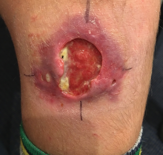
Mycobacterium ulcerans causes an infectious disease known internationally as Buruli ulcer, and also as Bairnsdale ulcer or Daintree ulcer in Australia. It causes severe destructive lesions of skin and soft tissue, resulting in significant morbidity, in attributable mortality and often in long term disability and cosmetic deformity.1 All age groups, including young children, are affected, and the emotional and psychological impact on patients and their carers is substantial (Box 1). Although treatment effectiveness has improved in recent years, with cure rates approaching 100% using combination antibiotic regimens such as rifampicin and clarithromycin,2 these antibiotics are not covered by the Pharmaceutical Benefits Scheme for this condition and are, therefore, expensive to patients. Moreover, these antibiotics have severe side effects in up to one-quarter of patients,1 and many people also require reparative plastic surgery, sometimes with prolonged hospital admissions. The disease thus results in substantial costs, averaging $14 000 per patient including direct3 and indirect costs (eg, transport, lost productivity and dressings) — it had an estimated cost to Victoria in 2016 of $2 548 000 (Paul Mwebaze, Research Scientist, Adaptive Urban and Social Systems, Land and Water, CSIRO, Australia, personal communication, June 2017).
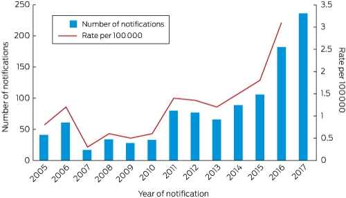
About 2000 Buruli ulcer cases per year are reported worldwide, most commonly from the tropical regions of West or Central Africa.4 In Australia, cases are frequently reported from the Daintree region (95 cases between 2009 and 2015)5 and, less commonly, the Capricorn Coast in Queensland, and occasionally from the Northern Territory, New South Wales and Western Australia. However, most cases are reported from the temperate south-eastern state of Victoria. Here, the community is facing a worsening epidemic, defined by cases rapidly increasing in number,6 becoming more severe in nature, and occurring in new geographic areas.7 In 2016, there were 182 new cases — the highest ever reported by 72%. Yet, cases reported until 11 November 2017 have further increased by 51% compared with the same period in 2016 (236 v 156 cases) (Box 2). Despite being recognised in Victoria since 1948, efforts to control the disease have been severely hampered because the environmental reservoir and mode of transmission to humans remain unknown. It is difficult to prevent a disease when it is not known how infection is acquired.
What is known about the epidemiology of M. ulcerans
The disease is highly focal, with endemic and non-endemic areas separated by only a few kilometres. It is usually associated with wetlands, especially those with slow flowing or stagnant waters.8 The organism can rarely be cultured from the environment, but PCR (polymerase chain reaction) testing of water, aquatic plants, soil and detritus from swamps can show evidence of M. ulcerans.8 In an outbreak in Phillip Island, Victoria, it was postulated that aerosols generated by spray irrigation using contaminated water may have disseminated M. ulcerans and infected humans via the respiratory tract, or through contamination of skin lesions and minor abrasions.9 Other outbreaks have been associated with environmental disturbance, such as flooding and road construction.9
Insects such as mosquitoes10 have been proposed as vectors of this disease because they have tested positive for M. ulcerans by PCR, and the use of insect repellent on exposed body surfaces and mosquito nets have been associated with a reduction in disease incidence.10 In Africa, M. ulcerans has been detected by PCR in aquatic insects known to bite humans, such as Naucoridae, and it has been suggested that transmission may occur via these bites.11 It has also been shown that if skin already contaminated with M. ulcerans is subjected to a puncturing injury in the form of a needle or a bite from a live mosquito, then M. ulcerans lesions can develop at the puncture site,12 suggesting mosquitoes may be involved in the disease transmission without being true vectors. In Victoria, native and domestic mammals including possums, dogs, cats and koalas have all developed the disease,13 but whether they are intimately involved in the transmission or are accidental hosts remains unclear. The strongest evidence for a zoonotic link comes from possums — both the common ringtail (Pseudocheirus peregrinus) and common brushtail possums (Trichosurus vulpecula). Research found that 19% of these animals in an endemic area (ie, Point Lonsdale in the Bellarine Peninsula) were found to have Buruli ulcers, and 14% were asymptomatic but had high levels of M. ulcerans DNA detected on PCR examination of their faeces. In addition, the location, proportion and concentration of M. ulcerans DNA in possum faeces strongly correlated with that of human M. ulcerans infection cases in at least two outbreaks, where it has been measured, but no M. ulcerans DNA was found in possum faeces in nearby areas with no human cases.13 However, with culture so far unsuccessful, it is unknown if the M. ulcerans DNA detected in possum faeces comes from viable bacteria.
The risk of infection appears to be seasonal, with an increased risk in the warmer months.14 Lesions most commonly occur on exposed body areas, suggesting that bites, environmental contamination or trauma may play a role in infection, and that clothing may protect against disease.14 Recent evidence indicates that human to human transmission does not occur, although cases are commonly clustered among families.15
The scientific questions that urgently need answering and how this will make a difference
We believe there are six critical questions that need to be answered:
-
what is the natural reservoir or source of M. ulcerans in endemic areas?
-
how is the pathogen transmitted to humans?
-
what role do possums, mosquitoes and other species play in transmission?
-
what environmental characteristics determine the presence and growth of M. ulcerans?
-
why is the disease incidence increasing in Victoria and spreading into new areas?; and
-
why are cases becoming more severe?
The key to success is to understand the risk factors for M. ulcerans infection and through this determine effective intervention points. Our knowledge is limited, with considerable unknowns and uncertainty, although we do know the underlying reasons for the outbreak are likely to be complex. To guard against false conclusions and ineffective interventions, we need to comprehensively analyse behavioural and environmental characteristics combined with climatic and geographical information system data to determine risk factors and provide the best chance to develop effective public health interventions. Such a risk analysis should include:
-
detailed environmental sampling of the residences of confirmed patients and of matched controls who do not have the disease, including the analysis of soil, water, insects and animal excreta;
-
molecular detection of M. ulcerans DNA and RNA to look for the presence of viable bacteria;
-
questionnaires examining human behavioural characteristics (eg, gardening habits, playing outside, BCG vaccination status) and environmental characteristics (eg, animal species encountered, frequency of insect bites, garden water sources); and
-
regional surveys examining population movements, housing development, alteration to foliage, landscape disturbance and the presence and type of water sources.
It is especially relevant that the two adjacent peninsulas where most cases in Victoria are acquired have diverging epidemics, with the Mornington Peninsula increasing and the Bellarine Peninsula decreasing in number of cases reported in the past 2 years — this may allow the pinpointing of risk factors that underlie the differing disease incidence patterns. Whole genome sequencing of M. ulcerans isolates would facilitate detailed molecular epidemiological analysis leading to improved understanding of case connectivity within endemic areas. Furthermore, whole genome sequencing of both host and pathogen should be performed to look for factors leading to the observed increased virulence of the bacteria and possible intrinsic genetic factors that make certain people more susceptible to this disease.
Once identified, more specific analyses can be performed to further assess the role of these risk factors, including transmission studies, monitoring the impact of targeted interventions (eg modification of human behaviour, vector control, changes to water use, and informed urban planning), or developing predictive models for new endemic areas and closely monitoring these areas for the emergence of the organism and human disease. Determining these risk factors will allow the definition of the source and transmission route of this pathogen. This knowledge will inform policy that will enable public health authorities to identify suitable intervention points, such as reducing human contact with sources, interrupting transmission routes and preventing disease establishment in new foci. It will also facilitate their role in promoting more effective community education and awareness campaigns, allowing people to protect themselves from this disease. This knowledge can hopefully also be applied globally to benefit individuals in overseas endemic areas.
Conclusions
As a community, we are facing a rapidly worsening epidemic of a severe disease without knowing how to prevent it. We therefore need an urgent response based on robust scientific knowledge acquired by a thorough and exhaustive examination of the environment, local fauna, human behaviour and characteristics, and the interactions between them. It is only when we are armed with this critical knowledge that we can hope to halt the devastating impact of this disease through the design and implementation of effective public health interventions. The time to act is now, and we advocate for local, regional and national governments to urgently commit to funding the research needed to stop Buruli ulcer.
Competing interests
No relevant disclosures.
References
- O’Brien DP, Friedman ND, Cowan R, et al. Mycobacterium ulcerans in the elderly: more severe disease and suboptimal outcomes. PLoS Negl Trop Dis 2015; 9: e0004253.
- Friedman ND, Athan E, Walton AL, O’Brien DP. Increasing experience with primary oral medical therapy for Mycobacterium ulcerans disease in an Australian cohort. Antimicrob Agents Chemother 2016; 60: 2692-2695.
- Pak J, O’Brien DP, Quek TY, Athan E. Treatment costs of Mycobacterium ulcerans in the antibiotic era. Int Health 2012; 4: 123-127.
- World Health Organization. Buruli ulcer — number of new cases of Buruli ulcer reported: 2016 [website]. http://apps.who.int/neglected_diseases/ntddata/buruli/buruli.html (viewed Nov 2017).
- Steffen CM, Freeborn H. Mycobacterium ulcerans in the Daintree 2009–2015 and the mini-epidemic of 2011. ANZ J Surg 2016.
- Centers for Disease Control and Prevention. Lesson 6: investigating an outbreak [website]. Atlanta: CDC; 2016. https://www.cdc.gov/ophss/csels/dsepd/ss1978/lesson6/section2.html (viewed Nov 2017).
- Tai A, Athan E, Friedman ND, et al. Increased severity and spread of Mycobacterium ulcerans, southeastern Australia. Emerg Infect Dis 2018. doi:10.3201/eid2401.171070. [Epub ahead of print].
- Williamson HR, Benbow ME, Nguyen KD, et al. Distribution of Mycobacterium ulcerans in Buruli ulcer endemic and non-endemic aquatic sites in Ghana. PLoS Negl Trop Dis 2008; 2: e205.
- Veitch MG, Johnson PD, Flood PE, et al. A large localized outbreak of Mycobacterium ulcerans infection on a temperate southern Australian island. Epidemiol Infect 1997; 119: 313-318.
- Quek TYJ, Athan E, Henry MJ, et al. Risk factors for Mycobacterium ulcerans infection, southeastern Australia. Emerg Infect Dis 2007; 13: 1661-1666.
- Portaels F, Elsen P, Guimaraes-Peres A, et al. Insects in the transmission of Mycobacterium ulcerans infection. Lancet 1999; 353: 986.
- Wallace JR, Mangas KM, Porter JL, et al. Mycobacterium ulcerans low infectious dose and mechanical transmission support insect bites and puncturing injuries in the spread of Buruli ulcer. PLoS Negl Trop Dis 2017; 11: e0005553.
- Fyfe JAM, Lavender CJ, Handasyde KA, et al. A major role for mammals in the ecology of Mycobacterium ulcerans. PLoS Negl Trop Dis 2010; 4: e791.
- Yerramilli A, Tay EL, Stewardson AJ, et al. The location of Australian Buruli ulcer lesions — implications for unravelling disease transmission. PLoS Negl Trop Dis 2017; 11: e0005800.
- O’Brien DP, Wynne JW, Buultjens AH, et al. Exposure risk for infection and lack of human-to-human transmission of Mycobacterium ulcerans disease, Australia. Emerg Infect Dis 2017; 23: 837-840.
Linked content
-
MJA Medical Education: A severe case of Mycobacterium ulcerans (Buruli ulcer) osteomyelitis requiring a below-knee amputation
-
MJA InSight: Buruli epidemic steps up; urgent funding needed
Provenance: Not commissioned; externally peer reviewed.
Costs of Delivering the Hep B PAST Model of Care in Remote Australia: A Retrospective Pre–Post Study
Hoa Nguyen, Anh Le Tuan Nguyen, Paula Binks, Melita McKinnon, Emily Vintour-Cesar, Karen Wills, Nicola Stephens, Benjamin Cowie, Joshua S. Davis, Yuejen Zhao, Peter Nihill, Julie A. Campbell, Andrew J. Palmer, Jane Davies, Barbara de Graaff
Buruli Ulcer Acquired From Ringtail Possum Rescue
Daniel P. O'Brien, Naomi Clarke, Sara Bonnici, Mohammad A. Hussain
Q Fever Visceral Abscesses After Contact With Native Australian Marsupials
Samantha J. Laws, Enzo Binotto, James Stewart, Jennifer Ho, Trent Yarwood, Josh Hanson, Simon Smith
Hospital-Admitted Injection-Related Infections Among Incarcerated People Who Inject Drugs in Australia: A Retrospective Cohort Study
Andrew Palmer, Matthew Carter, Jeremy Yeo, Cecilia Shim, Jason Connor, Jeremy Hayllar, Gerald Holtmann, Naomi Moy, Elliott G. Playford, Naomi Runnegar, Paul J. Clark
The Live Attenuated Influenza Vaccine in Australia: An Additional Tool for Influenza Prevention
Cyra Patel, Alexis Pillsbury, Tran Nguyen, Xia Wang, Helen E. Quinn, Clayton K. Chiu, Allen C. Cheng, Katie L. Flanagan, Zhicheng Wang
Reflex Testing for Hepatitis D Infection: A Unique Opportunity to Reduce Hepatitis D-Related Chronic Liver Disease Deaths in Australia
Jessica Howell, Lauren Andersson, Miriam T. Levy, James O'Beirne, Leon Adams, Katharine Irvine, Avik Majumdar, Golo Ahlenstiel, Kathy Jackson, Krispin Hajkowicz, Joseph Doyle, Jane Davies, Sarah Cherian, Wayne Dimech, Alexander J. Thompson