




Medical education
Lessons from practice
Volume 215 - Issue 9
Mucormycosis: early treatment is the key to survival
Authors: Timothy R Holmes, Jenny L Hepschke, Ian Jacobson and Anthony Maloof
Med J Aust 2021; 215 (9): 401-403. || doi: 10.5694/mja2.51290
Published online: 1 November 2021
Published online: 1 November 2021
A 67-year-old man presented to his rural GP with left facial pain
Clinical record
A 67‐year‐old man presented to his rural general practitioner with left facial pain. He had a background of insulin‐dependent type 2 diabetes, psoriatic arthritis, and sarcoidosis treated with regular methotrexate and golimumab. A recent flare of sarcoidosis was treated with prednisone, resulting in poor diabetic control (glycated haemoglobin, 10.7%; reference interval [RI], < 6.5%). He was initially treated for sinusitis with amoxicillin clavulanate, but was hospitalised 3 days later for orbital cellulitis and commenced on intravenous ceftriaxone and flucloxacillin. He was transferred to our institution following further deterioration.
On day 5 after symptom onset, orbital examination revealed no perception of light in the left eye, ptosis, proptosis, a non‐reactive pupil, chemosis, complete ophthalmoplegia, and paraesthesia in the left ophthalmic and maxillary nerve distributions (Box 1). Fundus examination identified a mildly swollen, pale optic nerve and mild diabetic retinopathy. Nasoendoscopy revealed subtle erythema of the lateral nasal wall and minor crusting on the middle turbinate which bled on contact.
Computed tomography (CT) showed inflammatory changes within the left orbit (Box 2). There was moderate opacification of the left ethmoid air cells and slight thickening of the maxillary sinus mucosa. Differential diagnoses included bacterial, fungal and viral infection and orbital sarcoidosis. He was commenced on daily intravenous liposomal amphotericin B (10 mg/kg). Repeat nasoendoscopy revealed a yellowish slough over the lateral nasal wall anteriorly, and pale mucosa over the middle turbinate which bled on contact. No pus, macroscopic fungus, eschar or dehiscence of the lateral nasal wall was evident.
Biopsy samples were taken of the superior orbital tissues. Intraoperatively, there was clinical ischaemia and necrosis of the medial rectus. The samples showed fat and arterial necrosis with numerous fungal hyphae, which were broad and ribbon‐like, suggestive of mucormycosis. Fungal polymerase chain reaction identified Rhizopus arrhizus. Neither swabs nor tissue cultures grew fungal species.
A left orbital exenteration with anterior and posterior ethmoidectomy, sphenoidotomy and excision of middle turbinate was performed, with aggressive debridement of necrotic mucosa to bleeding edges. Further debridement of the orbital apex was performed 2 days later, owing to residual necrosis (Box 3).
Medical management included withholding immunosuppressive medications and strict diabetic control. The patient developed acute renal toxicity, so the amphotericin dose was lowered.
Magnetic resonance imaging demonstrated progression into the left cavernous sinus and cisternal portion of the trigeminal nerve (Supporting Information). The amphotericin dose was increased, posaconazole was added, and haemodialysis commenced. Amphotericin‐soaked gauze was used to pack the orbit, and hyperbaric oxygen was attempted but was poorly tolerated due to claustrophobia.
The infection gradually responded. Amphotericin was ceased after 12 weeks and dialysis ceased 2 weeks later with residual moderate renal impairment (creatinine level, 145 µmol/L [RI, 60–110 µmol/L]; estimated glomerular filtration rate, 40 mL/min/1.73m2 [RI, > 90 mL/min/1.73m2]). Oral posaconazole was continued for 2 years. A forehead flap was used to cover the exenteration defect with good cosmetic outcome. At 3 years since presentation, the patient is well.
Discussion
Mucormycosis is a rare infection caused by the Mucorales order of fungi, and is characterised by rapid progression and vascular and perineural invasion, resulting in extensive tissue necrosis.1 It typically develops in immunocompromised patients, but sometimes in otherwise healthy people.2 Diabetes is an established risk factor, as is immune suppression from chemotherapy, immunotherapy, and solid organ and haematopoietic stem cell transplants.3 Mortality rates range from 40% to 80% depending on underlying conditions and sites of infection.
Rhino‐orbital cerebral infection usually originates from the paranasal sinuses, with subsequent invasion of the orbit and brain. Typical presentations include ophthalmoplegias, acute orbital apex syndrome and cavernous sinus syndrome. Diagnosis requires a high index of suspicion and delayed therapy is associated with higher mortality.4 Our patient demonstrated only subtle signs on imaging and nasoendoscopy at presentation, including mucosal thickening on CT. Bone destruction involving the sinuses and orbit is often absent on CT if performed sufficiently early.5 Diagnosis should be suspected on clinical grounds; a thorough examination of the orbit and nasal cavity should be performed, and the hard palate should be checked for eschar. CT and magnetic resonance imaging are required for excluding differential diagnoses, staging, identification of intracranial spread before symptom development, surgical treatment planning, prognostication, and monitoring treatment effect.
Guidelines recommend that any immunocompromised patient with suspected mucormycosis receive immediate high dose liposomal amphotericin B.1 Continuing attempts to establish a diagnosis should not delay therapy. Prompt, aggressive surgical intervention enables disease control and histopathological and microbiological diagnosis.
Treatment should be continued until resolution of imaging findings and causes of immunosuppression are reversed.1 Debridement should be repeated as needed. Renal toxicity from systemic amphotericin is usually reversible6 and must be tolerated to maximise the chance of survival. We also used amphotericin‐soaked gauze for topical delivery to a devascularised wound, which was easily accessible.7
This case highlights the importance of early treatment and aggressive surgical intervention. It demonstrates the complexity of management associated with this condition, and the need for multidisciplinary care.
Lessons from practice
- Mucormycosis should be considered in immunocompromised patients (including those with diabetes) presenting with sinonasal or orbital symptoms.
- Any immunocompromised patient with suspected mucormycosis should receive immediate treatment with high dose liposomal amphotericin B. Attempts to establish a diagnosis should continue but should not delay therapy.
- Diagnosis should be made clinically; a thorough examination of the orbit and nasal cavity should be performed, and the hard palate should be checked for eschar. It should be kept in mind that clinical, nasoendoscopic and imaging findings may be subtle. Histopathology and microbiological investigations should confirm the diagnosis.
- There may be a role for topical endonasal amphotericin in limiting disease spread following debridement.
Box 1 – Disease progression
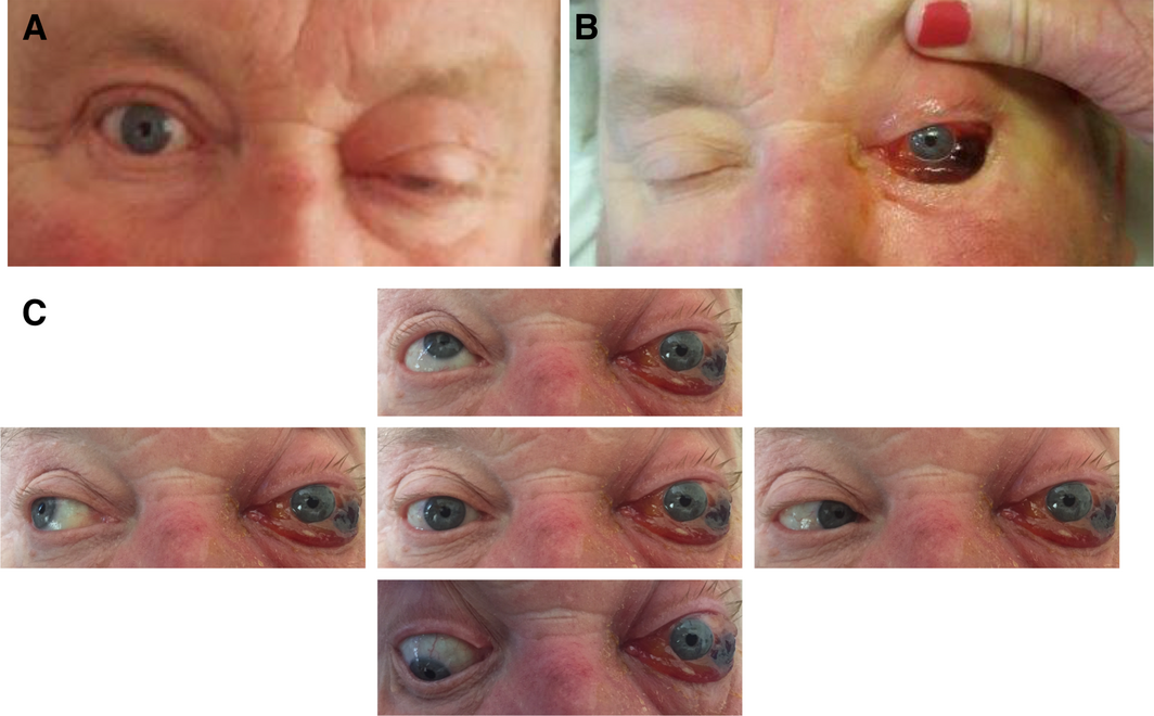
A: The patient at the time of admission to a rural hospital with diagnosed orbital cellulitis. B: After progression, showing worsening proptosis and haemorrhagic chemosis. C: At presentation to our institution, showing proptosis, chemosis, miosis and complete ophthalmoplegia.
Box 2 – Computed tomography scan on arrival at tertiary centre
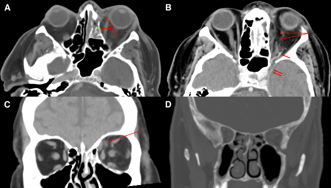
A and B: Axial venous phase images showing pre‐ and post‐septal fat stranding medial to the globe (long arrow, A) and extensive soft tissue density material within the left ethmoid cells (short arrow, A). There is a subperiosteal/extraconal collection between the globe and medial rectus (long arrows, B). The left superior ophthalmic vein is dilated (short arrow, B) compared with the right, and there is fat stranding around a tributary suspicious for thrombophlebitis. There is subtle thickening of the dura overlying the lateral aspect of the left cavernous sinus compared to the normal appearance on the right (double short arrows, B). C: Perivenular fat stranding is also visible in the coronal section (arrow). D: Coronal reconstructed image using bone window display settings showing mild generalised ethmoid and maxillary sinus mucosal disease.
Box 3 – Exenterated left orbit
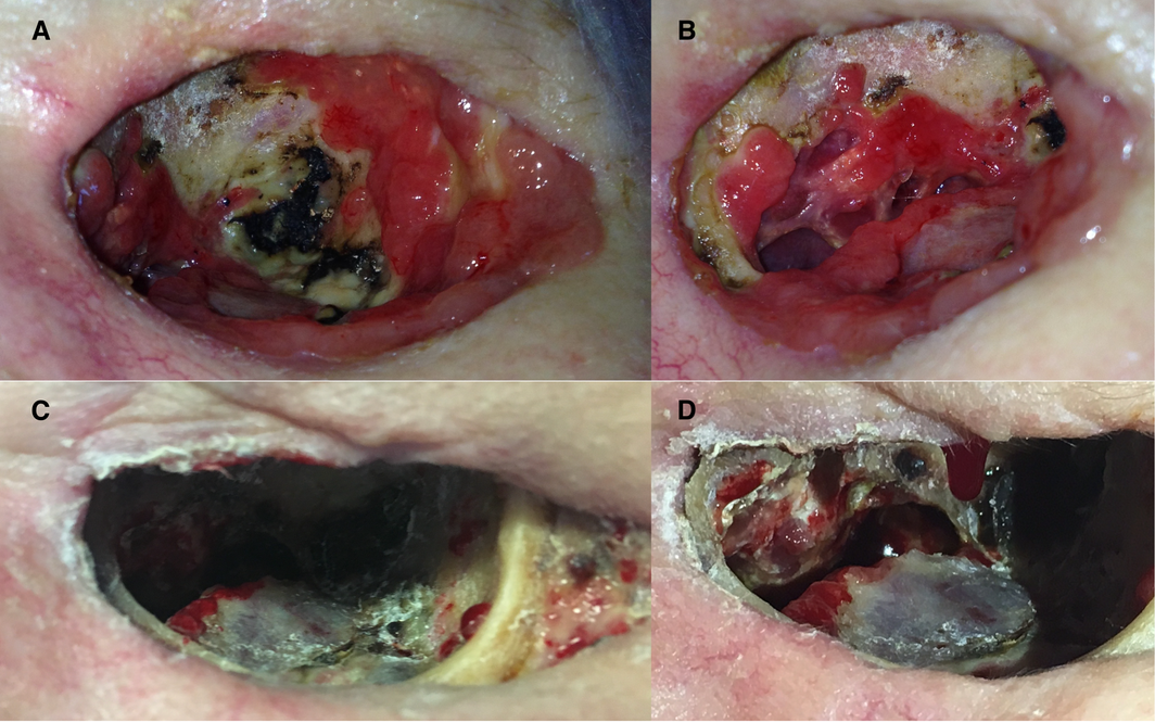
A: Early front‐on post‐operative image. Note the healthy granulation tissue peripherally with areas of black eschar surrounded by a necrotic slough. B: Oblique view of medial left orbital wall. The lamina papyracea has been removed and the ethmoid air cells are visible medially. C: Later front‐on image demonstrating resolution of the eschar and necrosis. D: Oblique view prior to discharge.
Competing interests
No relevant disclosures.
Acknowledgements
We thank the patient and his wife for their insights and help in preparing this article. Their fortitude and commitment throughout his illness were remarkable. We also acknowledge the many other clinicians involved in the patient's care, without whose contributions the outcome might have been quite different.
References
- Cornely OA, Alastruey‐Izquierdo A, Arenz D, et al. Global guideline for the diagnosis and management of mucormycosis: an initiative of the European Confederation of Medical Mycology in cooperation with the Mycoses Study Group Education and Research Consortium. Lancet Infect Dis 2019; 19: e405–e421.
- Vaughan C, Bartolo A, Vallabh N, Leong SC. A meta‐analysis of survival factors in rhino‐orbital‐cerebral mucormycosis‐has anything changed in the past 20 years? Clin Otolaryngol 2018; 43: 1454–1464.
- Prakash H, Ghosh AK, Rudramurthy SM, et al. A prospective multicenter study on mucormycosis in India: epidemiology, diagnosis, and treatment. Med Mycol 2019; 57: 395–402.
- Chamilos G, Lewis RE, Kontoyiannis DP. Delaying amphotericin B‐based frontline therapy significantly increases mortality among patients with hematologic malignancy who have zygomycosis. Clin Infect Dis 2008; 47: 503–509.
- Gamba JL, Woodruff WW, Djang WT, Yeates AE. Craniofacial mucormycosis: assessment with CT. Radiology 1986; 160: 207–212.
- Fanos V, Cataldi L. Amphotericin B‐induced nephrotoxicity: a review. J Chemother 2000; 12: 463–470.
- Saedi B, Sadeghi M, Seilani P. Endoscopic management of rhinocerebral mucormycosis with topical and intravenous amphotericin. Br J Laryngol Otol 2011; 125: 807–810.
Provenance: Not commissioned; externally peer reviewed.
Costs of Delivering the Hep B PAST Model of Care in Remote Australia: A Retrospective Pre–Post Study
Hoa Nguyen, Anh Le Tuan Nguyen, Paula Binks, Melita McKinnon, Emily Vintour-Cesar, Karen Wills, Nicola Stephens, Benjamin Cowie, Joshua S. Davis, Yuejen Zhao, Peter Nihill, Julie A. Campbell, Andrew J. Palmer, Jane Davies, Barbara de Graaff
Buruli Ulcer Acquired From Ringtail Possum Rescue
Daniel P. O'Brien, Naomi Clarke, Sara Bonnici, Mohammad A. Hussain
Q Fever Visceral Abscesses After Contact With Native Australian Marsupials
Samantha J. Laws, Enzo Binotto, James Stewart, Jennifer Ho, Trent Yarwood, Josh Hanson, Simon Smith
Hospital-Admitted Injection-Related Infections Among Incarcerated People Who Inject Drugs in Australia: A Retrospective Cohort Study
Andrew Palmer, Matthew Carter, Jeremy Yeo, Cecilia Shim, Jason Connor, Jeremy Hayllar, Gerald Holtmann, Naomi Moy, Elliott G. Playford, Naomi Runnegar, Paul J. Clark
The Live Attenuated Influenza Vaccine in Australia: An Additional Tool for Influenza Prevention
Cyra Patel, Alexis Pillsbury, Tran Nguyen, Xia Wang, Helen E. Quinn, Clayton K. Chiu, Allen C. Cheng, Katie L. Flanagan, Zhicheng Wang
Reflex Testing for Hepatitis D Infection: A Unique Opportunity to Reduce Hepatitis D-Related Chronic Liver Disease Deaths in Australia
Jessica Howell, Lauren Andersson, Miriam T. Levy, James O'Beirne, Leon Adams, Katharine Irvine, Avik Majumdar, Golo Ahlenstiel, Kathy Jackson, Krispin Hajkowicz, Joseph Doyle, Jane Davies, Sarah Cherian, Wayne Dimech, Alexander J. Thompson