




Medical education
Lessons from practice
Volume 215 - Issue 3
Isolated superior ophthalmic vein thrombosis
Authors: Douglas Dunn, Sebastian Brown, Sartaj Sandhu and Domit Azar
Med J Aust 2021; 215 (3): 106-107.e1. || doi: 10.5694/mja2.51172
Published online: 2 August 2021
Published online: 2 August 2021
A 62-year-old man presented with left eye swelling and 3 weeks of intermittent epiphora without visual disturbance
Clinical record
A 62‐year‐old man presented with left eye swelling and 3 weeks of intermittent epiphora without visual disturbance. He denied pain, photophobia or discharge. Medical history included Graves disease and dyslipidaemia for which he was taking rosuvastatin. His endocrinologist had ceased his thyroid medication 2 years ago following normal thyroid function test results.
On examination, his left eye was proptosed, his visual acuity was 6/4 right and 6/4‐1 left. Intraocular pressures were 12 mmHg right and 20 mmHg left (reference interval [RI], 10–21 mmHg). Exophthalmometry measured a left 6 mm axial proptosis. Ocular motility, anterior segment examination, fundoscopic examination, optic nerve function testing, optical coherence tomography and computerised perimetry results were normal. Several small corkscrew‐like vessels were noted in the bulbar conjunctiva of both eyes of unclear significance, although the left conjunctiva appeared slightly more chemotic than the right.
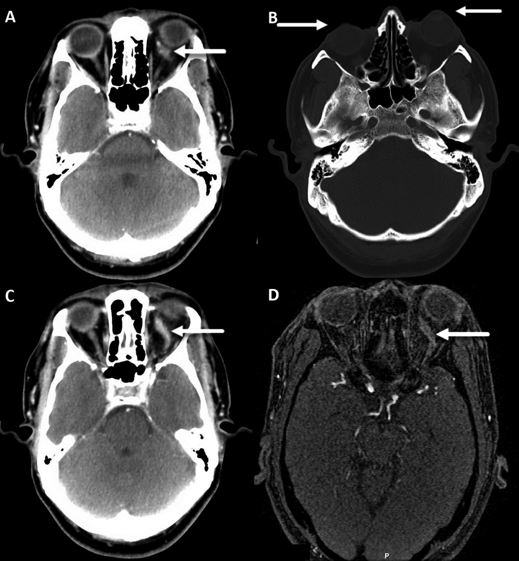
Computed tomography scans of the brain and orbits with contrast and cerebral venography showed superior ophthalmic vein (SOV) and superior rectus enlargement with adjacent fat stranding. The cavernous sinus was asymmetrical (larger on the left) and suspicious for carotid cavernous fistula (Box 1). However, digital subtraction angiography and magnetic resonance imaging of the brain excluded carotid cavernous fistula (Box 1). The patient was diagnosed with isolated SOV thrombosis.
An autoimmune screen was significant for a mildly elevated thyroid‐stimulating hormone receptor antibody level of 2.0 U/L (RI, < 1.0 U/L), consistent with his previous diagnosis of Graves disease. However, he did not have active thyrotoxicosis, as his other thyroid function tests were within normal limits. Antineutrophil cytoplasmic antibody levels were also mildly elevated (titre, 80 [RI, 0–19]. This was an atypical result which is non‐specific and can be associated with inflammatory conditions including Graves disease.
The patient was reviewed by the haematology team who completed a hypercoagulability screen which was unremarkable. Given the lack of functional impairment, it was decided not to proceed with anticoagulation because the risks outweighed the benefits.
Discussion
The estimated incidence of SOV thrombosis is 3–4 cases per million population per year.1 SOV thrombosis has previously been associated with intraorbital/periorbital inflammatory/infective states, hypercoagulable states, carotid cavernous fistula and thyroid eye disease.2,3,4,5 In isolated cases without other venous thrombosis, it has primarily been described in the context of Graves disease.3,4 Sorrentino and colleagues described isolated SOV thrombosis in a patient with symmetrical progressive bilateral proptosis and restricted eye movements in the context of active thyroid eye disease bilaterally.4 In comparison, our patient’s findings were unilateral proptosis and chemosis, both of which would be explained by SOV thrombosis or inflammatory thyroid eye disease, although there were no other indicators of inflammatory thyroid eye disease. Park and colleagues described thyroid eye disease‐associated SOV thrombosis combined with branch retinal vein occlusion.3 Our patient’s fundoscopic and visual examination results were normal. He was euthyroid, and did not demonstrate features of thrombosis elsewhere. Other than a history of Graves disease, our patient did not have any hypercoagulable risk factors. It is unclear in this case whether thyroid eye disease was the cause of the thrombosis, or whether this represented an idiopathic SOV thrombosis, given the lack of other thyroid eye disease signs. Regardless, this was a rare presentation in a patient with proptosis, and Graves disease should be considered in all patients presenting with unilateral proptosis.
Isolated superior rectus oedema, regardless of whether it is the cause of SOV thrombosis, is unusual and was another notable feature of our case. Hudson and colleagues explored the association between superior rectus inflammation and Graves exophthalmos.5 They reported associated SOV enlargement but no cases of thrombosis, and proposed a mechanism of orbital apex crowding leading to reduced venous outflow. Volpe and colleagues described cases of ocular muscular enlargement in patients with ocular misalignment and occult thyroid eye disease.6 They found that inferior rectus and superior rectus/levator complex involvement were most common, but did not report any cases of isolated superior rectus inflammation.
Isolated SOV thrombosis has functional and aesthetic implications for the patient. Diagnosis cannot be made clinically, and requires imaging studies including magnetic resonance imaging or angiography. Investigation should, within reason, exclude primary causes including orbital cellulitis or other periorbital infections, hypercoagulable states, an orbital mass lesion, arteriovenous malformation and carotid cavernous fistula.2 Following workup, an appropriate management and follow‐up plan will be required. The current literature lacks a structured approach to this management.
Management of SOV thrombosis should revolve around treating the underlying cause. For example, patients with SOV thrombosis secondary to orbital cellulitis should be treated with targeted antibiotics and managed symptomatically. However, for those with isolated SOV thrombosis, we propose the management plan set out in Box 2. Follow‐up should be guided by the clinical setting. Close clinical follow‐up (weekly to monthly) is recommended to ensure stability. Once clinically improving or stable, monthly to 6‐monthly follow‐up may be considered until resolution. Monitoring may include regular review of visual acuity, intraocular pressure, exophthalmomtery and cranial nerve examination. Patients should also be educated to seek medical attention if they develop symptoms of carotid cavernous fistula. Follow‐up imaging is also recommended to ensure resolution.
Lessons from practice
- Intraorbital venous thrombosis is an important differential for unilateral painless proptosis. Important aetiologies include thyroid eye disease, intraorbital infection, malignancy, hypercoagulable state, and carotid cavernous fistula.
- Diagnosis of intraorbital venous thrombosis requires orbital cross‐sectional imaging, such as computed tomography or magnetic resonance imaging.
- Management of superior ophthalmic vein thrombosis centres on treatment of the underlying condition; however, management of hypercoagulability and raised intraocular pressure is also an important consideration.
Box 1 – Computed tomography brain imaging showing enlargement of the left superior ophthalmic vein (SOV) with possible associated cavernous sinus enlargement on the left (A), significant proptosis (B) and left isolated SOV thrombosis (C), and magnetic resonance imaging showing SOV thrombosis (D)
Box 2 – Proposed management strategy for isolated superior ophthalmic vein thrombosis1,2,3
Pathological process |
Management approach | ||||||||||||||
Raised intraocular pressure (IOP) |
|
||||||||||||||
Hypercoagulability |
|
||||||||||||||
Competing interests
No relevant disclosures.
References
- Sotoudeh H, Shafaat O, Aboueldahab N, et al. Superior ophthalmic vein thrombosis: What radiologist and clinician must know? Eur J Radiol Open 2019; 6: 258–264.
- Rao R, Ali Y, Nagesh CP, Nair U. Unilateral isolated superior ophthalmic vein thrombosis. Indian J Ophthalmol 2018; 66: 155–157.
- Park SH, Gye JH, Kim MJ, Lee JY. A patient with branch retinal vein occlusion accompanied by superior ophthalmic vein thrombosis due to severe superior ophthalmic vein enlargement in a patient with Graves ophthalmopathy. J Craniofacial Surg 2014; 25: e322–e324.
- Sorrentino D, Taubenslag KJ, Bodily LM, et al. Superior ophthalmic vein thrombosis: a rare complication of Graves’ orbitopathy. Orbit 2018; 37: 175–178.
- Hudson HL, Levin L, Feldon SE. Graves exophthalmos unrelated to extraocular muscle enlargement. superior rectus muscle inflammation may induce venous obstruction. Ophthalmology 1991; 98: 1495–1499.
- Volpe NJ, Sbarbaro JA, Gendron Livingston K, et al. Occult thyroid eye disease in patients with unexplained ocular misalignment identified by standardized orbital echography. Am J Ophthalmol 2006; 142: 75–81.e1.
Provenance: Not commissioned; externally peer reviewed.
Splenic injury in severe cases of the zoonoses Q fever and rickettsial infection: diagnostic challenges
Ashleigh Drury, Philippa Harrison, Aiveen Bannan
Health and economic benefits of improving pre‐hospital identification of stroke in Australian women: a modelling study
Thomas Gadsden, Lei Si, Emily R Atkins, Cheryl Carcel, Xia Wang, Stephen Jan, Mark Woodward, Laura E Downey
Updating the diagnosis and management of elevated serum ferritin levels in the era of routine ferritin testing of blood donors by Australian Red Cross Lifeblood
Gary D Zhang, James Chen, Daniel M Johnstone, Martin B Delatycki, Katie Allen, John K Olynyk
Early cardiovascular collapse after envenoming by snakes in Australia, 2005–2020: an observational study (ASP‐31)
Geoffrey K Isbister, Katherine Z Isoardi, Angela L Chiew, Shane Jenkins, Nicholas A Buckley
Updated recommendations for warfarin reversal in the setting of four‐factor prothrombin complex concentrate
Danielle Robinson, James McFadyen, Eileen Merriman, Chee Wee Tan, Ross Baker, Huyen Tran
Updating the diagnosis and management of iron deficiency in the era of routine ferritin testing of blood donors by Australian Red Cross Lifeblood
Gary D Zhang, Daniel Johnstone, Michael F Leahy, John K Olynyk