




Medical education
Lessons from practice
Volume 211 - Issue 5
Disseminated histoplasmosis in a patient with Crohn's disease on dual immunosuppression
Authors: Michael B MacIsaac, Sasha R Fehily, Stephen Muhi, Linda Yang, Penelope A McKelvie, Cameron J Jeremiah and Barbara Demediuk
Med J Aust 2019; 211 (5): 206-207.e1. || doi: 10.5694/mja2.50298
Published online: 2 September 2019
Published online: 2 September 2019
A 76- year- old man from rural Victoria presented with 4 months of difficulty swallowing due to a painful, large, non-healing tongue ulcer
Clinical record
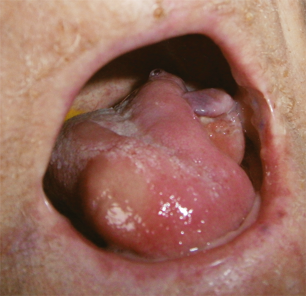
A 76‐year‐old man from rural Victoria presented with 4 months of difficulty swallowing due to a painful, large, non‐healing tongue ulcer (Box 1). He was diagnosed with Crohn's disease 20 years earlier, requiring previous small bowel resections and currently managed with azathioprine and infliximab. Associated symptoms included fatigue, fevers, night sweats and 5 kg weight loss. There was no relevant travel history or exposure to bird or bat guano.
Before admission, biopsies had been performed on the tongue lesion on two occasions, showing ulcerated mucosa with granulomatous inflammation and occasional fungal hyphae, but there was no evidence of malignancy and no organisms had been cultured (extended fungal culture was not performed due to a lack of diagnostic suspicion). A fluorodeoxyglucose positron emission tomography (FDG‐PET) scan demonstrated intense metabolic uptake of the left tongue, with extension into the sublingual space and cervical and retropharyngeal lymph nodes.
On examination, he was febrile with a large, ulcerated lesion on the left lateral tongue, crusted cutaneous lesions over his face, scalp and chest and a heliotrope rash around his eyelids. Blood analysis on admission revealed pancytopenia, with a haemoglobin level of 85 g/L (reference interval [RI], 130–180 g/L), white cell count of 3.0 × 10⁹/L (RI, 4.0–11.0 × 10⁹/L) and platelet count of 125 × 109/L (RI, 150–400 × 109/L). Inflammatory markers were raised, with a C‐reactive protein concentration of 170 mg/L (RI, < 5 mg/L) and erythrocyte sedimentation rate of 43 mm/h (RI, < 13 mm/h). Creatinine kinase concentration was normal (20 U/L; RI, < 170U/L). Antinuclear antibody was weakly positive (speckled). Myositis‐specific antibody panel was positive for anti‐Mi‐2β and anti‐nuclear matrix protein 2 antibodies.
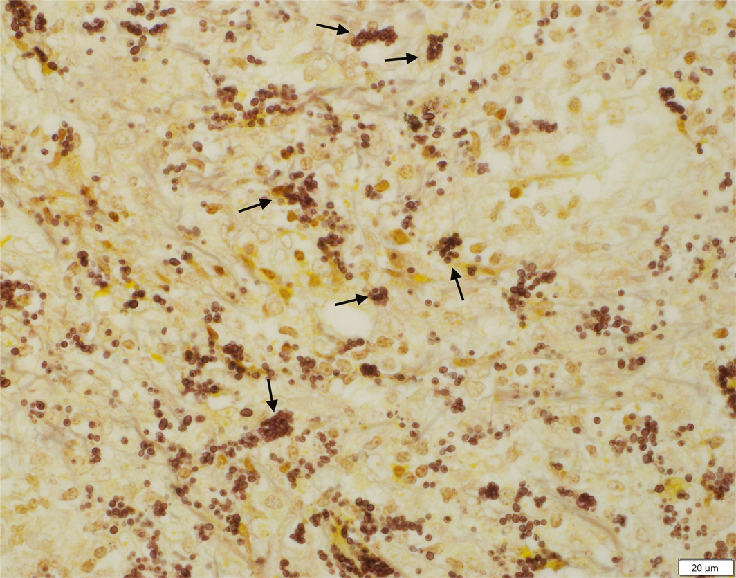
There was a strong suspicion of a primary oral cancer with associated paraneoplastic dermatomyositis. Neck to pelvis computed tomography showed an ill defined irregular region in the left tongue, enlarged cervical lymph nodes bilaterally (maximum size, 6 mm), and focal consolidation and centrilobular nodules throughout both lungs. A repeat FDG‐PET scan was again suspicious for a primary oral cancer. A further tongue biopsy was performed, with periodic acid–Schiff (PAS) and silver methenamine staining (Box 2) showing intracellular organisms in the cytoplasm of macrophages, suggestive of microsporidia. Retrospective review of other biopsy sites (periorbital rash, duodenal ulcer) also revealed PAS‐positive intracytoplasmic organisms. Although therapy with albendazole was commenced, the patient died shortly thereafter.
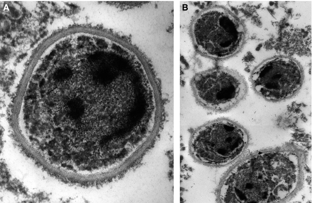
Posthumous examination of electron microscopy samples revealed no internal features of microsporidia. Microsporidium polymerase chain reaction (PCR) testing was negative. Further evaluation with panfungal PCR was positive for Histoplasma capsulatum. A final diagnosis of disseminated histoplasmosis was supported by the clinical presentation and subsequent molecular investigations (Box 3).
Discussion
Histoplasma capsulatum is a dimorphic soil fungus that is endemic to Northern and Central America, parts of Europe and South‐East Asia, but is rarely observed in Australia.1 Less than 70 cases have been reported in Australia, with only four cases described in Victoria — one locally acquired and three imported.1,2 This is therefore the second reported case of histoplasmosis acquired in Victoria and the first Australian case in a patient receiving anti‐tumour necrosis factor (TNF)‐α therapy.
Most immunocompetent patients exposed to H. capsulatum are asymptomatic, although a mild, self‐limiting pneumonitis may occur with greater inoculation. Disseminated disease is more frequently seen in immunosuppressed patients, particularly those with human immunodeficiency virus (CD4 count < 150/μL).1 Disseminated disease is increasingly recognised in patients receiving anti‐TNF‐α therapy, due to the critical role of TNF‐α in the host immune response to H. capsulatum.3
Disseminated histoplasmosis manifests with a broad range of symptoms including fever, cough, weight loss, hepatosplenomegaly, lymphadenopathy and pancytopenia.4 Oropharyngeal ulceration is well recognised, particularly in immunosuppressed patients. Although our patient was pancytopenic, a bone marrow biopsy was considered to be of low diagnostic yield and therefore not performed. Liver enzyme levels were mildly raised, but there was no demonstrated liver or spleen involvement on imaging.
Few reports exist of disseminated histoplasmosis in patients with dermatomyositis, with most already receiving immunosuppressive therapy at the time of presentation.5
Diagnosis of histoplasmosis is difficult, requiring a combination of fungal culture and histopathology. Fungal culture can take up to 6 weeks and must be specifically requested. Extended culture was not done in this case but should be considered in similar cases in the future. Histopathological identification requires methenamine silver or PAS stains.4 Newer diagnostic techniques including serology and PCR may be helpful but are available at reference laboratories only, and serology is unreliable in immunocompromised hosts. Urinary antigen is unavailable in Australia. Treatment is indicated for disseminated disease in immunosuppressed patients with intravenous amphotericin followed by oral itraconazole in severe cases, or itraconazole alone for mild manifestations.
Disseminated histoplasmosis is rare and has a broad range of clinical manifestations, and culture requires a long incubation period. With increasing use of anti‐TNF‐α therapy, clinicians should be aware of histoplasmosis when investigating for opportunistic infections.
Lessons from practice
- Histoplasma capsulatum is endemic to Australia. Despite clinical infection occurring infrequently, it is an important opportunistic pathogen to recognise.
- Patients with progressive disseminated histoplasmosis present with diverse clinical features, hindering early diagnosis and timely initiation of therapy.
- Oropharyngeal infection has been well described as a manifestation of disseminated histoplasmosis, particularly in immunocompromised patients.
- Anti‐tumour necrosis factor‐α therapy is increasingly used for a range of autoimmune and inflammatory conditions and is associated with an increased risk of opportunistic infections, including histoplasmosis.
Box 1 – Clinical photograph showing left‐sided tongue ulceration, following biopsies performed as part of the diagnostic evaluation
Competing interests
No relevant disclosures.
References
- McLeod DS, Mortimer RH, Perry‐Keene DA, et al. Histoplasmosis in Australia: report of 16 cases and literature review. Medicine 2011; 90: 61–68.
- Muhi S, Crowe A, Daffy J. Acute pulmonary histoplasmosis outbreak in a documentary film crew travelling from Guatemala to Australia. Trop Med Infect Dis 2019; 4: 25.
- Culter JE, Deepe Jr GS, Klein BS. Advances in combating fungal disease: vaccines on the threshold. Nat Rev Microbiol 2007; 5: 13–28.
- Kauffman CA. Histoplasmosis: a clinical and laboratory update. Clin Microbiol Rev 2007; 20: 115–132.
- Dussouil AS, Allardet‐Servent J, Dunogeant L, et al. Disseminated histoplasmosis partially mimicking a dermatomyositis in a patient with rheumatoid arthritis. Br J Dermatol 2015; 173: 797–800.
Provenance: Not commissioned; externally peer reviewed.
Costs of Delivering the Hep B PAST Model of Care in Remote Australia: A Retrospective Pre–Post Study
Hoa Nguyen, Anh Le Tuan Nguyen, Paula Binks, Melita McKinnon, Emily Vintour-Cesar, Karen Wills, Nicola Stephens, Benjamin Cowie, Joshua S. Davis, Yuejen Zhao, Peter Nihill, Julie A. Campbell, Andrew J. Palmer, Jane Davies, Barbara de Graaff
Buruli Ulcer Acquired From Ringtail Possum Rescue
Daniel P. O'Brien, Naomi Clarke, Sara Bonnici, Mohammad A. Hussain
Q Fever Visceral Abscesses After Contact With Native Australian Marsupials
Samantha J. Laws, Enzo Binotto, James Stewart, Jennifer Ho, Trent Yarwood, Josh Hanson, Simon Smith
Hospital-Admitted Injection-Related Infections Among Incarcerated People Who Inject Drugs in Australia: A Retrospective Cohort Study
Andrew Palmer, Matthew Carter, Jeremy Yeo, Cecilia Shim, Jason Connor, Jeremy Hayllar, Gerald Holtmann, Naomi Moy, Elliott G. Playford, Naomi Runnegar, Paul J. Clark
The Live Attenuated Influenza Vaccine in Australia: An Additional Tool for Influenza Prevention
Cyra Patel, Alexis Pillsbury, Tran Nguyen, Xia Wang, Helen E. Quinn, Clayton K. Chiu, Allen C. Cheng, Katie L. Flanagan, Zhicheng Wang
Reflex Testing for Hepatitis D Infection: A Unique Opportunity to Reduce Hepatitis D-Related Chronic Liver Disease Deaths in Australia
Jessica Howell, Lauren Andersson, Miriam T. Levy, James O'Beirne, Leon Adams, Katharine Irvine, Avik Majumdar, Golo Ahlenstiel, Kathy Jackson, Krispin Hajkowicz, Joseph Doyle, Jane Davies, Sarah Cherian, Wayne Dimech, Alexander J. Thompson