




Medical education
Lessons from practice
Volume 209 - Issue 9
Menagerie: Bartonella henselae chorioretinitis in the setting of diverse animal exposure
Authors: Fumitaka Nonaka, Susan M Carden, Coen Butters and Christine Chen
Med J Aust 2018; 209 (9): 284-286. || doi: 10.5694/mja18.00205
Published online: 5 November 2018
Published online: 5 November 2018
A previously healthy 12-year-old girl presented to a rural hospital with 10 days of intermittent fevers
Clinical record
A previously healthy 12-year-old girl presented to a rural hospital with 10 days of intermittent fevers. On Day 19 of fever, the patient was referred to a tertiary paediatric centre. On arrival, she described lethargy, headache, mild photophobia, and pain affecting her neck and back. Her medical history was remarkable for diverse animal exposure, including chickens, rabbits, horses, seven cats, one kitten, a green-cheeked conure and a pet cow. Examination revealed isolated 5 mm left-sided cervical lymphadenopathy. There was mild lumbar spinal tenderness on palpation. Cardiac examination was normal. Ophthalmoscopy was not performed at presentation due to photophobia.
Testing revealed a high C-reactive protein level and erythrocyte sedimentation rate, together with a positive antinuclear antibody (Box 1). The differential diagnoses included subacute bacterial endocarditis, occult osteomyelitis and a range of zoonoses (Q fever, cat scratch fever and rickettsial disease). Urine culture and serial blood cultures were subsequently negative.
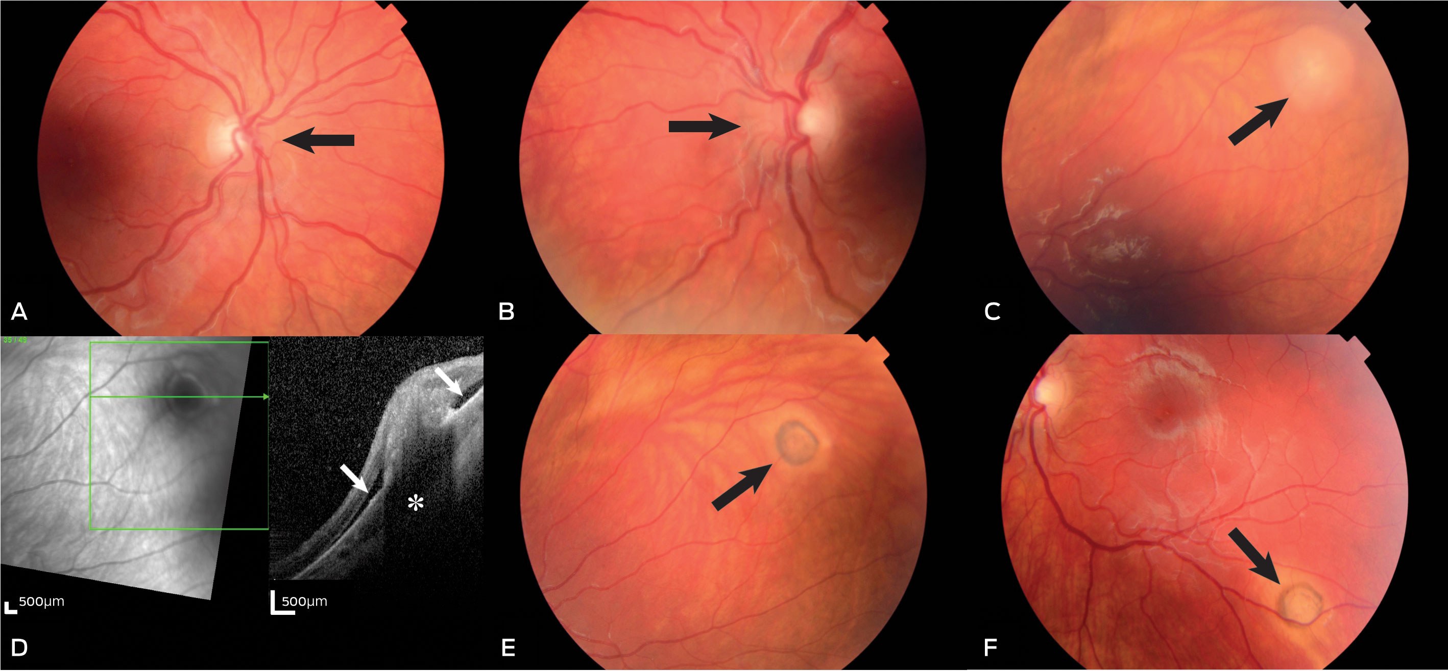
Vague lower back pain and headache suggested possible malignancy or discitis and prompted magnetic resonance imaging (MRI) scan of the brain and spine. While the MRI scan was unremarkable, the patient continued to spike fevers ≥ 39°C. Persisting photophobia and concern for zoonotic infection led to formal ophthalmology assessment. On Day 21, her uncorrected visual acuity was 6/6 in the right eye and 6/5 in the left eye. The anterior segments were normal. The optic disc margins were blurred. The left eye showed mild vitritis and two chorioretinal lesions: the first in the superotemporal retina and the second in the inferotemporal retina (Box 2). This raised the possibility of endocarditis with embolic phenomenon, and treatment was deferred while retinal lesions were documented and transthoracic echocardiography arranged.
Echocardiography confirmed normal cardiac anatomy and no vegetation. Doxycycline was commenced on Day 27, with the provisional diagnosis of Bartonella henselae infection. The patient had rapid improvement in symptoms and resolution of fever. On Day 37, serology was available, confirming bartonellosis with total antibody titre ≥ 2028 (Box 1). Rickettsial serology (spotted fever and typhus groups) showed low positive titres, which was attributed to antibody cross-reactivity. The patient successfully completed 6 weeks of oral antibiotic therapy. At 7 weeks, the patient was asymptomatic with visual acuity of 6/6 in each eye. Ophthalmoscopy showed mild retinal scarring with partial involution of the retinal lesions and pigmentary changes (Box 2, E and F).
B artonella henselae is a small Gram-negative intracellular organism first described in 1983. It is most readily cultured from under the nails of kittens, giving rise to the term “cat scratch disease” or “cat scratch fever”.1 It has also been cultured from dogs and Australian foxes. The spectrum of disease is broad, from isolated pyrexia and classic cat scratch fever, consisting of fever, malaise and regional lymphadenopathy, to rarer complications, including encephalitis, glomerulonephritis, endocarditis and osteomyelitis.1 B. henselae is an important cause of pyrexia of unknown origin, implicated in an estimated 5% of paediatric cases.2
Eye involvement in bartonellosis is thought to occur in about 5% of cases.1 Features include follicular conjunctivitis, keratitis, uveitis, retinal or choroidal lesions, neuroretinitis and retinal vessel occlusion.3 Retinal and choroidal lesions are under-recognised but may be a key diagnostic pointer. It is essential to exclude alternate infectious causes such as toxoplasmosis, syphilis and tuberculosis, and non-infectious causes, such as sarcoidosis and leukaemia. While ophthalmoscopy in children may be challenging, as this case clearly demonstrates, it is critical in all cases of pyrexia of unknown origin.
Many patients with bartonellosis do not recall an animal scratch; hence, it is important to explore potential home and occupational exposures. Serum antibody titres aid diagnosis but have several limitations. IgM levels may be low in early disease and are raised for only 3 months from the time of exposure. There is also well described cross-reactivity between antibody for Rickettsiae and Bartonella species as well as Coxiella burnetii.4 Polymerase chain reaction by a variety of methods has established specificity but disappointing sensitivity.1
There is a paucity of data to guide treatment. Fortunately, most infections are self-limiting and require symptomatic management only. For neuroretinitis, doxycycline may be preferable due to its oral bioavailability, activity against intracellular organisms and distribution within neuroretinal tissue.1 Doxycycline is generally considered safe in children older than 8 years. There is no evidence to support removal of healthy animals from households. Domestic animals should receive regular health checks and treatment for arthropod infections.
This case highlights B. henselae as a cause of undifferentiated fever in children. As Bartonella and Rickettsiae antibody response lags behind clinical presentation, there may be a role for a therapeutic trial of oral doxycycline when clinical suspicion is high.Lessons from practice
Bartonella henselae infection is an under-recognised cause of undifferentiated fever, responsible for about 5% of cases in children.
Retinal or choroidal lesions are an important clinical manifestation of infection with B. henselae and many non-infectious causes of fever. An early ophthalmic examination should be considered in all cases of undifferentiated fever in children.
Doxycycline may be used as a therapeutic trial while awaiting serological diagnosis in patients who have a high pre-test probability of zoonotic infection due to Rickettsia or Bartonella species.
There is no evidence to support removal of healthy animals from households; however, domestic animals should receive regular health checks and treatment for arthropod infections.
Box 1 – Laboratory tests performed during admissions, between Day 10 and Day 35
Test items |
Reference intervals |
First admission* (Day 10–19) |
Second admission† (Day 19–35) |
||||||||||||
General | |||||||||||||||
Full blood examination |
WNL |
WNL |
|||||||||||||
Urea, electrolytes and creatinine |
WNL |
WNL |
|||||||||||||
Liver function tests |
WNL |
WNL |
|||||||||||||
Coagulation profile |
WNL |
||||||||||||||
C-reactive protein, mg/L (Day) |
0–5 |
82 (10) |
64 (24) |
||||||||||||
96 (13) |
43 (27) |
||||||||||||||
63 (18) |
23 (31) |
||||||||||||||
Erythrocyte sedimentation rate, mm/h (Day) |
0–7 |
44 (12) |
77 (20) |
||||||||||||
61 (15) |
65 (34) |
||||||||||||||
HLA-B27 |
Negative |
Negative |
|||||||||||||
Autoantibodies | |||||||||||||||
Rheumatoid factor |
Negative |
Negative |
|||||||||||||
Antinuclear antibodies |
Negative (titre < 160) |
Negative |
Positive (160) |
||||||||||||
ANCA |
|||||||||||||||
Myeloperoxidase antibodies (p-ANCA), RU/mL |
Negative (titre < 20) |
Negative |
|||||||||||||
Proteinase 3 antibodies (c-ANCA), RU/mL |
Negative (titre < 20) |
Negative |
|||||||||||||
Antidouble-stranded DNA, IU/mL |
Negative (< 29) |
Negative |
|||||||||||||
Anti-extractable nuclear antigen antibodies |
Negative |
Negative |
|||||||||||||
Infectious disease screening | |||||||||||||||
Cytomegalovirus serology |
Negative |
IgM negative |
|||||||||||||
IgG positive | |||||||||||||||
Epstein–Barr serology |
Negative |
Negative |
|||||||||||||
Mycoplasma serology |
Negative |
Negative |
|||||||||||||
Brucella serology |
Negative |
Negative |
|||||||||||||
Streptococcal serology |
|||||||||||||||
Antistreptolysin O antibodies, IU/mL |
< 535 |
Lower level (134) |
|||||||||||||
Anti-DNase B testing, IU/mL |
< 300 |
400 |
|||||||||||||
Cryptococcal antigen |
Negative |
Negative |
|||||||||||||
Q fever serology |
Negative (titre < 25) |
Negative |
|||||||||||||
Toxoplasma serology |
Negative |
Negative |
|||||||||||||
Ross River virus antibodies |
Negative |
Negative |
|||||||||||||
Bartonella serology |
|||||||||||||||
Bartonella henselae |
Negative (titre < 128) |
Positive (≥ 2028) |
|||||||||||||
Rickettsial serology |
|||||||||||||||
Spotted fever group |
Negative (titre < 128) |
Positive (128–256) |
|||||||||||||
Typhus group |
Negative (titre < 128) |
Positive (128–256) |
|||||||||||||
Scrub typhus group |
Negative (titre < 128) |
Negative |
|||||||||||||
ANCA = antineutrophil cytoplasmic antibodies. HLA-B27 = human leukocyte antigen B27. WNL = within normal limit. * First admission: rural hospital. † Second admission: tertiary paediatric centre. | |||||||||||||||
Box 2 – A and B: Fundus photographs of the right (A) and left (B), showing mild blurriness of the optic disc margins (arrows). C: Fundus photographs of the left, showing a whitish chorioretinal plaque in the superotemporal retina (arrow). D: Ocular coherence tomography image of the corresponding area, representing a focus mainly located in the subretinal area, casting an acoustic shadow (asterisk), and being surrounded by subretinal fluid (white arrows). E and F: Fundus photographs of the left, showing two lesions in the superotemporal retina (E, arrow, same as the lesion in C) and in the inferotemporal retina (F, arrow, found on review) after 2 weeks of treatment
Competing interests
No relevant disclosures.
References
- Florin TA, Zaoutis TE, Zaoutis LB. Beyond cat scratch disease: widening spectrum of Bartonella henselae infection. Pediatrics 2008; 121: e1413-e1425.
- Jacobs RF, Schutze GE. Bartonella henselae as a cause of prolonged fever and fever of unknown origin in children. Clin Infect Dis 1998; 26: 80-84.
- Amer R, Tugal-Tutkun I. Ophthalmic manifestations of bartonella infection. Curr Opin Ophthalmol 2017; 28: 607-612.
- La Scola B, Raoult D. Serological cross-reactions between Bartonella quintana, Bartonella henselae, and Coxiella burnetii. J Clin Microbiol 1996; 34: 2270-2274.
A Diagnostic Headache
Thomas Glynn, Michelle T. Leech, Stacy K. Goergen, Emily Lin, Nadeem Toodayan, Ralph Junckerstorff
Neisseria meningitidis: an uncommon cause of conjunctivitis
Chloe Story, Tina Marinelli, Simeon Crawford, Amrita Ronnachit
Dismantling barriers to research and clinical care for individuals with a vision impairment
Eden G Robertson, Kate Hetherington, Meredith Prain, Julia Hall, Leighton Boyd AM, Rosemary Boyd OAM, Emily Shepard, Hollie Feller, Sally Karandrews, Fleur O'Hare, Kanae Yamamoto, Matthew P Simunovic, Robyn V Jamieson, Alan Ma, Lauren Ayton AM, Anai Gonzalez‐Cordero
The New South Wales Pharmacy Trial for herpes zoster: on the nose?
Christian P Pappas, Timothy R Holmes, Minas T Coroneo
Skin fragility disorder misdiagnosed as child abuse: a cautionary tale
Heather G Mack, Fred K Chen, John R Grigg
Health economic aspects of inherited retinal diseases: looking for cost‐effective treatments
Benjamin Kamien, Rachael Heath Jeffery, Fred K Chen