




Medical education
Lessons from practice
Volume 209 - Issue 7
Hunting for a cause of painful diplopia
Authors: Sarah L Stevens and Daniel Schweitzer
Med J Aust 2018; 209 (7): 293-294. || doi: 10.5694/mja18.00002
Published online: 1 October 2018
Published online: 1 October 2018
A fit, active 80-year-old man presented to the emergency department with double vision
Clinical record
A fit, active 80-year-old man presented to the emergency department (ED) with double vision. He had no relevant past medical history.
He had woken one week earlier with pain over his left brow and behind his left eye; diplopia developed a day later. The headache resolved after 4 days but he had ongoing double vision on presentation to the ED. There was no prior history of trauma or infections. He had no temporal pain or jaw claudication. He was otherwise systemically well, without constitutional symptoms.
On examination, he looked well. He was afebrile and normotensive. His diplopia resolved with closure of either eye in keeping with binocular diplopia. Although we could not easily discern any ophthalmoplegia in the ED, his description of the diplopia pointed to the offending muscles: he described normal vision with rightward gaze, vertical diplopia with downward gaze and horizontal diplopia with leftward gaze. The presence of both vertical and horizontal diplopia suggested that more than one extra-ocular muscle was affected; in this case, the left lateral rectus and left inferior rectus muscles. This was verified by specialist ophthalmology testing. Visual acuity was preserved and fundoscopic evaluation was unremarkable. Neurological examination was otherwise normal; specifically, he had no facial sensory loss and no temporal tenderness. In summary, his examination revealed left partial third and sixth nerve palsy.
Blood tests, which included full blood count, electrolytes and liver function tests, erythrocyte sedimentation rate and C-reactive protein, and tests for diabetes, sarcoidosis, vasculitis, Treponema pallidum, tuberculosis and syphilis, were within normal limits. Brain computed topography (CT) was also normal.
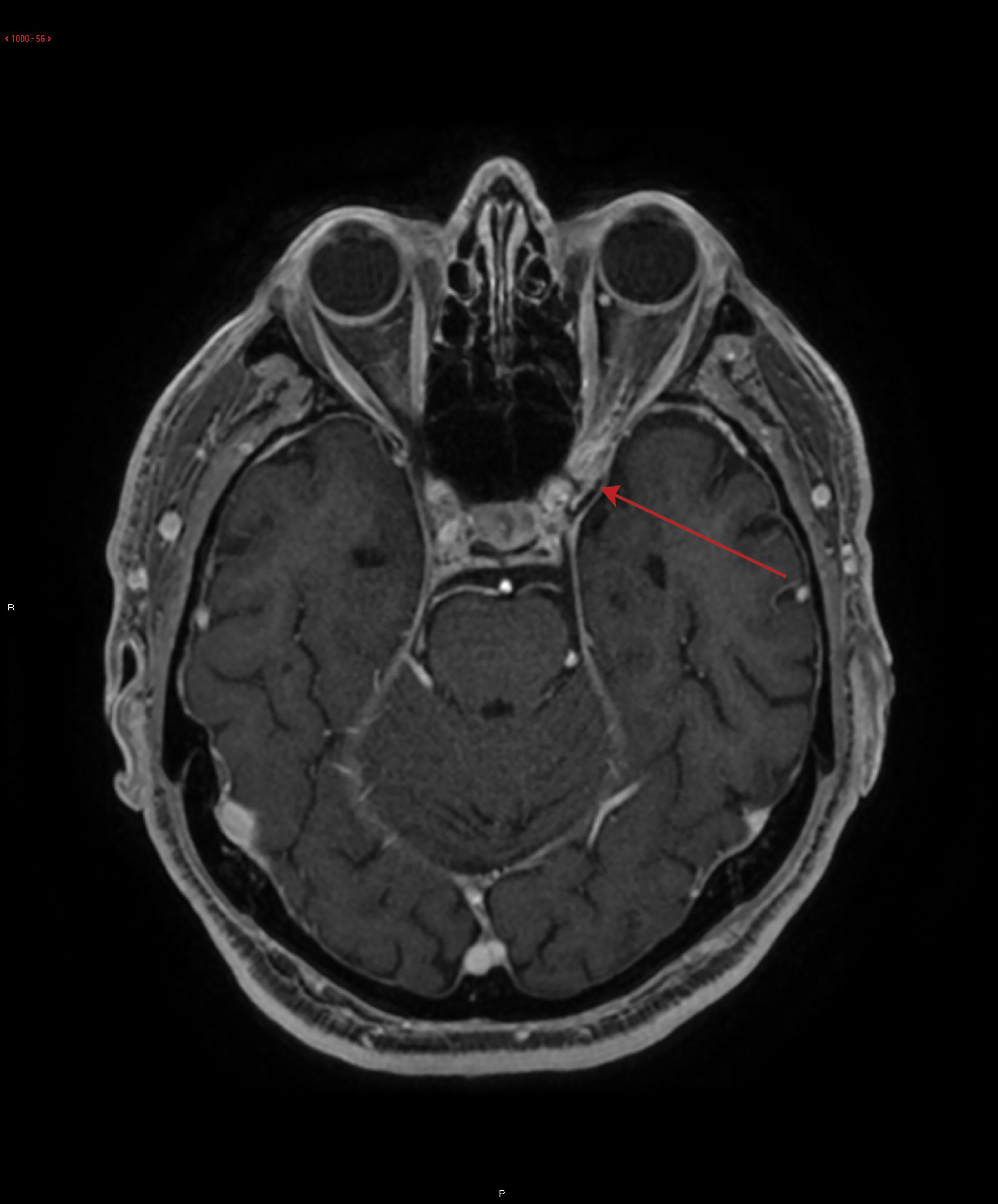
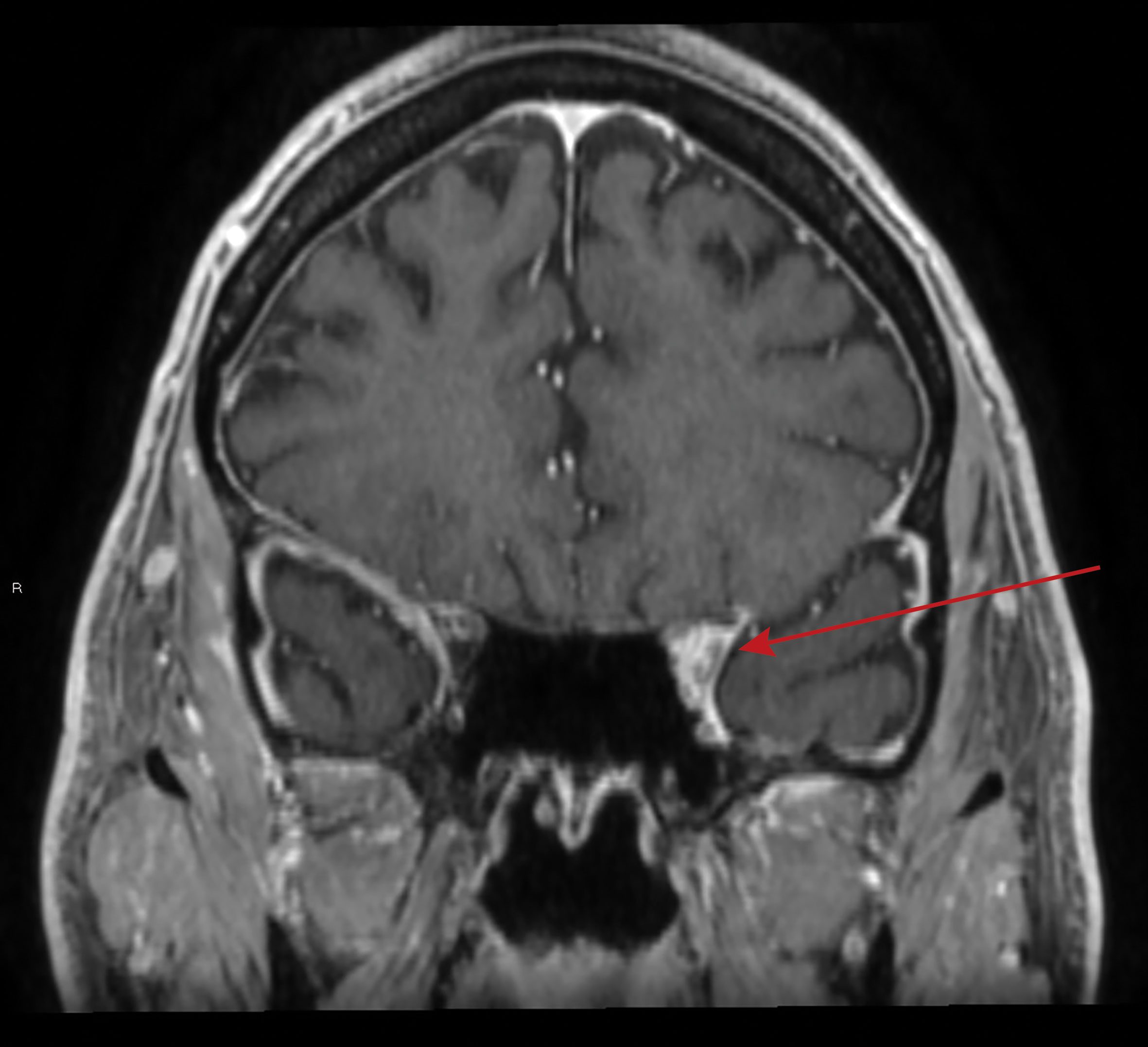
A magnetic resonance imaging (MRI) brain scan revealed an ill-defined soft tissue enhancement and oedema from the orbital apex towards the cavernous sinus, consistent with Tolosa–Hunt syndrome (Box 1 and Box 2).
He was prescribed oral prednisolone; initially 50 mg daily, but then increased to 100 mg daily when he failed to respond after 2 days. Prednisolone 100 mg was continued for a total of 7 days without a tapering course. His diplopia resolved and he continued to remain well at his 6-month review.
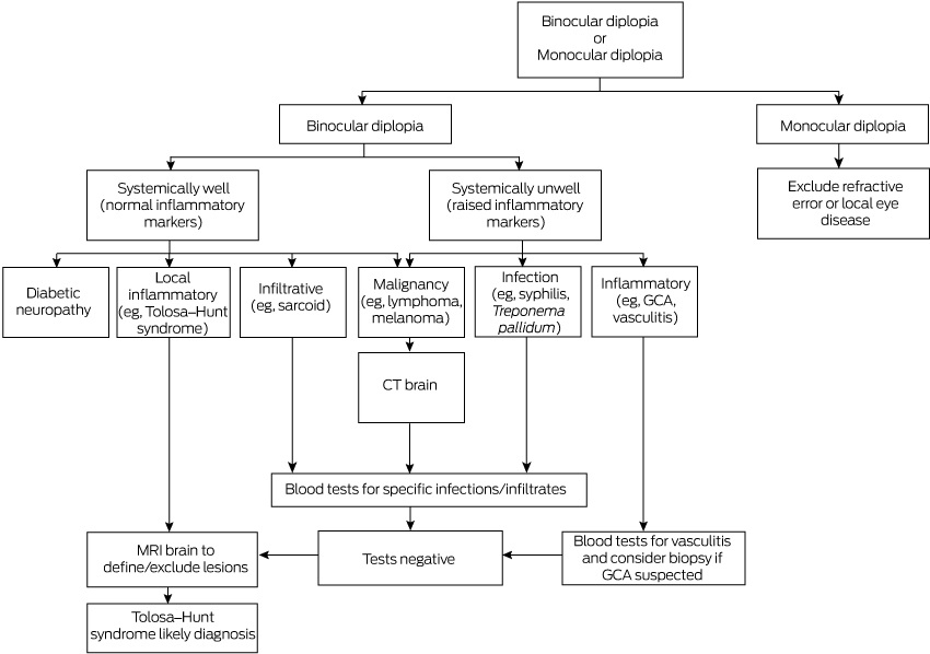
Distinguishing between monocular and binocular diplopia is the first step in determining the diagnosis of double vision. Monocular diplopia can be differentiated from binocular diplopia by asking the patient to close one eye. If the double vision resolves, this indicates binocular diplopia, suggesting that the underlying cause is ocular malalignment. This is usually caused by ophthalmoplegia. Differential diagnosis includes neoplasms such as lymphoma, infiltrative causes, inflammatory disease and infective causes (Box 3). Causes of monocular diplopia include refractive error and local eye disease.1
It is important to exclude giant cell arteritis (GCA), which, if left untreated, can lead to permanent visual loss and death. It is the most common systemic vasculitis and occurs in people aged over 50 years. GCA typically presents with a new headache and abrupt onset of visual changes, most commonly transient visual loss. Jaw claudication can occur. Presentations can be variable but, generally, patients have constitutional symptoms such as fever and malaise and have elevated inflammatory markers such as erythrocyte sedimentation rate and C-reactive protein. The gold standard for diagnosis is a temporal artery biopsy.2
In view of the normal inflammatory markers in our patient, a diagnosis of vasculitis and GCA was less likely. Similarly, we could find no evidence of infection or infiltrative process (eg, sarcoidosis). Diabetic ischaemic neuropathy was considered but the patient was normoglycaemic with a normal glycated haemoglobin level. Malignancy and large space-occupying lesions were grossly ruled out with CT brain scan.
After the initial investigations, the differential diagnosis narrowed to local inflammatory conditions without a systemic illness, which, based on the neuroimaging findings, included Tolosa–Hunt syndrome, sclerosing pseudotumour and IgG4-related disease.3
The MRI brain scan was ordered to determine if there was a compressive lesion affecting cranial nerves III and VI. It was invaluable in ruling out a neoplasm and collection and vascular causes for his symptoms (eg, cavernous sinus thrombosis). Sclerosing pseudotumours typically present bilaterally and extend into the sinuses. IgG4-related disease has a predilection for lacrimal glands. Tolosa–Hunt syndrome usually presents unilaterally and extends into the cavernous sinus and, hence, was most consistent with the clinical presentation and MRI findings.3
Tolosa–Hunt syndrome is caused by an inflammatory process of unknown aetiology affecting the cavernous sinus. It is an uncommon cause of unilateral headache and ophthalmoplegia (incidence approximately one in 1 million). It can present at any age and affects both men and women equally.4
The resultant ophthalmoplegia most commonly affects cranial nerves’ III and VI due to local oedema, as in this case; however, given that the cavernous sinus contains cranial nerves III, IV, V1, V2 and VI, it can affect any of these nerves. The natural history of Tolosa–Hunt syndrome is spontaneous resolution after 8 weeks and it is known to be very sensitive to glucocorticoid therapy.4,5
Lessons from practice
-
Determining whether diplopia is monocular or binocular is important in establishing the cause of double vision.
-
Binocular diplopia is usually caused by extra-ocular muscle dysfunction.
-
Monocular diplopia is most likely due to local eye disease or refractive error.
-
For binocular diplopia, the direction (vertical, horizontal, diagonal) may indicate which muscles are involved, especially if the signs are subtle.
-
For people aged over 50 years, it is important to consider giant cell arteritis as a cause of new onset headache or visual disturbances.
-
Tolosa–Hunt syndrome is a rare cause of painful diplopia and typically responds well to oral corticosteroids.
Box 1 – Magnetic resonance imaging axial T1 post-contrast: ill-defined lesion of the left orbital apex extending into the cavernous sinus (arrow)
Competing interests
No relevant disclosures.
References
- Bienfang DC. Overview of diplopia [website]. UpToDate; 2017. https://www.uptodate.com/contents/overview-of-diplopia?search=diplopia&source=search_result&selectedTitle=1∼150&usage_type=default&display_rank=1 (viewed Jan 2018).
- William PD. Diagnosis of giant cell arteritis [website]. UpToDate; 2017. https://www.uptodate.com/contents/diagnosis-of-giant-cell-temporal-arteritis (viewed Feb 2018).
- Schuknecht B, Sturm V, Huisman TA, Landau K. Tolosa–Hunt syndrome: MR imaging features in 15 patients with 20 episodes of painful ophthalmoplegia. Eur J Radiol 2009; 69: 445-453.
- Shindler KS. Tolosa–Hunt syndrome [website]. UpToDate; 2016. https://www.uptodate.com/contents/tolosa-hunt-syndrome?search=Tolosa%20Hunt&source=search_result&selectedTitle=1∼6&usage_type=default&display_rank=1 (viewed Dec 2017).
- Kline LB, Hoyt WF. The Tolosa–Hunt syndrome. J Neurol Neurosurg Psychiatry 2001; 71: 577-582.
Linked content
-
MJA Letter: Hunting for a cause of painful diplopia
Provenance: Not commissioned; externally peer reviewed.
A Diagnostic Headache
Thomas Glynn, Michelle T. Leech, Stacy K. Goergen, Emily Lin, Nadeem Toodayan, Ralph Junckerstorff
Neisseria meningitidis: an uncommon cause of conjunctivitis
Chloe Story, Tina Marinelli, Simeon Crawford, Amrita Ronnachit
Dismantling barriers to research and clinical care for individuals with a vision impairment
Eden G Robertson, Kate Hetherington, Meredith Prain, Julia Hall, Leighton Boyd AM, Rosemary Boyd OAM, Emily Shepard, Hollie Feller, Sally Karandrews, Fleur O'Hare, Kanae Yamamoto, Matthew P Simunovic, Robyn V Jamieson, Alan Ma, Lauren Ayton AM, Anai Gonzalez‐Cordero
The New South Wales Pharmacy Trial for herpes zoster: on the nose?
Christian P Pappas, Timothy R Holmes, Minas T Coroneo
Skin fragility disorder misdiagnosed as child abuse: a cautionary tale
Heather G Mack, Fred K Chen, John R Grigg
Health economic aspects of inherited retinal diseases: looking for cost‐effective treatments
Benjamin Kamien, Rachael Heath Jeffery, Fred K Chen