




Volume 209 - Issue 7
The gut microbiota: cause and cure of gut diseases
Authors: Lauren S White, Johan Van den Bogaerde and Michael Kamm
Med J Aust 2018; 209 (7): 312-317. || doi: 10.5694/mja17.01067
Published online: 1 October 2018
Published online: 1 October 2018
The gut microbiota is a key cause, and potential cure, of modern gut disorders including inflammatory bowel disease, liver diseases, metabolic syndrome and obesity
Summary
- The gastrointestinal microbiota is emerging as a central factor in the pathogenesis of a range of gastrointestinal and hepatic disorders. Epidemiological studies, and experimental studies in animals and humans, have highlighted a likely causative role of this microbial community in the modern global epidemics of inflammatory bowel disease, non-alcoholic fatty liver disease, non-alcoholic steato-hepatitis, obesity and metabolic syndrome.
- New techniques for microbial culture and gene sequencing are enabling the identification of specific pathogens and protective organisms in these conditions.
- Factors that change the microbiota are being defined: dietary pattern, specific foods, food additives in processed food and drinks, such as emulsifiers and non-sugar sweeteners, and antibiotics. Microbiota changes in early life appear critical to the later development of a range of inflammatory disorders.
- For many of these conditions, the treatment paradigm will change, at least in part, from immune suppression and drug therapy to treatments that reshape the microbiota or restore its integrity. These treatments include dietary changes, specific microbial manipulation and faecal microbiota transplantation.
- A dialogue is needed regarding population strategies that target disease prevention. This will include how food is produced, what additives it contains, and how it is processed. Widespread use of antibiotics, from agricultural and veterinary to medicinal settings, needs more attention.
- At the individual level, microbial profiles may be able to predict who is at risk of disease when subjected to particular environmental influences, and what microbial restoration is needed to minimise risk.
The gut microbiota is recognised increasingly to be of central importance to our wellbeing, disease pathogenesis, and our ability to treat many common conditions. This microbial community inhabiting the human gastrointestinal tract comprises a vast ecosystem of bacteria, viruses, fungi and archaebacteria,1 living within a chemical environment of nutrients, proteins and transmitters.
The differentiation between extrinsic pathogens and commensal organisms, the latter previously believed to be harmless, is changing. Commensals can cause disease when gut homoeostasis changes. Key examples are Helicobacter pylori in the genesis of gastro-duodenal ulcer disease and cancer, and the transformation of Clostridium difficile into a toxin-producing dominant organism when antibiotics change the enteric microbial balance.
Most gut bacteria have not been amenable to traditional culture, although this is changing with new techniques.2 The 3 million bacterial genes present in our gut vastly outnumber the human genome.3,4 Recent metagenomic analysis has provided insights into the composition of this community and its relationship to disease processes. Combining novel culture techniques with 16S rRNA analysis and shotgun sequencing may allow up to 95% of gut organisms with a concentration above 0.1% to be characterised.5
This review considers how the gut microbiota might be central to a range of gut-related diseases,6-8 such as inflammatory bowel disease (IBD), Crohn's disease and ulcerative colitis, non-alcoholic fatty liver disease (NAFLD), non-alcoholic steato-hepatitis (NASH), obesity and diabetes; how it is shaped by environmental factors; and its modification to prevent and treat these disorders.
References were sourced after searching English language publications in PubMed for gastrointestinal microbiota, and selected for inclusion on the basis of relevance and new insights.
This review does not consider gastrointestinal cancers, functional gut disorders, or areas of medicine outside gastroenterology, although the gut microbiota may play a role in these conditions.
Recent advances in understanding the main features of microbiota
Studies of the bacterial 16S rDNA genes tell us “who’s there”, while functional metagenomics answers the question “what are they doing?”1
Seventy per cent of colonic bacteria belong to the main phyla Bacteriodetes, Firmicutes, Actinobacteria and Proteobacteria1,9 Progressing caudally along the gut, the microbial population increases in quantity and diversity and transitions from predominantly aerobes to anaerobes.6 Changes in microbial populations at the mucosal level may be more important for gut diseases which involve microbial–epithelial–immune interactions,6 such as IBD, while disorders related to food processing and harvesting, such as obesity and diabetes, may relate to altered luminal populations.
The gut microbiota involvement in growth and maturation starts in utero. Bacteria have been detected in the placenta of healthy mothers. The individual microbiome relates to mode of delivery, breastfeeding, illness, diet and antibiotics in childhood.10 The first years of life, especially the first year after cessation of breastfeeding,11 shape the microbial fingerprint. By the age of 3 years it resembles that of adults.10 It then remains stable but can change with diet, antibiotics, illness and age.12
The gut microbiota protects against pathogenic organisms through immune and metabolic processes.6,10 For example, the pathogenic emergence of C. difficile after antibiotics appears to relate to elimination of the competitive C. scindens,13 bile acid changes that enhance C difficile spore germination,14 and increased levels of succinate which enhance C. difficile proliferation.15
Chemical production by the microbiota — metabolomics — reveals one aspect of microbial functional activity.1 In addition to local effects, small-molecule microbial metabolites can cross the epithelium and play a systemic role, creating a microbial endocrine “organ”.10
Gut microbiota and the changing disease landscape
The prevalence of IBD, metabolic syndrome, NAFLD, NASH and obesity have all increased markedly over the past 50 years in Western populations16-18 and 25 years in Asian populations.19 The gut microbiota appears to play a role in the pathogenesis of these conditions.20
IBDs are modern diseases,19,21 previously rare in developing countries but now representing a substantial health care burden.22
More than 200 genetic changes have been associated with the development of IBD but each confers only a slightly increased risk, and they are often present in non-affected individuals. Many of these genes relate to host antimicrobial defence mechanisms, but differ between Caucasian, Asian and Afro-Caribbean populations.23
The gut microbiota is the antigenic drive in IBD.16 Environmental factors that have an impact on the microbiota most likely cause these epidemiological changes.16 In China, for example, the past 25 years have witnessed the end of China’s isolation, with increased exposure to Western microbiota through Chinese travelling abroad and Westerners visiting China. The staple Chinese diet has changed from home-grown to food intensively farmed and transported, and the incorporation of processed Western foods with a range of additives. Antibiotic exposure is now ubiquitous in the food chain and water supply, prescribed in Western-style medicine, and sold over the counter. These environmental factors have paralleled the shift from rural to urban habitation. In Asia, traditional protective factors for IBD — such as breastfeeding, rural habitation and contact with domestic animals — have diminished, whereas permissive factors such as exposure to Westerners and Western diets have increased.19
Environmental exposure to factors that influence the microbiome at a young age is a precursor to developing IBD later. Asian adults who migrate from countries of low prevalence to countries of high prevalence do not have an increased risk of developing IBD, but their children experience the IBD incidence of their new country of residence.24,25
The recent increasing incidence of obesity, metabolic syndrome, NAFLD and NASH in the West and developing countries likely relates to similar environmental changes. Specific environmental factors influencing an individual’s particular microbiota and genetic factors are likely to determine which disorder is expressed in a particular person.26
Microbiota in disease pathogenesis
Whether microbial differences between disease and health are cause or effect is addressed by intervention and experimental studies. For example, bacterial diversity is reduced in active ulcerative colitis compared with health, is increased by faecal microbiota transplantation (FMT) in all subjects, but increases to a greater degree in patients in whom FMT induces remission.27 Prospective longitudinal studies are identifying specific bacteria predictive of disease course, and which may have pathophysiological significance. For example, the presence of Proteus species at the time of Crohn's disease resection is associated with early disease recurrence, while the presence of Faecalibacterium prauznitzii is protective against recurrence.28 The persistence of Fusobacterium is associated with lack of response to FMT in ulcerative colitis.29
Inflammatory bowel disease
The mucosa-associated microbiota in IBD30 is less diverse, has decreased abundance of Firmicutes and Bacterioidetes, and increased abundance of Proteobacteria and Actinobacteria.31 At the species level in Crohn's disease, the “protective” F. prauznitzii, and butyrate-producing Roseburia, are reduced, while Escherichia coli populations are increased.31 However, these cross-sectional observational differences do not establish causality.
The mechanisms by which organisms such as F. prauznitzii protect against Crohn's disease may provide direction for the development of specific new therapies. An anti-inflammatory protein produced by F. prauznitzii has been identified.32
Specific microbial associations with disease phenotypes are emerging. In children with newly diagnosed Crohn's disease, Ruminococcus has been associated with subsequent stricturing disease and Veillonella has been associated with penetrating disease.33
Diet has a profound impact on the microbial profile.34,35 The impact on short chain fatty acid production may be particularly important in shaping disease expression. A decrease in Firmicutes-driven butyrogenesis36 in Crohn's disease may contribute to disease activity.
The success of FMT in treating ulcerative colitis suggests that the gut microbiota drives inflammation in ulcerative colitis.27 Specific bacteria have been associated with both treatment failure and success.29
While most attention has focused on bacteria in the gut, the virome (viruses) and mycobiome (fungi) in IBD6,37,38 remain relatively underexplored.
Microbiota role in obesity and the metabolic syndrome
The gut microbiota appears to be directly implicated in the dramatic global increase in metabolic syndrome prevalence,39,40 manifesting as obesity, hypertension, glucose intolerance and dyslipidaemia.41 The blood glucose level is a key pathophysiological factor; maintaining blood glucose levels within a narrow range prevents the development of the metabolic syndrome.42
The gut microbiota is involved in energy harvesting. FMT from lean or obese humans to germ-free mice causes mice to develop the phenotype of their human faecal donor.43
Patients with type 2 diabetes have a microbial profile similar to that of obese patients.44 Microbial profiling can predict a type 2 diabetes mellitus — permissive metabolism and disease behaviour.44
Microbiota in liver disease
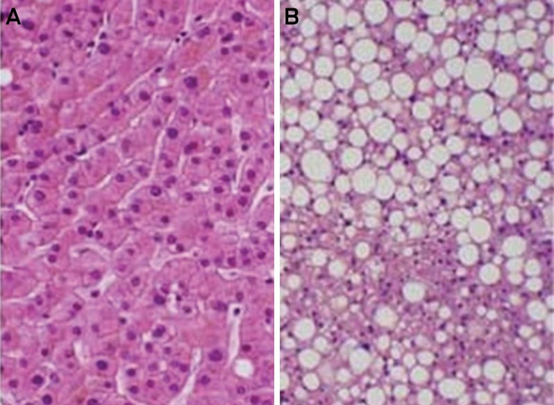
NAFLD, the hepatic manifestation of the metabolic syndrome, affects a quarter of the global population, and is the most common liver abnormality in Western and some Asian countries (Box 1).45 A microbial compositional shift towards energy harvesting and production of volatile organic compounds, including ethanol, is associated with obesity-related NAFLD.18 However, these microbial changes are also observed in non-obese individuals with NAFLD.46 Germ-free mice exposed to the microbiome of mice with NAFLD develop obesity and NAFLD.47
Bile salts are likely involved in this microbiota-related liver pathology. They act as signalling molecules that regulate hepatic glucose and lipid metabolism and inflammation,48 and modulate the intestinal microbiota via their detergent effects on bacterial cell membranes.49
A proportion of patients with NAFLD develop inflammation (NASH), and a proportion of these progress to liver fibrosis. NASH is now the most common indication for liver transplant in the Western world. NASH with advanced fibrosis is associated with a faecal microbiome-derived metagenomic signature50 and increased endotoxaemia.51
The gut microbiota may also play a role in the variable susceptibility to alcoholic liver disease. The microbiota differs between patients with severe alcoholic hepatitis and those who maintain normal hepatic function despite similar alcohol intake.7 Transfer of microbiota from each cohort to germ-free mice reproduces their donor phenotype. Further, amelioration of hepatitis occurred after FMT from patients without hepatitis and with a high alcohol intake to mice in which hepatitis had been induced by FMT from patients with hepatitis and a high alcohol intake.7
In summary, the human gut microbiota and its metabolites affect mucosal immune homoeostasis, as evidenced in IBD. Both mucosal and remote effects, mediated by metabolites or non-mucosal immunity, may be involved in the inflammation associated with insulin resistance, obesity, NAFLD, NASH and alcoholic liver disease.
What causes microbiota differences?
Diversity of the microbiome in adults is related to socio-economic status, diet, age, geography, drugs and other environmental substances.
Microbial composition differs geographically and between vegans, vegetarians and omnivores.52,53 Vegans, vegetarians and omnivores demonstrate a positive correlation between short chain fatty acid production, fibre-degrading bacteria and levels of fruit, vegetable and legume intake.54 Dietary manipulation leads to an increase in bile-tolerant organisms and decreased Firmicutes, a pro-inflammatory microbiota phenotype.55 Western diets, and diets in Western and non-Western lower socio-economic populations, are both associated with a more inflammatory microbiota phenotype.56-58
Strong evidence is emerging that a key link between dietary changes and the susceptibility to modern gut diseases is the introduction of processed foods and their dietary additives.
An ulcerative colitis-like colitis can be induced in animals by carrageenan gum, a thickener and stabiliser, at concentrations used in common food products.59
Emulsifiers are emerging as potent pro-inflammatory modifiers of the microbiota. These detergent-like molecules are present in a wide range of common processed foods, homogenising their composition and preventing separation of fat-soluble and water-soluble ingredients. They radically alter bacterial–mucous–epithelial interactions. In mouse models, emulsifiers, in concentrations normally found in processed foods, alter the microbiota towards a pro-inflammatory composition, inducing low grade inflammation, obesity, NAFLD and metabolic syndrome (Box 2).60,61 Pro-inflammatory microbiota leads to erosion of the protective mucous layer lining the gut, increased gut permeability, and microbial epithelial adherence and translocation.60 FMT from emulsifier-fed mice results in the same process in germ-free mice, demonstrating that these changes are microbiota dependent.
Non-caloric artificial sweeteners (saccharin, sucralose or aspartame) are also concerning. Among the most widely used food additives globally, they were originally introduced to reduce caloric intake.62 A wide range of processed foods, including breakfast cereals, and “diet” or “sugar-free” products, contain these substances. Most non-caloric artificial sweeteners pass through the gut without being absorbed or metabolised, ultimately reaching the colonic microbiota.63 The ingestion of non-caloric artificial sweeteners, at levels corresponding to the equivalent daily intake in humans, drives glucose intolerance in lean and obese mice independent of caloric intake or energy expenditure. This glucose intolerance is mediated by alteration of gut microbiota,64 and is transferrable by FMT from non-caloric artificial sweetener-fed mice to germ-free mice.64
The presence of metabolic syndrome appears to be related to non-caloric artificial sweetener intake even when corrected for body mass index.64 In a small study, typical non-caloric artificial sweetener intake resulted in impaired glycaemic responses in four of seven individuals, with associated microbiota compositional changes. FMT from these subjects to germ-free mice resulted in similar impaired glycaemic control to the donors.64
Antibiotics are another potent cause of microbiota alterations, potentially associated with “modern” diseases. Exposure to antibiotics is ubiquitous in developed and developing countries. Urine analysis in Shanghai school children detected a wide range of antibiotics and a relationship between antibiotic presence and obesity.65
In a nationwide study of 577 627 Danish children born between 1995 and 2003, the relative risk for those who had received antibiotics for developing IBD was 1.9; for Crohn's disease, 3.4; and for Crohn's disease after receiving seven or more courses of antibiotics, 7.3.66
In a UK study of more than a million subjects, antibiotic exposure in childhood was associated with an increased risk of subsequently developing IBD, proportional to the number of courses of antibiotics.67
A causal relationship also appears to exist between antibiotic exposure and the subsequent development of obesity, insulin resistance and NAFLD (Box 3).39,68-70 The development of NAFLD and insulin resistance in mice receiving a high fat diet is increased when combined with low dose antibiotics.69 Antibiotic exposure in children has also been associated with the development of obesity later in childhood, independent of covariates such as socio-economic status, maternal and sibling obesity.68,71
Microbiota manipulation as a treatment
The elimination of H. pylori, originally considered a harmless commensal organism, changed the natural history of peptic ulcer disease.72,73
FMT has been shown to be the most effective therapy for recurrent C. difficile infection.74
Four controlled trials have evaluated FMT as a treatment for active ulcerative colitis, three of which have shown a consistent beneficial effect (Box 4).27,75-77 FMT will enter the therapeutic armamentarium for this condition, shifting the treatment paradigm from chemical therapy and immune suppression to changing the antigenic drive. FMT involves no assumption about which part of the microbiota is responsible for driving disease, instead changing the entire microbial and chemical luminal environment.
Many refinements of FMT therapy will now emerge. Faecal filtrate which excludes whole bacteria was successful in curing C. difficile infection in a small group of patients, raising the possibility that full FMT may not be required for this indication.78
In FMT for ulcerative colitis, pooled donor stool provides greater microbial diversity than single stool infusions,77,79 although it is unknown if this is therapeutically important. Ruminococcus gnavus in donor stool is associated with failed FMT,80 while Fusobacterium in the recipient is associated with a lower chance of achieving remission.27 One-year follow-up of sustained remission after FMT in ulcerative colitis demonstrated ongoing high levels of butyrate producers and relapse associated with higher abundance of Proteobacteria and Bacteroidetes.80 The optimal intensity and frequency of treatment, the value of maintenance therapy, and the identification of “good” donors and patients likely to respond have yet to be defined.27,75 Mixtures of key bacteria are the logical next step in this kind of therapy.
FMT has not been evaluated sufficiently in other diseases to confirm benefit. A single trial in obesity demonstrated promising results with FMT from lean to obese humans, lowering triglyceride levels and increasing insulin sensitivity.81 FMT resulted in an increase in faecal butyrate concentration and the abundance of the butyrate producer Roseburia intestinalis.81
Dietary therapy may also profoundly affect the microbiota. Enteral formulated liquid nutrition diets, with food exclusion, induce remission in Crohn's disease, an effect thought to be mediated by microbial change.82 Wholefood anti-inflammatory diets exclude food items that are postulated to degrade the mucus layer, increase intestinal permeability or induce microbial changes, and include wholefoods such as certain fruits and vegetables, resistant starch and certain animal proteins.82 An anti-inflammatory diet combined with liquid formulated enteral nutrition has shown promising results for induction of clinical remission in Crohn's disease,82 and controlled trials are currently in progress.
Dietary manipulation in other inflammatory-driven diseases, including obesity, NAFLD and diabetes, is currently a focus of research. Personalised nutrition based on an individual’s gut microbiota, anthropometric data, dietary habits, blood glucose monitoring and physical activity has demonstrated improved post-prandial glucose control.42
Probiotics (live bacteria thought to confer health benefits) have not shown benefit in the majority of inflammatory-driven conditions.40,83,84 There is some evidence for benefit in pouchitis,85 and in ulcerative colitis maintenance.86
Conclusion
The microbiota is central to the development of a wide range of gut and liver diseases in Western and non-Western populations. Although the precise causative microbial changes for most or all of these emerging diseases have not yet been fully defined, data are rapidly emerging. Changes in the microbiota result in altered gut barrier and metabolic functions, resulting in systemic inflammation and dissemination of bacterial products.
The nature of food production and the widespread use of antibiotics in agricultural, veterinary and medicinal settings are now public health issues. At an individual level, microbial profiles may be able to predict who is at risk of disease when subjected to particular environmental influences, and what microbial restoration is needed to minimise risk. Microbial manipulation is an effective therapy, likely to have broadening indications.26
Box 1 – Liver histology (haematoxylin–eosin stain), showing normal liver (A) and non-alcoholic fatty liver disease (B)
Box 2 – Bioluminescence imaging showing the effect of emulsifiers on mouse gut microbiota and resulting destruction of the protective mucous layer
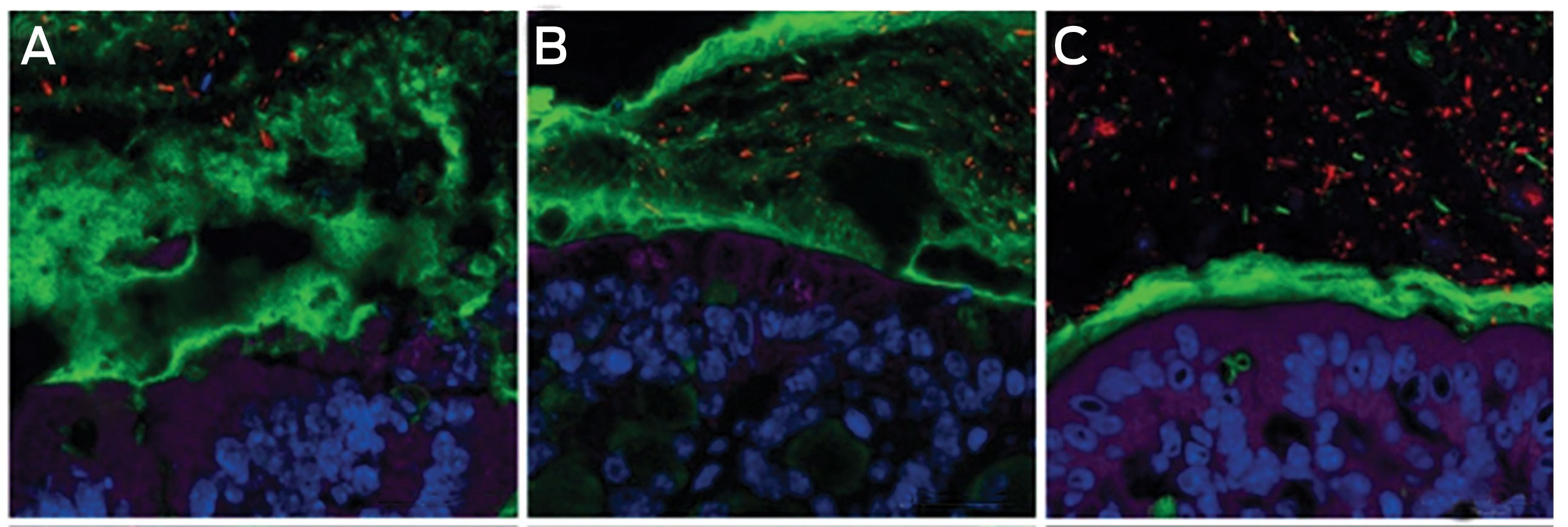
A: Normal thick mucous layer (green) between epithelium below (purple and blue) and luminal content above. B and C: Emulsifiers (carboxymethyl cellulose [B] and P-80 [C]) given to mice in the same concentrations found in human food cause changes in the gut microbiota (red), resulting in destruction of the protective gut mucus layer. Luminal contents reach the epithelium, causing epithelial inflammation. Mice subjected to emulsifiers developed obesity and non-alcoholic fatty liver disease, in contrast to their unexposed counterparts.60 Reprinted with permission from Chassaing B, Koren O, Goodrich JK, et al Dietary emulsifiers impact the mouse gut microbiota promoting colitis and metabolic syndrome. Nature 2015; 519: 92–96.
Box 3 – Effect of penicillin on obesity in mice, mediated via changes in the microbiota70
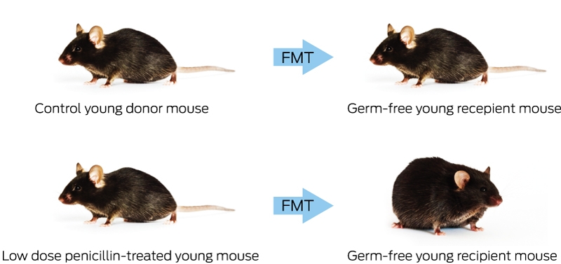
FMT = faecal microbiota transplantation.
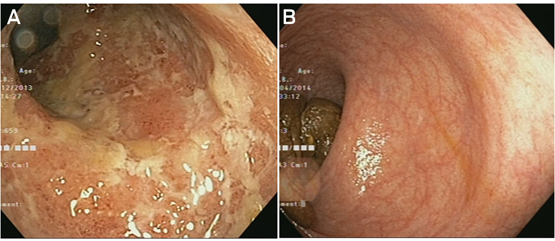
Competing interests
No relevant disclosures.
Acknowledgements
We acknowledge the support for this work from the Helmsley Charitable Trust.
References
- Wang WL, Xu SY, Ren ZG, et al. Application of metagenomics in the human gut microbiome. World J Gastroenterol 2015; 21: 803-814.
- Lagkouvardos I, Overmann J, Clavel T. Cultured microbes represent a substantial fraction of the human and mouse gut microbiota. Gut Microbes 2017; 8: 493-503.
- Qin J, Li Y, Cai Z, et al. A metagenome-wide association study of gut microbiota in type 2 diabetes. Nature 2012; 490: 55-60.
- Gilbert JA, Quinn RA, Debelius J, et al. Microbiome-wide association studies link dynamic microbial consortia to disease. Nature 2016; 535: 94-103.
- Lau JT, Whelan FJ, Herath I, et al. Capturing the diversity of the human gut microbiota through culture-enriched molecular profiling. Genome Med 2016; 8: 72.
- Forbes JD, Van Domselaar G, Bernstein CN. The gut microbiota in immune-mediated inflammatory diseases. Front Microbiol 2016; 7: 1081.
- Llopis M, Cassard AM, Wrzosek L, et al. Intestinal microbiota contributes to individual susceptibility to alcoholic liver disease. Gut 2016; 65: 830-839.
- Curtis MM, Hu Z, Klimko C, et al. The gut commensal Bacteroides thetaiotaomicron exacerbates enteric infection through modification of the metabolic landscape. Cell Host Microbe 2014; 16: 759-769.
- Tap J, Mondot S, Levenez F, et al. Towards the human intestinal microbiota phylogenetic core. Environ Microbiol 2009; 11: 2574-2584.
- Tilg H, Moschen AR. Food, immunity, and the microbiome. Gastroenterology 2015; 148: 1107-1119.
- Backhed F, Roswall J, Peng Y, et al. Dynamics and stabilization of the human gut microbiome during the first year of life. Cell Host Microbe 2015; 17: 690-703.
- Marti JM, Martinez-Martinez D, Rubio T, et al. Health and disease imprinted in the time variability of the human microbiome. mSystems 2017; 2: e00144-16.
- Buffie CG, Bucci V, Stein RR, et al. Precision microbiome reconstitution restores bile acid mediated resistance to Clostridium difficile. Nature 2015; 517: 205-208.
- Weingarden AR, Dosa PI, DeWinter E, et al. Changes in colonic bile acid composition following fecal microbiota transplantation are sufficient to control Clostridium difficile germination and growth. PLoS One 2016; 11: e0147210.
- Ferreyra JA, Wu KJ, Hryckowian AJ, et al. Gut microbiota-produced succinate promotes C. difficile infection after antibiotic treatment or motility disturbance. Cell Host Microbe 2014; 16: 770-777.
- Lee D, Albenberg L, Compher C, et al. Diet in the pathogenesis and treatment of inflammatory bowel diseases. Gastroenterology 2015; 148: 1087-1106.
- Hildebrandt MA, Hoffmann C, Sherrill-Mix SA, et al. High-fat diet determines the composition of the murine gut microbiome independently of obesity. Gastroenterology 2009; 137: 1716-1724.
- Raman M, Ahmed I, Gillevet PM, et al. Fecal microbiome and volatile organic compound metabolome in obese humans with nonalcoholic fatty liver disease. Clin Gastroenterol Hepatol 2013; 11: 868-875.
- Ng SC, Tang W, Ching JY, et al. Incidence and phenotype of inflammatory bowel disease based on results from the Asia-pacific Crohn’s and colitis epidemiology study. Gastroenterology 2013; 145: 158-165.
- Koh JC, Loo WM, Goh KL, et al. Asian consensus on the relationship between obesity and gastrointestinal and liver diseases. J Gastroenterol Hepatol 2016; 31: 1405-1413.
- Lophaven SN, Lynge E, Burisch J. The incidence of inflammatory bowel disease in Denmark 1980-2013: a nationwide cohort study. Aliment Pharmacol Ther 2017; 45: 961-972.
- Ng SC SH, Hamidi N, Underwood FE, et al. Worldwide incidence and prevalence of inflammatory bowel disease in the 21st century: a systematic review of population-based studies. Lancet 2018; 390: 2769-2778.
- Fofanova TY, Petrosino JF, Kellermayer R. Microbiome-epigenome interactions and the environmental origins of inflammatory bowel diseases. J Pediatr Gastroenterol Nutr 2016; 62: 208-219.
- Ko Y, Kariyawasam V, Karnib M, et al. Inflammatory bowel disease environmental risk factors: a population-based case-control study of Middle Eastern migration to Australia. Clin Gastroenterol Hepatol 2015; 13: 1453-1463.
- Benchimol EI, Manuel DG, To T, et al. Asthma, type 1 and type 2 diabetes mellitus, and inflammatory bowel disease amongst South Asian immigrants to Canada and their children: a population-based cohort study. PLoS One 2015; 10: e0123599.
- Zmora N, Zeevi D, Korem T, et al. Taking it personally: personalized utilization of the human microbiome in health and disease. Cell Host Microbe 2016; 19: 12-20.
- Paramsothy S, Kamm MA, Kaakoush NO, et al. Multidonor intensive faecal microbiota transplantation for active ulcerative colitis: a randomised placebo-controlled trial. Lancet 2017; 389: 1218-1228.
- Wright EK, Kamm MA, Wagner J, et al. Microbial factors associated with postoperative Crohn’s disease recurrence. J Crohn’s Colitis 2017; 11: 191-203.
- Paramsothy S, Kamm M, Nielsen S, et al. OP019 In faecal microbiota transplantation (FMT) for ulcerative colitis, Fusobacterium is associated with lack of remission, while metabolic shifts to starch degradation and short chain fatty acid production are associated with remission (FOCUS study) [abstract]. J Crohn’s Colitis 2018; 12 (1 Suppl): S013-S014.
- Gevers D, Kugathasan S, Denson LA, et al. The treatment-naive microbiome in new-onset Crohn’s disease. Cell Host Microbe 2014; 15: 382-392.
- Shen Z, Zhu C, Quan Y, et al. Update on intestinal microbiota in Crohn’s disease 2017: mechanisms, clinical application, adverse reactions and outlook. J Gastroenterol Hepatol 2017; 32: 1804-1812.
- Quevrain E, Maubert MA, Michon C, et al. Identification of an anti-inflammatory protein from Faecalibacterium prausnitzii, a commensal bacterium deficient in Crohn’s disease. Gut 2016; 65: 415-425.
- Kugathasan S, Denson LA, Walters TD, et al. Prediction of complicated disease course for children newly diagnosed with Crohn’s disease: a multicentre inception cohort study. Lancet 2017; 389: 1710-1718.
- David LA, Maurice CF, Carmody RN, et al. Diet rapidly and reproducibly alters the human gut microbiome. Nature 2014; 505: 559-563.
- Nikolaus S, Schulte B, Al-Massad N, et al. Increased tryptophan metabolism is associated with activity of inflammatory bowel diseases. Gastroenterology 2017; 153: 1504-1516.
- Tanca A, Abbondio M, Palomba A, et al. Potential and active functions in the gut microbiota of a healthy human cohort. Microbiome 2017; 5: 79.
- El Mouzan M, Wang F, Al Mofarreh M, et al. Fungal microbiota profile in newly diagnosed treatment-naive children with Crohn’s disease. J Crohn’s Colitis 2017; 11: 586-592.
- Lopes S, Andrade P, Conde S, et al. Looking into enteric virome in patients with IBD: defining guilty or innocence? Inflamm Bowel Dis 2017; 23: 1278-1284.
- Le Chatelier E, Nielsen T, Qin J, et al. Richness of human gut microbiome correlates with metabolic markers. Nature 2013; 500: 541-546.
- Boulange CL, Neves AL, Chilloux J, et al. Impact of the gut microbiota on inflammation, obesity, and metabolic disease. Genome Med 2016; 8: 42.
- Grundy SM. Pre-diabetes, metabolic syndrome, and cardiovascular risk. J Am Coll Cardiol 2012; 59: 635-643.
- Zeevi D, Korem T, Zmora N, et al. Personalized nutrition by prediction of glycemic responses. Cell 2015; 163: 1079-1094.
- Ridaura VK, Faith JJ, Rey FE, et al. Gut microbiota from twins discordant for obesity modulate metabolism in mice. Science 2013; 341: 1241214.
- Karlsson FH, Tremaroli V, Nookaew I, et al. Gut metagenome in European women with normal, impaired and diabetic glucose control. Nature 2013; 498: 99-103.
- Fan JG, Farrell GC. Epidemiology of non-alcoholic fatty liver disease in China. J Hepatol 2009; 50: 204-210.
- Wang B, Jiang X, Cao M, et al. Altered fecal microbiota correlates with liver biochemistry in nonobese patients with non-alcoholic fatty liver disease. Sci Rep 2016; 6: 32002.
- Le Roy T, Llopis M, Lepage P, et al. Intestinal microbiota determines development of non-alcoholic fatty liver disease in mice. Gut 2013; 62: 1787-1794.
- Mouzaki M, Wang AY, Bandsma R, et al. Bile acids and dysbiosis in non-alcoholic fatty liver disease. PLoS One 2016; 11: e0151829.
- Begley M, Gahan CG, Hill C. The interaction between bacteria and bile. FEMS Microbiol Rev 2005; 29: 625-651.
- Loomba R, Seguritan V, Li W, et al. Gut microbiome-based metagenomic signature for non-invasive detection of advanced fibrosis in human nonalcoholic fatty liver disease. Cell Metab 2017; 25: 1054-1062.e5.
- Pang J, Xu W, Zhang X, et al. Significant positive association of endotoxemia with histological severity in 237 patients with non-alcoholic fatty liver disease. Aliment Pharmacol Ther 2017; 46: 175-182.
- De Filippo C, Cavalieri D, Di Paola M, et al. Impact of diet in shaping gut microbiota revealed by a comparative study in children from Europe and rural Africa. Proc Natl Acad Sci U S A 2010; 107: 14691-14696.
- Zimmer J, Lange B, Frick JS, H, et al. A vegan or vegetarian diet substantially alters the human colonic faecal microbiota. Eur J Clin Nutr 2012; 66: 53-60.
- De Filippis F, Pellegrini N, Vannini L, et al. High-level adherence to a Mediterranean diet beneficially impacts the gut microbiota and associated metabolome. Gut 2016; 65: 1812-1821.
- Ley RE, Turnbaugh PJ, Klein S, Gordon JI. Microbial ecology: human gut microbes associated with obesity. Nature 2006; 444: 1022-1023.
- Agus A, Denizot J, Thevenot J, et al. Western diet induces a shift in microbiota composition enhancing susceptibility to adherent-invasive E. coli infection and intestinal inflammation. Sci Rep 2016; 6: 19032.
- Martinez-Medina M, Denizot J, Dreux N, et al. Western diet induces dysbiosis with increased E coli in CEABAC10 mice, alters host barrier function favouring AIEC colonisation. Gut 2014; 63: 116-124.
- Miller GE, Engen PA, Gillevet PM, et al. Lower neighborhood socioeconomic status associated with reduced diversity of the colonic microbiota in healthy adults. PLoS One 2016; 11: e0148952.
- Munyaka PM, Sepehri S, Ghia JE, Khafipour E. Carrageenan gum and adherent invasive Escherichia coli in a piglet model of inflammatory bowel disease: impact on intestinal mucosa-associated microbiota. Front Microbiol 2016; 7: 462.
- Chassaing B, Koren O, Goodrich JK, et al. Dietary emulsifiers impact the mouse gut microbiota promoting colitis and metabolic syndrome. Nature 2015; 519: 92-96.
- Singh RK, Wheildon N, Ishikawa S. Food additive P-80 impacts mouse gut microbiota promoting intestinal inflammation, obesity and liver dysfunction. SOJ Microbiol Infect Dis 2016: 4; doi: 10.15226/sojmid/4/1/00148.
- Gardner C, Wylie-Rosett J, Gidding SS, et al. Nonnutritive sweeteners: current use and health perspectives: a scientific statement from the American Heart Association and the American Diabetes Association. Diabetes Care 2012; 35: 1798-1808.
- Roberts A, Renwick AG, Sims J, Snodin DJ. Sucralose metabolism and pharmacokinetics in man. Food Chem Toxicol 2000; 38 Suppl 2: S31-S41.
- Suez J, Korem T, Zeevi D, et al. Artificial sweeteners induce glucose intolerance by altering the gut microbiota. Nature 2014; 514: 181-186.
- Wang H, Wang N, Wang B, et al. Antibiotics in drinking water in Shanghai and their contribution to antibiotic exposure of school children. Environ Sci Technol 2016; 50: 2692-2699.
- Hviid A, Svanstrom H, Frisch M. Antibiotic use and inflammatory bowel diseases in childhood. Gut 2011; 60: 49-54.
- Kronman MP, Zaoutis TE, Haynes K, et al. Antibiotic exposure and IBD development among children: a population-based cohort study. Pediatrics 2012; 130: e794-e803.
- Scott FI, Horton DB, Mamtani R, et al. Administration of antibiotics to children before age 2 years increases risk for childhood obesity. Gastroenterology 2016; 151: 120-129.
- Mahana D, Trent CM, Kurtz ZD, et al. Antibiotic perturbation of the murine gut microbiome enhances the adiposity, insulin resistance, and liver disease associated with high-fat diet. Genome Med 2016; 8: 48.
- Jess T. Microbiota, antibiotics, and obesity. N Engl J Med 2014; 371: 2526-2528.
- Blaser MJ. Antibiotic use and its consequences for the normal microbiome. Science 2016; 352: 544-545.
- Marshall BJ, Goodwin CS, Warren JR, et al. Prospective double-blind trial of duodenal ulcer relapse after eradication of Campylobacter pylori. Lancet 1988; 2: 1437-1442.
- Forbes GM, Glaser ME, Cullen DJ, et al. Duodenal ulcer treated with Helicobacter pylori eradication: seven-year follow-up. Lancet 1994; 343: 258-260.
- Moayyedi P, Yuan Y, Baharith H, Ford AC. Faecal microbiota transplantation for Clostridium difficile-associated diarrhoea: a systematic review of randomised controlled trials. Med J Aust 2017; 207: 166-172.
- Moayyedi P, Surette MG, Kim PT, et al. Fecal microbiota transplantation induces remission in patients with active ulcerative colitis in a randomized controlled trial. Gastroenterology 2015; 149: 102-109.
- Rossen NG, Fuentes S, van der Spek MJ, et al. Findings from a randomized controlled trial of fecal transplantation for patients with ulcerative colitis. Gastroenterology 2015; 149: 110-118 e4.
- Costello S, Walters O, Bryant R, et al. OP036 Short duration, low intensity pooled faecal microbiota transplantation induces remission in patients with mild-moderately active ulcerative colitis: a randomised controlled trial [abstract]. J Crohn’s Colitis 2017; 11 (1 Suppl): S23.
- Ott SJ, Waetzig GH, Rehman A, et al. Efficacy of sterile fecal filtrate transfer for treating patients with Clostridium difficile infection. Gastroenterology 2017; 152: 799-811.e7.
- Paramsothy S, Paramsothy R, Rubin DT, et al. Faecal microbiota transplantation for inflammatory bowel disease: a systematic review and meta-analysis. J Crohn’s Colitis 2017; 11: 1180-1199.
- Fuentes S, Rossen NG, van der Spek MJ, et al. Microbial shifts and signatures of long-term remission in ulcerative colitis after faecal microbiota transplantation. ISME J 2017; 11: 1877-1889.
- Vrieze A, Van Nood E, Holleman F, et al. Transfer of intestinal microbiota from lean donors increases insulin sensitivity in individuals with metabolic syndrome. Gastroenterology 2012; 143: 913-916.
- Sigall Boneh R, Sarbagili-Shabat C, et al. Dietary therapy with the Crohn’s disease exclusion diet is a successful strategy for induction of remission in children and adults failing biological therapy. J Crohn’s Colitis 2017; 11: 1205-1212.
- Jones ML, Martoni CJ, Prakash S. Cholesterol lowering and inhibition of sterol absorption by Lactobacillus reuteri NCIMB 30242: a randomized controlled trial. Eur J Clin Nutr 2012; 66: 1234-1241.
- Marchesi JR, Adams DH, Fava F, et al. The gut microbiota and host health: a new clinical frontier. Gut 2016; 65: 330-339.
- Abraham BP, Quigley EMM. Probiotics in inflammatory bowel disease. Gastroenterol Clin North Am 2017; 46: 769-782.
- Kruis W, Fric P, Pokrotnieks J, et al. Maintaining remission of ulcerative colitis with the probiotic Escherichia coli Nissle 1917 is as effective as with standard mesalazine. Gut 2004; 53: 1617-1623.
Provenance: Not commissioned; externally peer reviewed.
Early-Onset Colorectal Cancer With Liver-Only Metastases: A Retrospective Cohort Study Integrating Prospectively Collected Real-World Clinical and Molecular Data From an Australian National Database (2009–2024) to Guide Treatment Planning
Savio G. Barreto, Christos S. Karapetis, Shahid Ullah, Matthew Burge, Susan Caird, Angus Campbell, Azim Jalali, Ross Jennens, Muhammad A. Khattak, Belinda Lee, Stephanie H. Lim, Shehara Mendis, Louise Nott, Timothy J. Price, Jeremy D. Shapiro, Jeanne Tie, Javier Torres, Colin Williams, Rachel Wong, Vanessa Wong, Peter Gibbs
Australian Pathways for Specialist Pain Management and Early Palliative Care for People With Pancreatic Cancer: Developed Using a Community Consensus Approach
Jennifer Philip, Melanie R. Lovell, Kylee Bellingham, Gail Garvey, Gregory B. Crawford, Nicole M. Rankin, Kara Burns, Isabel Young, Vivienne Milch, Dorothy Keefe, Katrina Anderson, James Lawson, Meinir Krishnasamy
Cause‐specific mortality among Queensland people with cirrhosis, by cirrhosis aetiology and decompensation status, 2007–22: a retrospective cohort study
Vikas Bhasker, Jessica R Fong, Paul J Clark, Gunter F Hartel, Richard Skoien, James O’Beirne, Elizabeth E Powell, Patricia C Valery
Clinical practice guidelines for hepatocellular carcinoma surveillance for people at high risk in Australia: summary of recommendations
Jacob George, Nicole L Allard, Stuart K Roberts, Leon A Adams, Jane Davies, Behzad Hajarizadeh, Jennifer H MacLachlan, Suzanne E Mahady, Rosalie Altus, Catherine Brown, David C Fry, Belinda Greenwood‐Smith, Natali Smud, Patricia C Valery, Nafisa Yussf, Kate Broun, Denise Campbell, Karen Canfell, Chelsea Carle Harrison, Victoria Freeman, Paul Grogan, Catherine Holliday, Suzanne Hughes, Anna Kelly, Cathelijne Kemenade, Claire Latumahina, Amanda McAtamney, Megan Varlow, Joachim Worthington, Susan Yuill, Eleonora Feletto
Assessment of metabolic dysfunction‐associated fatty liver disease in primary care: a consensus statement summary
Leon A Adams, William W Kemp, Kate R Muller, Elizabeth E Powell, Stuart K Roberts, Luis Calzadilla Bertot, Stephanie Best, Gary Deed, Jon D Emery, Samantha L Hocking, Graham R Jones, John S Lubel, Sinead Sheils, Stephen M Twigg, Gerald F Watts, Jacob George
Treatment outcomes for people with hepatitis C referred to tertiary care in Victoria, 2021–22: a retrospective observational study
Elly Layton, Nicole Matthews, Brendan Quinn, Nasra Higgins, Gabrielle Lindeman, Mielle Abbott, Jennifer MacLachlan, Elizabeth Birbilis, Margaret E Hellard, Joseph Doyle, Benjamin C Cowie, Mark Stoové