




Medical education
Lessons from practice
Volume 208 - Issue 6
A case of adult-onset Kawasaki disease
Authors: Alexandra Gehrmann, Karen Morwood, David Gillis, Terence Coleman and Shradha Subedi
Med J Aust 2018; 208 (6): 250-251. || doi: 10.5694/mja17.00949
Published online: 2 April 2018
Published online: 2 April 2018
A previously healthy 30-year-old man of European ancestry presented with a one-week history of fevers and vomiting …
Clinical record
A previously healthy 30-year-old man of European ancestry presented to hospital in August 2017 with a one-week history of fevers and vomiting followed by painful mouth and throat and erythema and swelling of his hands and feet. His general practitioner treated him with oral penicillin for presumed streptococcal pharyngitis, with minimal improvement. His symptoms progressed to non-suppurative bilateral conjunctivitis; desquamation of his scrotum, face and fingertips; and arthralgias in both hands.
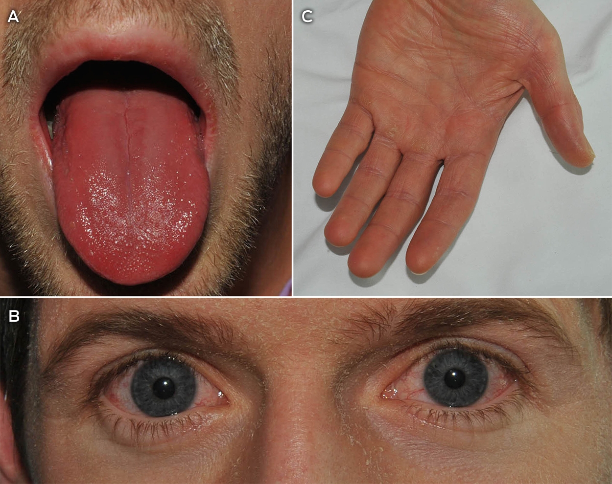
On admission, he was febrile, with a temperature of 38.3°C; his heart rate was 90 beats per minute; blood pressure was 120/60 mmHg; respiratory rate was 18; and oxygen saturations were 97% on room air. Physical examination revealed a strawberry red tongue and oral mucosa, dry cracked lips, conjunctival chemosis, palpable cervical lymphadenopathy, swollen hands and feet and desquamating rash on his neck, face, shoulders and scrotum (Box 1).
Initial blood tests showed a raised white cell count, neutrophilia, eosinophilia, anaemia and thrombocytosis (Box 2). He also had elevated levels of C-reactive protein, γ-glutamyltransferase, alkaline phosphatase, alanine transaminase and aspartate transaminase, and total bilirubin was 37 μmol/L. Urinalysis showed aseptic leukocyturia. He received empirical intravenous benzylpenicillin and oral doxycycline to cover streptococcal pharyngitis and other atypical infections.
A pharyngeal swab grew normal respiratory flora. Two sets of blood cultures as well as serological testing for HIV, syphilis, Epstein–Barr virus, cytomegalovirus, group A Streptococcus, Q fever, Ross River and Barmah Forest viruses, leptospirosis, Mycoplasma pneumoniae, hepatitis B and C viruses, and auto-immune diseases screen were negative. The nasopharyngeal swab, which was initially positive for adenovirus by multiplex polymerase chain reaction, was negative on repeat testing. Adenovirus polymerase chain reaction on conjunctival swab and blood were negative. The initial positive adenovirus polymerase chain reaction on nasopharyngeal swab was later determined to be a false positive result. A diagnosis of complete Kawasaki disease was made on the basis of clinical criteria (Box 3) and exclusion of infection and other auto-immune causes.
A transthoracic echocardiogram showed no evidence of coronary artery aneurysms. The patient was treated with a single 2 g/kg dose of intravenous immunoglobulin on Day 12 of his illness and with high dose aspirin (600 mg four times daily) for 2 weeks, and 100 mg daily for a further 6 weeks. His symptoms improved with intravenous immunoglobulin and he was discharged home. A follow-up computed tomography coronary angiography 4 weeks later showed no coronary artery aneurysms.
Kawasaki disease, or mucocutaneous lymph node syndrome, is a systemic vasculitis affecting the medium and small sized vessels, with predilection for the coronary arteries. Kawasaki disease predominantly affects children between the ages of 6 months and 5 years, and is rare in adults. A case of Kawasaki disease in an adult has not been reported in Australia; and until 2015, only 100 cases of adult-onset Kawasaki disease had been reported worldwide.1
The exact pathogenesis of Kawasaki disease is unknown, but is thought to occur from exposure to an undefined infectious trigger2 in a genetically susceptible host.3 The genetic theory is supported by its high prevalence in East Asia, particularly Japan; the annual incidence of Kawasaki disease in children aged under 5 years in Japan is 265 per 100 000, compared with 9 per 100 000 in Australia.4 Only five cases of adult-onset Kawasaki disease have originated in Asia, with the majority reported in North America and Europe.3
Less common symptoms of Kawasaki disease include arthritis, jaundice, hepatitis, pericarditis, diarrhoea, anterior uveitis and aseptic meningitis. Common laboratory changes include raised inflammatory markers, hepatic cytolysis, delayed thrombocytosis, hyponatraemia, eosinophilia and aseptic leukocyturia,5 most of which were present in our patient. The largest study of adult-onset Kawasaki disease, by Fraison and colleagues,5 reviewed 43 cases in France and found that, compared with childhood Kawasaki disease, adults are more likely to have liver function anomalies and arthralgias, and are less likely to have thrombocytosis, cheilitis and meningitis.3,5 Coronary artery aneurysms are a devastating complication of this disease, and the rate of coronary complications in both adult-onset and childhood Kawasaki disease is similar.5
Features of Kawasaki disease linked to a higher risk of coronary aneurysms include: male sex, age < 12 months or > 8 years, albumin < 35 g/L, C-reactive protein > 200 mg/dL, platelets < 35 × 109/L, delay in initiation of intravenous immunoglobulin or lower dose of intravenous immunoglobulin and recurrence of Kawasaki disease.2
In the acute phase of the disease, perivascular inflammation leads to panvasculitis and subsequent coronary fibrosis, aneurysms, thrombosis and, in severe cases, myocardial infarction and sudden death. Intravenous immunoglobulin has been proven to inhibit this process and is recommended within 10 days of symptoms.6 Due to its natural history and rarity, adult-onset Kawasaki disease is often misdiagnosed, with only 28% of patients treated within 10 days of presentation.5
Treatment with intravenous immunoglobulin reduces the risk of coronary aneurysms in childhood Kawasaki disease from 30% to 3%.7 While Fraison and colleagues5 found that early intravenous immunoglobulin treatment reduced the duration of fever, it did not significantly reduce the number of cardiac complications. A larger study assessing the role of intravenous immunoglobulin in adult-onset Kawasaki disease is needed.
The recommended treatment regime for Kawasaki disease is a single 2 g/kg infusion of intravenous immunoglobulin, which can be repeated if the fever persists or recurs after 36 hours7 — this is defined as treatment-resistant Kawasaki disease.8 If fever persists after the second dose of intravenous immunoglobulin, intravenous methylprednisolone is used at 30 mg/kg up to 1000 mg daily for up to 3 consecutive days. High dose aspirin is recommended while the patient is febrile, as it theoretically reduces vascular inflammation and thrombosis, although there is no direct evidence that it reduces coronary artery lesions. Doses range from 30 mg/kg to 100 mg/kg daily in four divided doses, which can be reduced 48 hours after the fever resolves to a lower dose of 3–5 mg/kg daily.7 In childhood Kawasaki disease, echocardiography is recommended at 6 weeks after fever resolution, at which time, low dose aspirin can be ceased if no coronary artery lesions are evident.7 However, due to limitations of echocardiography in viewing the coronary vessels in adults, computed tomography coronary angiography or cardiac magnetic resonance imaging should be considered.5 Recurrent Kawasaki disease occurs in 0.8–3% of patients, and has a higher rate of coronary complications.8
While adult-onset Kawasaki disease is rare, clinicians should be familiar with the diagnostic criteria to consider it in the differential diagnosis of patients presenting with undifferentiated febrile illness. Early treatment with intravenous immunoglobulin should be instituted to prevent cardiac sequelae.
Lessons from practice
-
Adult-onset Kawasaki disease is rare and often misdiagnosed, and clinicians should be familiar with the diagnostic criteria to provide early treatment.
-
Intravenous immunoglobulin prevents cardiac sequelae and is recommended within 10 days of symptoms.
-
Follow-up with computed tomography coronary angiography or cardiac magnetic resonance imaging at 6 weeks is preferred to echocardiography in patients with adult-onset Kawasaki disease.
Box 1 – Strawberry red tongue and dry cracked lips (A), desquamating rash on the face (B), and desquamation of skin on the palms (C)
Box 2 – Laboratory results
|
|
Value (RI) |
||||||||||||||
|
|
|||||||||||||||
|
Haemoglobin (g/L) |
131 (135–180) |
||||||||||||||
|
Haematocrit (g/L) |
0.38 (0.39–0.52) |
||||||||||||||
|
WCC (109/L) |
16.1 (4.0–11.0) |
||||||||||||||
|
Neutrophils (109/L) |
13.3 (2.0–8.0) |
||||||||||||||
|
Platelets (109/L) |
484 (140–400) |
||||||||||||||
|
Eosinophils (109/L) |
2.45 (< 0.60) |
||||||||||||||
|
CRP (mg/L) |
242 (< 2.0) |
||||||||||||||
|
Sodium (mmol/L) |
136 (135–145) |
||||||||||||||
|
Creatinine (μmol/L) |
77 (60–110) |
||||||||||||||
|
Albumin (g/L) |
25 (35–50) |
||||||||||||||
|
GGT (U/L) |
110 (< 55) |
||||||||||||||
|
ALP (U/L) |
134 (30–110) |
||||||||||||||
|
ALT (U/L) |
72 (< 45) |
||||||||||||||
|
AST (U/L) |
42 (< 35) |
||||||||||||||
|
Bilirubin, total (μmol/L) |
37 (< 20) |
||||||||||||||
|
Bilirubin, conjugated (μmol/L) |
17 (< 4) |
||||||||||||||
|
|
|||||||||||||||
|
ALP = alkaline phosphatase. ALT = alanine transaminase. AST = aspartate transaminase. CRP = C-reactive protein. GGT = γ-glutamyltransferase. RI = reference interval. WCC = white cell count. |
|||||||||||||||
Box 3 – Diagnostic criteria for Kawasaki disease
Unexplained fever for 5 or more days, plus at least four of the following:- polymorphous exanthema
- erythema and oedema followed by desquamation of the extremities
- cervical lymphadenopathy > 1.5 cm diameter
- bilateral non-exudative conjunctival injection
- oropharyngeal changes, such as injected or fissured lips, strawberry tongue or injected pharynx
Competing interests
No relevant disclosures.
References
- Kontopoulou T, Kontopoulos DG, Vaidakis E, Mousoulis GP. Adult Kawasaki disease in a European patient: a case report and review of the literature. J Med Case Rep 2015; 9: 75.
- Galeotti C, Bayry J, Kone-Paut I, Kaveri SV. Kawasaki disease: aetiopathogenesis and therapeutic utility of intravenous immunoglobulin. Autoimmun Rev 2010; 9: 441-448.
- Garrido-García LM, López-Amézquita M, Villaverde-Rosas R. Kawasaki disease in a healthcare provider: an adult case in a paediatric resident. Cardiol Young 2016; 26: 976-978.
- Singh S, Vignesh P, Burgner D. The epidemiology of Kawasaki disease: a global update. Arch Dis Child 2015; 100: 1084-1088.
- Fraison JB, Sève P, Dauphin C, et al. Kawasaki disease in adults: observations in France and literature review. Autoimmun Rev 2016; 15: 242-249.
- Wolff AE, Hansen KE, Zakowski L. Acute Kawasaki disease: not just for kids. J Gen Intern Med 2007; 22: 681-684.
- Therapeutic Guidelines. Kawasaki disease (revised Mar 2017) [website]. Melbourne: eTG; 2017. https://www.tg.org.au (viewed Nov 2017).
- Osman NM. Recurrent Kawasaki disease resistant to initial treatment with intravenous immunoglobulin. Sudan J Paediatr 2012; 12: 65-69.
Provenance: Not commissioned; externally peer reviewed.
Rhabdomyolysis After COVID-19
Navya Kataria, Abel Devadass, Lana Sundac, Laura De Souza, Nabeel Sheikh, Samantha Ng, Scott Campbell, Yishen Wang, Ross S. Francis
Technological advances in the search for a CURE for asthma
Sara Quon, Timothy M Johanson, Ridhima Wadhwa, Alen Faiz, Anthony Flynn, Gary P Anderson, Amanda J Cox, Nicholas P West, Michael P Menden, Rhys S Allan
The CURE Asthma roadmap
Gary P Anderson, Anthony Flynn, Phil G Bardin, John D Blakey, Shyamali C Dharmage, Paul Foster, Peter G Gibson, Adam Jaffe, Alan James, Christine R Jenkins, Sundram Sivamalai, Peter D Sly, Guy B Marks, Vanessa M McDonald, Judy Wetttenhall
The first live term birth following uterus transplantation in Australia
Rebecca Deans, Brigitte Gerstl, Antonia W Shand, Sarah Lyons, Aaron Budden, Helen L Barrett, Grant Luxton, Mangalee Fernando, Kenneth Yong, Karen Keung, Kaushalya Arulpragasam, Henry Pleass, King Man Wan, Eva Kehag, Jana‐Emily Pittman, Mianna Lotz, Maria Fenn, Erin Nesbitt‐Hawes, Lily Byun, Katrina Tang, Mats Brannstrom, Jason Abbott
The public health impacts of mining in Australia
Javier Cortes‐Ramirez, Ruby N Michael, Leisa‐Maree Toms, Melissa Haswell
Lingual Raynaud phenomenon
Michael Taggart, Mina John