




Medical education
Lessons from practice
Volume 207 - Issue 4
An unusual pain in the gut
Authors: Ben Gerhardy, Robyn A Nagel, Ross Sellars and Aliki Andreou
Med J Aust 2017; 207 (4): 145-146. || doi: 10.5694/mja16.01196
Published online: 21 August 2017
Published online: 21 August 2017
An atypical infective colitis masquerading as autoinflammatory colitis
Clinical record
A 63-year-old non-Indigenous Australian woman was referred for colonoscopy and gastroenterologist opinion for a 2-month history of increasing constipation and abdominal pain, without change in dietary habits or systemic features, such as fever or weight loss.
Her medical history included osteoarthritis, dermatofibrosarcoma protruberans and a single miscarriage. Her only medications were vitamin supplements. She had no known allergies, was a lifelong non-smoker, did not drink alcohol and was physically active. Her family history included chronic renal failure of unclear aetiology. She had no known exposure to tuberculosis.
Her travel history included 7 weeks in Africa in the previous 8 years, and 2 weeks in Thailand in the previous 5 years; both trips involved rural environments. Her childhood on a dairy farm included unpasteurised milk consumption.
The patient’s first colonoscopy in 2008 revealed multiple small, shallow ulcers in the proximal transverse colon, with biopsies showing active chronic inflammation. Olsalazine was started with a provisional diagnosis of Crohn disease. After 4 months of treatment, she reported symptomatic improvement.
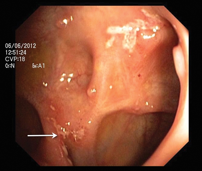
A colonoscopy in 2010 showed a small area of deep ulceration with partial stricturing at the hepatic flexure. Biopsies indicated focal active chronic colitis with granulomas consistent with Crohn disease; however, the pathologist suggested that Yersinia species and mycobacterial infection should be excluded. She was changed to mesalazine, and a colonoscopy in 2012 showed no significant change, with no symptoms (Box 1).
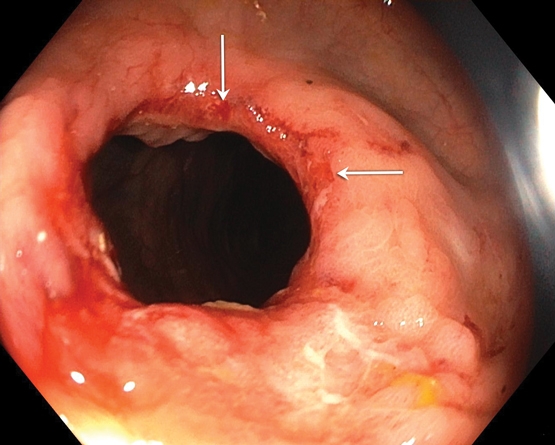
A 2014 colonoscopy showed endoscopic progression with transverse colon stricturing, which was coincident with symptom recurrence (Box 2). An immunosuppression screen was taken and mercaptopurine was initiated. The screen revealed a strongly positive interferon-γ release assay. A review of these initial 2014 biopsies suggested a single acid-fast bacilli organism in one slide, and repeat biopsies were performed for histology and culture. Her mercaptopurine and mesalazine therapy was ceased.
Repeat colonic biopsies tested positive for Mycobacterium tuberculosis protein 64 antigen, indicating M. tuberculosis complex growth. She was referred to the regional tuberculosis control unit and antimycobacterial chemotherapy with rifampicin, isoniazid, pyrazinamide and ethambutol was initiated until a biopsy specimen cultured M. bovis. Pyrazinamide was ceased and the treatment was extended to 9 months: 2 months of ethambutol, isoniazid and rifampicin, followed by 7 months of isoniazid and rifampicin.
The patient reported symptom resolution following antimycobacterial chemotherapy. Repeat endoscopies at treatment completion showed a healed ulceration with scarring and no new ulcer or stricture formation.
We present an atypical infective colitis masquerading as autoinflammatory colitis. Mycobacterium bovis is an M. tuberculosis complex (MTBC) species (MTBC also includes M. tuberculosis, M. kansasii, M. marinum etc) that typically infects animals and is thought to be the progenitor species of M. tuberculosis. Worldwide, it accounts for about 2% of all MTBC infections, and the primary risk factors are animal husbandry and unpasteurised dairy product consumption from infected livestock.1 Australia averages only 1–2 cases per year of human M. bovis infection.2
Milk pasteurisation became widespread in Australia after World War II. In 1970, Australia introduced a program to eliminate M. bovis from livestock, and it was declared bovine tuberculosis free in 1997. At present, the surveillance program continues due to wild M. bovis vectors, including pigs and buffalo, and the most recent detected livestock case was in 2002.3
Human M. bovis infection is clinically indistinguishable from M. tuberculosis. It can be pulmonary or extra-pulmonary, and the disease can be a primary infection or a reactivation from latency.4 Symptoms vary with site: pulmonary disease manifests as cough, fever, night sweats, dyspnoea and haemoptysis, while extra-pulmonary disease may present as a rash, gastric ulceration, lymphadenopathy or central nervous system infection.
Tuberculosis is diagnosed with a combination of microscopy, nucleic acid amplification and culture. Interferon-γ release assays are used to test for latent infection by exposing patient serum to MTBC antigens — therefore, not specific for M. tuberculosis — and assessing T cell response. Individual species identification occurs after culture by analysing biochemical characteristics, such as the ability to reduce nitrate.5 Identification is not required before commencing chemotherapy, but M. bovis has intrinsic pyrazinamide resistance.5 Moreover, there are epidemiological implications to species identification, including contact tracing.
This case illustrates the importance of latent infection screening before immunosuppressive therapy. The link between antitumour necrosis factor-α agents and tuberculosis reactivation is clear through both mechanism and experience, yet the use of other immunosuppressants, such as corticosteroids, thiopurines and calcineurin inhibitors, are also risk factors.6 The 2010 Australian Rheumatology Association guidelines on screening for latent tuberculosis infection before use of biological agents recommends appropriate history and examination with either a tuberculin skin test or an interferon-γ release assay followed by a chest x-ray for all patients.7
In detailing an atypical infective colitis, this case demonstrates the importance of latent infection screening in all patients requiring immunosuppression.Lessons from practice
All patients starting immunosuppression therapies require a latent infection screening, including tuberculosis, hepatitis B and C, HIV and, where appropriate, Strongyloides stercoralis, Histoplasma capsulatum and Entamoeba hystolytica.
Consumption of unpasteurised dairy products is a risk factor for infection including Salmonella, Campylobacter, Escherichia coli, Listeria and, rarely, Brucella species or mycobacterial infection.
Mycobacterium bovis infection is an uncommon mycobacterial infection that should be considered in patients with extra-pulmonary disease and who are involved in animal husbandry, consume unpasteurised dairy products or have a mycobacterial infection with pyrazinamide mono-resistance.
M. bovis infection requires multidrug therapy for 9 months, or up to 12 months with meningeal involvement. Failure to identify the correct diagnosis is implicated in M. bovis infection having a worse prognosis than M. tuberculosis infection.
Competing interests
No relevant disclosures.
References
- Müller B, Dürr S, Alonso S, et al. Zoonotic Mycobacterium bovis-induced tuberculosis in humans. Emerg Infect Dis 2013; 19: 898-908.
- Department of Health. Zoonotic tuberculosis: on the decline. Canberra: Commonwealth of Australia; 2010. http://www.health.gov.au/internet/main/publishing.nsf/Content/cda-cdi3403m.htm (accessed July 2016).
- Department of Agriculture and Fisheries. Bovine tuberculosis overview. Brisbane: Queensland Government; 2012. https://www.daf.qld.gov.au/animal-industries/animal-health-and-diseases/surveillance/tuberculosis (accessed July 2017).
- Grange JM. Mycobacterium bovis infection in human beings. Tuberculosis (Edinb) 2001; 81: 71-77.
- Hannan MM, Desmond EP, Morlock GP, et al. Pyrazimamide-monoresistant Mycobacterium tuberculosis in the United States. J Clin Microbiol 2001; 39: 647-650.
- Orlicka K, Barnes E, Culver EL. Prevention of infection caused by immunosuppressive drugs in gastroenterology. Ther Adv Chronic Dis 2013; 4: 167-185.
- Australian Rheumatology Association. Screening for latent tuberculosis infection (LTBI) before use of biological agents in Australia. Sydney: ARA; 2010. https://rheumatology.org.au/downloads/April2010-FINAL-SCREENINGFORLATENTTUBERCULOSISINFECTION.pdf (accessed Feb 2016).
Provenance: Commissioned; externally peer reviewed.
Early-Onset Colorectal Cancer With Liver-Only Metastases: A Retrospective Cohort Study Integrating Prospectively Collected Real-World Clinical and Molecular Data From an Australian National Database (2009–2024) to Guide Treatment Planning
Savio G. Barreto, Christos S. Karapetis, Shahid Ullah, Matthew Burge, Susan Caird, Angus Campbell, Azim Jalali, Ross Jennens, Muhammad A. Khattak, Belinda Lee, Stephanie H. Lim, Shehara Mendis, Louise Nott, Timothy J. Price, Jeremy D. Shapiro, Jeanne Tie, Javier Torres, Colin Williams, Rachel Wong, Vanessa Wong, Peter Gibbs
Australian Pathways for Specialist Pain Management and Early Palliative Care for People With Pancreatic Cancer: Developed Using a Community Consensus Approach
Jennifer Philip, Melanie R. Lovell, Kylee Bellingham, Gail Garvey, Gregory B. Crawford, Nicole M. Rankin, Kara Burns, Isabel Young, Vivienne Milch, Dorothy Keefe, Katrina Anderson, James Lawson, Meinir Krishnasamy
Cause‐specific mortality among Queensland people with cirrhosis, by cirrhosis aetiology and decompensation status, 2007–22: a retrospective cohort study
Vikas Bhasker, Jessica R Fong, Paul J Clark, Gunter F Hartel, Richard Skoien, James O’Beirne, Elizabeth E Powell, Patricia C Valery
Clinical practice guidelines for hepatocellular carcinoma surveillance for people at high risk in Australia: summary of recommendations
Jacob George, Nicole L Allard, Stuart K Roberts, Leon A Adams, Jane Davies, Behzad Hajarizadeh, Jennifer H MacLachlan, Suzanne E Mahady, Rosalie Altus, Catherine Brown, David C Fry, Belinda Greenwood‐Smith, Natali Smud, Patricia C Valery, Nafisa Yussf, Kate Broun, Denise Campbell, Karen Canfell, Chelsea Carle Harrison, Victoria Freeman, Paul Grogan, Catherine Holliday, Suzanne Hughes, Anna Kelly, Cathelijne Kemenade, Claire Latumahina, Amanda McAtamney, Megan Varlow, Joachim Worthington, Susan Yuill, Eleonora Feletto
Assessment of metabolic dysfunction‐associated fatty liver disease in primary care: a consensus statement summary
Leon A Adams, William W Kemp, Kate R Muller, Elizabeth E Powell, Stuart K Roberts, Luis Calzadilla Bertot, Stephanie Best, Gary Deed, Jon D Emery, Samantha L Hocking, Graham R Jones, John S Lubel, Sinead Sheils, Stephen M Twigg, Gerald F Watts, Jacob George
Treatment outcomes for people with hepatitis C referred to tertiary care in Victoria, 2021–22: a retrospective observational study
Elly Layton, Nicole Matthews, Brendan Quinn, Nasra Higgins, Gabrielle Lindeman, Mielle Abbott, Jennifer MacLachlan, Elizabeth Birbilis, Margaret E Hellard, Joseph Doyle, Benjamin C Cowie, Mark Stoové