




Volume 207 - Issue 4
Coeliac disease: review of diagnosis and management
Authors: Marjorie M Walker, Jonas F Ludvigsson and David S Sanders
Med J Aust 2017; 207 (4): 173-178. || doi: 10.5694/mja16.00788
Published online: 21 August 2017
Published online: 21 August 2017
Coeliac disease is an immune-mediated systemic disease triggered by exposure to gluten
Summary
- Coeliac disease is an immune-mediated systemic disease triggered by exposure to gluten, and manifested by small intestinal enteropathy and gastrointestinal and extra-intestinal symptoms. Recent guidelines recommend a concerted use of clear definitions of the disease.
- In Australia, the most recent estimated prevalence is 1.2% in adult men (1:86) and 1.9% in adult women (1:52). Active case finding is appropriate to diagnose coeliac disease in high risk groups. Diagnosis of coeliac disease is important to prevent nutritional deficiency and long term risk of gastrointestinal malignancy.
- The diagnosis of coeliac disease depends on clinico-pathological correlation: history, presence of antitransglutaminase antibodies, and characteristic histological features on duodenal biopsy (when the patient is on a gluten-containing diet). Human leucocyte antigen class II haplotypes DQ2 or DQ8 are found in nearly all patients with coeliac disease, but are highly prevalent in the general population at large (56% in Australia) and testing can only exclude coeliac disease for individuals with non-permissive haplotypes.
- Adhering to a gluten free diet allows duodenal mucosal healing and alleviates symptoms. Patients should be followed up with a yearly review of dietary adherence and a health check.
- Non-coeliac gluten or wheat protein sensitivity is a syndrome characterised by both gastrointestinal and extra-intestinal symptoms related to the ingestion of gluten and possibly other wheat proteins in people who do not have coeliac disease or wheat allergy recognised by diagnostic tests.
Coeliac disease is a common immune-mediated systemic condition, manifested by small intestinal enteropathy triggered by exposure to gluten (a complex of water insoluble proteins in wheat, rye and barley). Genes play a key role in this condition; almost all patients with coeliac disease harbour human leucocyte antigen (HLA) — DQ haplotypes DQ2 and DQ8.1-3 There are a number of recent guidelines for the management of coeliac disease in adults1,4-8 and children,3 and for the transition of paediatric patients to adult care.9 In this article, we have reviewed current international guidelines and the recent literature, and recommend an approach for Australian practitioners.
Epidemiology
In Australia, the most recent estimated prevalence of coeliac disease is 1.2% in adult men (1:86) and 1.9% in adult women (1:52).10 In New Zealand, a population screening study using antibody testing and subsequent biopsy confirmation identified a prevalence of 1.2% in adults, which is three times more cases than were detected by clinical suspicion alone.11
The prevalence of coeliac disease in Europe is about 1% of both children and adults12 and, in clinical settings, it is up to 2% in adults.13 Moreover, the prevalence varies by age, race and geographic location.14,15 One tertiary centre reported that, while most patients present at a young age, 2.6% of adults with newly diagnosed coeliac disease were aged 65 years or over.16 Estimates suggest that 80% or more of Australians with coeliac disease remain undiagnosed.17 Recent reports have now indicated the global occurrence of coeliac disease in North Africa (0.5%), the Middle East (1%), India (1–1.4%) and Pakistan (case reports), and that permissive HLA haplotyping DQ2 and DQ8 is not rare in China.5
Definitions
In the past, the terminology applied has been confusing, but the Oslo guidelines have provided clarity, and the suggested use of agreed terms should facilitate research and clinical management, while some terms should be deleted (Box 1).1 Following the Oslo terminology:
-
classical and non-classical coeliac disease separates patients with and without signs and symptoms of malabsorption;
-
symptomatic coeliac disease includes both gastrointestinal and extra-intestinal symptoms;
-
asymptomatic coeliac disease applies to patients without symptoms even on direct questioning; and
-
subclinical coeliac disease comprises patients with coeliac disease below the threshold of clinical detection.
Potential coeliac disease includes patients with positive coeliac serology, but with normal duodenal biopsy. These patients may develop villous atrophy, and a study of patient follow-up found that, if symptomatic, these adult patients showed significant improvement with a gluten free diet.18
Clinical presentation
Many people with coeliac disease are asymptomatic and remain undiagnosed. Patients may present with either gastrointestinal or extra-intestinal symptoms or both.3,4,6 The National Institute for Health and Clinical Excellence guidelines8 (Box 2) are an up-to-date source of advice on serology testing for coeliac disease in these differing clinical situations. The guidelines include tests in patients with both classical and non-classical symptoms and first degree relatives of patients with coeliac disease.4 Associated symptoms and autoimmune diseases, such as thyroid disease and type 1 diabetes mellitus, are listed in Box 2. The pooled prevalence of coeliac disease among first degree relatives is 7.5%.19 Recent studies show that children with diabetes mellitus type 1 and Down syndrome have a high prevalence of coeliac disease, which may present with abdominal pain and constipation rather than classical diarrhoea20 — also pointing to case finding in this group. Although current evidence is not sufficient to support mass screening for coeliac disease, active case finding is appropriate in high risk groups.12
The importance of diagnosing coeliac disease
Long term complications in coeliac disease — in patients not adhering to a gluten free diet and with severe enteropathy — include refractory coeliac disease (RCD), enteropathy-associated T cell lymphoma and small intestinal adenocarcinoma.21 Even with mild enteropathy, patients may have nutritional deficiencies, including iron deficiency anaemia and B12 or folate deficiency.22 Calcium malabsorption may result in reduced bone mineral density, with increased fracture risks in patients with coeliac disease.23,24 A delay in diagnosis is a risk factor for developing RCD and related cancer.25
Coeliac disease in children
Earlier concerns over the influence of breastfeeding and of the introduction of gluten into the infant diet on the development of coeliac disease have not been supported by recent evidence, even though the concerns are still listed by the European Society for Paediatric Gastroenterology, Hepatology and Nutrition.26 Recent United States data show that the timing of the introduction of gluten has little impact on risk and does not reduce the incidence of coeliac disease in later childhood. The duration of breastfeeding did not affect the outcome either.27
Tests to use for diagnosis
Serology tests for diagnosis in coeliac disease
The serological tests recommended are total IgA and IgA tissue transglutaminase (tTG) as the first choice. If the IgA tTG test is weakly positive, consider using the IgG deamidated gliadin peptide test to confirm the diagnosis, or if the IgA level is deficient, then use the IgG tTG test. When using enzyme-linked immunosorbent assay tests to determine tTG antibodies, it is important to note that the commercial tests may perform differently depending on the tTG antigen quality, purity, method of extraction and confirmation, thus yielding differing numbers of false positives and false negatives according to the test used.12 Therefore, it is necessary to consult with local laboratories to agree on the test cut-off values. IgA endomysial antibody (EMA) typing is not universally used as a test in Australian and US laboratories,4 although it is readily accessible in Europe.3,6
Most importantly, the guidelines also advise that patients undergoing investigations for coeliac disease should be instructed that any test is accurate only if a gluten-containing diet is consumed.4,6 Patients should not start a gluten free diet until the diagnosis is confirmed by a specialist, even if serology is positive.8
Human leucocyte antigen testing
HLA class II haplotypes DQ2 and DQ8 are found in nearly all patients with coeliac disease.5 These haplotypes are essential for the recognition of gliadin epitopes by antigen-presenting cells.28 If a patient is negative for both HLA DQ types, then coeliac disease is very unlikely, as the negative predictive value is more than 99%.4 However, 30–40% of individuals in the Western population express at least one of these HLA susceptibility genes, which is reported to be as high as 56% in Australia,29 and most people with permissive haplotypes will never develop coeliac disease.
The guidelines recommend that HLA testing (HLA DQ2 [DQ2.2 and DQ2.5] and DQ8) should not be routinely used in the initial tests for coeliac disease, but it is useful in evaluation of patients with discrepant serology and histology, and in individuals where the diagnosis remains in question, such as patients on a gluten free diet with no prior tests and those with Down syndrome.4 Moreover, it may be a frontline test in children if a biopsy is not to be considered.3
In general practice, a further role for HLA testing may be to exclude coeliac disease in patients who self-report symptoms when they consume gluten. Such patients may already be on a gluten free diet and may be unwilling or unable to tolerate a gluten challenge. The absence of HLA DQ2 or DQ8 in such individuals effectively excludes coeliac disease.
In Europe, there is a move to diagnose coeliac disease by serology and HLA status without biopsy in children as a non-invasive practice; however, this is dependent on stringent criteria.3 In symptomatic children and adolescents with signs suggestive of coeliac disease and high anti-tTG antibody titres — with levels over ten times the upper limits of normal rates — the likelihood of histological coeliac enteropathy with villous atrophy is very high.3 It is then recommended that in discussion with a paediatric gastroenterologist and the parents and the patient (as appropriate for age) there is an option of performing further laboratory tests, such as testing patients for EMA in an experienced laboratory and performing HLA typing in order to make the diagnosis of coeliac disease without biopsies. If EMA testing is not available, then a biopsy must be performed. Patients should also be subsequently monitored for symptomatic improvement and reduction in antibody levels. However, while the European Society for Paediatric Gastroenterology, Hepatology and Nutrition adopted the no biopsy policy,3 this is not yet a widely accepted practice in either the US or Australia (although it is being considered by some specialists when access to both EMA and HLA typing is available) and, therefore, this approach must be viewed with caution.9
Biopsy diagnosis in coeliac disease
The guidelines for diagnosis of adult coeliac disease universally recommend taking a biopsy sample to confirm coeliac enteropathy.4-7 While there are macroscopic clues to diagnosis at endoscopy (eg, loss of folds, mosaic pattern, scalloping, nodularity and fissuring),30 the sensitivity of the endoscopy is variable (59–94%), yet with high specificity (92–100%). Multiple biopsy samples of the duodenum, including one or two samples of the bulb (either from the 9 or 12 o’clock position) and at least four samples of the second part of the duodenum are adequate for diagnosis (Box 3, A).4,6 The importance of taking biopsy samples of the bulb is illustrated by a recent report on ultrashort coeliac disease, where the enteropathy may be limited to the duodenal bulb, with a mild clinical phenotype and infrequent nutritional deficiencies.31
If a coeliac diagnosis is suggested by histology and serology data are lacking, serology should be carried out by the physician receiving the report.6 Histology diagnosis is based on the classifications of Marsh2 or Marsh modified (Oberhuber),32 or on the more recent, simplified Corazza classification.33 The histological diagnosis is not solely reliant on villous atrophy (flat, non-detectable or partial; villous–crypt ratio < 3:1) but includes normal villous architecture with = 25/100 intra-epithelial lymphocytes (IELs) — lymphocytic duodenosis (Box 3, A). This norm was verified in a study correlating IELs to serological indicators of coeliac disease in a general population study.13 Therefore, to diagnose coeliac disease, it is paramount to correlate clinical, serological and pathological information.4
While biopsy may confirm coeliac disease, there are mimics and pitfalls in histological diagnosis.6 There are two categories described in histology: villous atrophy without increased IELs and normal villous architecture with increased IELs (lymphocytic duodenosis). These categories are associated with infection (eg, Giardia lamblia), immune disease, inflammatory bowel disease and drug use.6 Prospective investigation of lymphocytic duodenosis showed that the most common association was drugs (21%); followed by infection, including Helicobacter pylori (19%); and immune disorders (4%).34 Olmesartan, an angiotensin II receptor antagonist, has recently been associated with an enteropathy that mimics coeliac disease.35
Biopsies and serology should be performed when the patient is on a gluten-containing diet. This may be a problem given that many patients are on a gluten free diet at presentation — as patients may limit their gluten intake without a medical diagnosis of coeliac disease.17 A gluten challenge is traditionally defined as 8–10 g of gluten (~ 4 slices of wheat bread) per day for 6–8 weeks.4 However, a recent study showed that when ingesting only 3 g of gluten (~ 2 slices of wheat bread per day) for 2 weeks, diagnostic changes are seen in most patients, and if symptomatic at this stage, a biopsy may be performed at this earlier time point.6,36 In a minority of people, sensitivity to gluten may vary, and if asymptomatic at 2 weeks, the gluten challenge should be extended in these patients to ensure a robust biopsy diagnosis.36 In children, a gluten challenge should only be performed under medical supervision and discouraged before the child is 5 years old or during the pubertal growth spurt, unless the child lacks permissive HLA or has been placed on a gluten free diet without medical supervision.3
Management and follow-up
The treatment for coeliac disease is a gluten free diet, which requires substantial patient education, motivation, access to a dietitian and follow-up.4 While dietary advice to patients may include consumption of gluten free oats — which should contain no more than 20 parts per million of gluten37 — currently, this food labelling is not available in Australia; and because the Australian Food Standards Code prohibits the use of a “gluten free” claim on oat-containing products, oats cannot be recommended.38 Coeliac Australia provides excellent advice on dietary adherence (http://www.coeliac.org.au/faqs), which can be monitored by a reduction in symptoms, serology and dietary assessment by a dietitian.6
The National Institute for Health and Clinical Excellence guidelines recommend offering patients an annual review,8 to include symptoms review, weight and height measurements and dietary assessment to consider the need for specialist dietetic and nutritional advice. Follow-up should also include bone density measurements after one year of a gluten free diet in patients with additional risk factors for osteoporosis or who are aged 55 years or over. A gluten free diet is a core management strategy for osteoporosis prevention. Newly diagnosed patients should additionally receive the pneumococcal vaccine due to their increased risk of hyposplenism.6
The diagnosis must be robust as even though the gluten free diet is successful in alleviating symptoms and halts mucosal damage, it is a lifelong commitment, despite sometimes being unpalatable and socially restrictive. Moreover, malignancies are a common concern of patients with coeliac disease. In the largest mortality study to date, the proportion of deaths from malignancy (30.4%) was higher than in the general population (23.6%), and was the second most common cause of death in patients with coeliac disease after cardiovascular disease.39 The overall risk of incident malignancy is similar in patients with coeliac disease and the general population, with the exception of lymphoproliferative malignancy and gastrointestinal cancer.40 This excess risk seems to be particularly high in individuals with persistent villous atrophy, often linked to poor dietary adherence on control biopsy.41 The increased risk of gastrointestinal tract cancer, however, may be due to surveillance bias and reverse causation, as the relative risk beyond the first year after a coeliac disease diagnosis is close to one.42 Moreover, in a Swedish cancer registry,43 women with coeliac disease seem to be at lower risk of reproductive cancer (ie, ovarian, endometrial and breast cancer) than the general population. The absolute risk of breast cancer in women with coeliac disease was 90 out of 100 000 person-years, compared with 106 out of 100 000 person-years in controls (hazard ratio [HR] for breast cancer, 0.85; 95% confidence interval [CI], 0.72–1.01; HR for endometrial cancer, 0.60; 95% CI, 0.41–0.86; HR for ovarian cancer 0.89; 95% CI, 0.59–1.34).43 Whenever patients with coeliac disease are informed about cancer, it should be emphasised that although the relative risks for certain cancers may be increased, this reflects only minimal increases in the absolute risk.43
Prognosis
Mucosal healing in coeliac disease takes a considerable time: studies show that this can take up to 2 years in children and up to 8 years in adults.44 A recent Australian study showed that adherence to a gluten free diet improves both mucosal healing (85% of patients showed improvement and 53% showed remission) and consequences of nutritional deficiency at 5 years, while serology is poorly predictive of duodenal mucosal damage.45 Non-responsive coeliac disease can be defined as primary if there is no response to a gluten free diet at 12 months, and secondary if, after an initial response, the symptoms relapse.46
Recent guidelines on the transition of paediatric patients to adult care offer advice on the gradual assumption of responsibility for self-care in adolescence (adhering to a gluten free diet), while realising that parental responsibilities are still important.9 A biopsy to confirm diagnosis is not generally needed if the guidelines have been followed, but may be considered in patients in whom paediatric diagnostic criteria have not been fulfilled or who are symptomatic despite a gluten free diet or the diagnosis is in doubt.9
Patients who have persistent symptoms, subject to dietary review, should undergo a biopsy to exclude RCD (Box 1).1,4,6,7 In Finland, the prevalence of RCD is 0.31% in patients with coeliac disease.47 In symptomatic patients with ongoing enteropathy and RCD, it is recommended that, to exclude coeliac-related malignancies and disorders that mimic coeliac disease, small bowel imaging should be performed in any patient with abdominal pain, fever, obstruction, anaemia, gastrointestinal bleeding or unexplained weight loss.6 In particular, it is recommended to rule out hyperthyroidism, exocrine pancreatic insufficiency and small intestinal bacterial overgrowth, and to perform a biopsy to exclude pathogens (including G. lamblia) and microscopic colitis.6
Patients with RCD should be referred to a tertiary centre to optimise their management.6,25 RCD can be further categorised to type I and type II: in type I, IELs of usual phenotype are increased to levels of untreated coeliac disease, and in type II, intra-epithelial T cells exhibit an abnormal immunophenotype, losing expression of normal CD8 cell surface differentiation markers. This can be determined by immunohistochemistry, flow cytometry or by the identification of clonal T cell receptor gene rearrangement by molecular analysis.4 Type I RCD generally follows a benign course and is treated with nutritional support and immunosuppressive therapy. By contrast, type II RCD is considered a low grade, no mass lymphoma. Treatment focuses on the prevention of progression to enteropathy-associated T cell lymphoma — a lymphoma with poor outcome.48
Non-coeliac gluten or wheat protein sensitivity
The Salerno guidelines49 address non-coeliac gluten sensitivity, defined as a syndrome characterised by both gastrointestinal and extra-intestinal symptoms related to the ingestion of food containing gluten in individuals who do not have either coeliac disease or wheat allergy. These guidelines propose diagnosis by combining a symptom questionnaire (a modified version of the Gastrointestinal Symptom Rating Scale) and a double-blind placebo-controlled gluten challenge, which is difficult to follow in practice. Moreover, there is a proposed algorithm to differentiate wheat allergy, coeliac disease and non-coeliac gluten sensitivity (Box 3, B).50
Wheat allergy is an IgE-mediated food allergy to wheat proteins affecting the skin, gastrointestinal tract or respiratory tract, and causing wheat-dependent, exercise-induced anaphylaxis; occupational asthma (baker’s asthma) and rhinitis; and contact urticaria. A comprehensive review51 noted that a concerted effort should be made to find alternative causes of symptoms in self-reported gluten sensitivity, such as other food intolerance or irritable bowel syndrome.
The validity of the term non-coeliac gluten sensitivity has been questioned and the term non-coeliac wheat protein sensitivity should be used in practice, as this does not attribute effects solely to gluten, because other wheat proteins, such as amylase-trypsin inhibitors, may evoke an innate immune response. This term also excludes symptoms induced by fructan, rye and barley.52 In addition, it is important to inform patients that we have a limited current understanding of non-coeliac gluten sensitivity and non-coeliac wheat protein sensitivity. HLA typing can exclude coeliac disease and patients can be followed up with dietary guidance to control symptoms.
Box 1 – Oslo definitions for coeliac disease and related terms1
|
|
|||||||||||||||
|
Asymptomatic coeliac disease |
Not accompanied by symptoms even in response to direct questioning at initial diagnosis. |
||||||||||||||
|
If minor symptoms, such as fatigue, resolve after the introduction of a gluten free diet, these patients should be reclassified to subclinical coeliac disease. |
|||||||||||||||
|
Classical coeliac disease |
Signs and symptoms of malabsorption. |
||||||||||||||
|
Diarrhoea, steatorrhoea, weight loss or growth failure required. |
|||||||||||||||
|
Non-classical coeliac disease |
Presents without signs and symptoms of malabsorption (eg, no weight loss, constipation or abdominal pain). |
||||||||||||||
|
Subclinical coeliac disease |
Coeliac disease below the threshold of clinical detection — without signs or symptoms sufficient to trigger coeliac disease testing in routine practice with extra-intestinal manifestations or laboratory signs (eg, enamel defects, incidental endoscopic features, osteoporosis, iron deficiency anaemia and abnormalities in liver function tests). |
||||||||||||||
|
Symptomatic coeliac disease |
Clinically evident gastrointestinal or extra-intestinal symptoms attributable to gluten intake. |
||||||||||||||
|
Refractory coeliac disease |
Persistent or recurrent malabsorption symptoms and signs with histological villous atrophy, despite a strict gluten free diet for more than 12 months. |
||||||||||||||
|
Potential coeliac disease |
People with a normal small intestinal mucosa who are at increased risk of developing coeliac disease, indicated by positive coeliac disease serology. |
||||||||||||||
|
Biopsy results should be histologically normal, with no increased IELs, = 25/100 IELs and adequate in number and site. |
|||||||||||||||
|
Genetically at risk of coeliac disease |
While the term is limited to family members of patients with coeliac disease who test positive for HLA DQ2 or DQ8, people who harbour these genes are also at risk of developing coeliac disease. |
||||||||||||||
|
Gluten-related disorders |
Used to describe conditions related to gluten: coeliac disease, gluten ataxia, dermatitis herpetiformis, and NCGS. |
||||||||||||||
|
Non-coeliac gluten sensitivity |
Relates to one or more of a variety of immunological, morphological or symptomatic manifestations that are precipitated by the ingestion of gluten in people in whom coeliac disease has been excluded. |
||||||||||||||
|
Coeliac disease serology |
Endomysium, transglutaminase, deamidated gliadin antibodies, and in small children, also gliadin antibodies for the assessment of coeliac disease. |
||||||||||||||
|
It is preferable to specify the antibody used, as tests vary in specificity and sensitivity. |
|||||||||||||||
|
Gluten ataxia |
Idiopathic sporadic ataxia with positive antigliadin antibodies. |
||||||||||||||
|
Dermatitis herpetiformis |
Cutaneous manifestation of small intestinal immune-mediated enteropathy precipitated by exposure to dietary gluten. |
||||||||||||||
|
Terms not recommended |
Preferred term |
||||||||||||||
|
Typical coeliac disease |
“Typical” implies that this is the most frequently encountered form of coeliac disease, but as presentation has changed over time, it is preferable to use a defined term that encompasses symptoms and investigative criteria. |
||||||||||||||
|
Atypical coeliac disease |
Some patients fulfil non-classical coeliac disease characteristics (also see the definition for typical coeliac disease). |
||||||||||||||
|
Silent coeliac disease |
Asymptomatic coeliac disease. |
||||||||||||||
|
Overt coeliac disease |
Symptomatic coeliac disease. |
||||||||||||||
|
Latent coeliac disease |
The literature reveals five definitions; apply the most suitable from preferred terms. |
||||||||||||||
|
Gluten intolerance |
Gluten-related disorders. |
||||||||||||||
|
Gluten sensitivity |
Non-coeliac gluten or wheat sensitivity. |
||||||||||||||
|
|
|||||||||||||||
|
HLA = Human leucocyte antigen. IELs = intra-epithelial lymphocytes. NCGS = non-coeliac gluten sensitivity. |
|||||||||||||||
Box 2 – National Institute for Health and Clinical Excellence guidelines for serological testing 20158
|
|
|||||||||||||||
|
Offer serological testing for coeliac disease to patients with the following symptoms: |
|||||||||||||||
|
|
|||||||||||||||
|
Consider serological testing for coeliac disease in people with any of the following: |
|||||||||||||||
|
|
|||||||||||||||
|
|
|||||||||||||||
|
|
|||||||||||||||
Box 3 – Multiple duodenal biopsies required for diagnosis
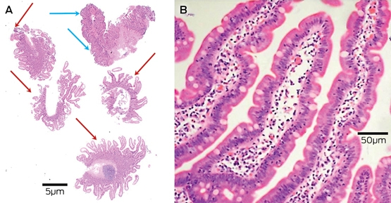
Five duodenal biopsy samples were taken at endoscopy (A), but only the sample from the duodenal bulb shows villous atrophy (blue arrows). The remaining four samples show normal architecture, but with increased intra-epithelial lymphocytes, up to 30/100 enterocytes (lymphocytic duodenosis) (red arrows). Lymphocytic duodenosis (B) with normal architecture and increased intra-epithelial lymphocytes, up to 30/100 enterocytes.
Competing interests
No relevant disclosures.
References
- Ludvigsson JF, Leffler DA, Bai JC, et al. The Oslo definitions for coeliac disease and related terms. Gut 2013; 62: 43-52.
- Marsh MN. Gluten, major histocompatibility complex, and the small intestine. A molecular and immunobiologic approach to the spectrum of gluten sensitivity (‘celiac sprue’). Gastroenterology 1992; 102: 330-354.
- Husby S, Koletzko S, Korponay-Szabo IR, et al. European Society for Pediatric Gastroenterology, Hepatology, and Nutrition guidelines for the diagnosis of coeliac disease. J Pediatr Gastroenterol Nutr 2012; 54: 136-160.
- Rubio-Tapia A, Hill ID, Kelly CP, et al. ACG clinical guidelines: diagnosis and management of celiac disease. Am J Gastroenterol 2013; 108: 656-676.
- Bai JC, Fried M, Corazza GR, et al. World Gastroenterology Organisation global guidelines on celiac disease. J Clin Gastroenterol 2013; 47: 121-126.
- Ludvigsson JF, Bai JC, Biagi F, et al. Diagnosis and management of adult coeliac disease: guidelines from the British Society of Gastroenterology. Gut 2014; 63: 1210-1228.
- Makharia GK, Mulder CJ, Goh KL, et al. Issues associated with the emergence of coeliac disease in the Asia-Pacific region: a working party report of the World Gastroenterology Organization and the Asian Pacific Association of Gastroenterology. J Gastroenterol Hepatol 2014; 29: 666-677.
- National Institute for Health and Clinical Excellence. Coeliac disease: recognition, assessment and management. London: NICE; 2015. https://www.nice.org.uk/guidance/ng20/resources/coeliac-disease-recognition-assessment-and-management-pdf-1837325178565 (accessed June 2017).
- Ludvigsson JF, Agreus L, Ciacci C, et al. Transition from childhood to adulthood in coeliac disease: the Prague consensus report. Gut 2016; 65: 1242-1251.
- Anderson RP, Henry MJ, Taylor R, et al. A novel serogenetic approach determines the community prevalence of celiac disease and informs improved diagnostic pathways. BMC Med 2013; 11: 188.
- Cook HB, Burt MJ, Collett JA, et al. Adult coeliac disease: prevalence and clinical significance. J Gastroenterol Hepatol 2000; 15: 1032-1036.
- Ludvigsson JF, Card TR, Kaukinen K, et al. Screening for celiac disease in the general population and in high-risk groups. United European Gastroenterol J 2015; 3: 106-120.
- Walker MM, Murray JA, Ronkainen J, et al. Detection of celiac disease and lymphocytic enteropathy by parallel serology and histopathology in a population-based study. Gastroenterology 2010; 139: 112-119.
- Mustalahti K, Catassi C, Reunanen A, et al. The prevalence of celiac disease in Europe: results of a centralized, international mass screening project. Ann Med 2010; 42: 587-595.
- Kang JY, Kang AH, Green A, et al. Systematic review: worldwide variation in the frequency of coeliac disease and changes over time. Aliment Pharmacol Ther 2013; 38: 226-245.
- Tortora R, Zingone F, Rispo A, et al. Coeliac disease in the elderly in a tertiary centre. Scand J Gastroenterol 2016; 51: 1179-1183.
- Anderson RP. Coeliac disease is on the rise. Med J Aust 2011; 194: 278-279.
- Volta U, Caio G, Giancola F, et al. Features and progression of potential celiac disease in adults. Clin Gastroenterol Hepatol 2016; 14: 686-693.e1.
- Singh P, Arora S, Lal S, et al. Risk of celiac disease in the first- and second-degree relatives of patients with celiac disease: a systematic review and meta-analysis. Am J Gastroenterol 2015; 110: 1539-1548.
- Costa Gomes R, Cerqueira Maia J, Fernando Arrais R, et al. The celiac iceberg: from the clinical spectrum to serology and histopathology in children and adolescents with type 1 diabetes mellitus and Down syndrome. Scand J Gastroenterol 2016; 51: 178-185.
- Tio M, Cox MR, Eslick GD. Meta-analysis: coeliac disease and the risk of all-cause mortality, any malignancy and lymphoid malignancy. Aliment Pharmacol Ther 2012; 35: 540-551.
- Leffler D, Vanga R, Mukherjee R. Mild enteropathy celiac disease: a wolf in sheep’s clothing? Clin Gastroenterol Hepatol 2013; 11: 259-261.
- West J, Logan RF, Card TR, et al. Fracture risk in people with celiac disease: a population-based cohort study. Gastroenterology 2003; 125: 429-436.
- Ludvigsson JF, Michaelsson K, Ekbom A, Montgomery SM. Coeliac disease and the risk of fractures - a general population-based cohort study. Aliment Pharmacol Ther 2007; 25: 273-285.
- Eigner W, Bashir K, Primas C, et al. Dynamics of occurrence of refractory coeliac disease and associated complications over 25 years. Aliment Pharmacol Ther 2017; 45: 364-372.
- Szajewska H, Shamir R, Mearin L, et al. Gluten introduction and the risk of coeliac disease: a position paper by the European Society for Pediatric Gastroenterology, Hepatology, and Nutrition. J Pediatr Gastroenterol Nutr 2016; 62: 507-513.
- Lebwohl B, Murray JA, Verdu EF, et al. Gluten introduction, breastfeeding, and celiac disease: back to the drawing board. Am J Gastroenterol 2016; 111: 12-14.
- Abadie V, Sollid LM, Barreiro LB, Jabri B. Integration of genetic and immunological insights into a model of celiac disease pathogenesis. Annu Rev Immunol 2011; 29: 493-525.
- Tye-Din JA, Cameron DJ, Daveson AJ, et al. Appropriate clinical use of human leukocyte antigen typing for coeliac disease: an Australasian perspective. Intern Med J 2015; 45: 441-450.
- Oxentenko AS, Murray JA. Celiac disease: ten things that every gastroenterologist should know. Clin Gastroenterol Hepatol 2015; 13: 1396-1404; quiz e127-e129.
- Mooney PD, Kurien M, Evans KE, et al. Clinical and immunologic features of ultra-short celiac disease. Gastroenterology 2016; 150: 1125-1134.
- Oberhuber G. Histopathology of celiac disease. Biomed Pharmacother 2000; 54: 368-372.
- Corazza GR, Villanacci V, Zambelli C, et al. Comparison of the interobserver reproducibility with different histologic criteria used in celiac disease. Clin Gastroenterol Hepatol 2007; 5: 838-843.
- Aziz I, Evans KE, Hopper AD, et al. A prospective study into the aetiology of lymphocytic duodenosis. Aliment Pharmacol Ther 2010; 32: 1392-1397.
- Marietta EV, Nadeau AM, Cartee AK, et al. Immunopathogenesis of olmesartan-associated enteropathy. Aliment Pharmacol Ther 2015; 42: 1303-1314.
- Leffler D, Schuppan D, Pallav K, et al. Kinetics of the histological, serological and symptomatic responses to gluten challenge in adults with coeliac disease. Gut 2013; 62: 996-1004.
- La Vieille S, Pulido OM, Abbott M, et al. Celiac disease and gluten-free oats: a Canadian position based on a literature review. Can J Gastroenterol Hepatol 2016; 2016: 1870305.
- Food Standards Australia New Zealand. Review of gluten claims with specific reference to oats and malt: final assessment report, proposal P264. Canberra: FSANZ; 2004.
- Ludvigsson JF, Montgomery SM, Ekbom A, et al. Small-intestinal histopathology and mortality risk in celiac disease. JAMA 2009; 302: 1171-1178.
- Ludvigsson JF. Mortality and malignancy in celiac disease. Gastrointest Endosc Clin N Am 2012; 22: 705-722.
- Lebwohl B, Granath F, Ekbom A, et al. Mucosal healing and risk for lymphoproliferative malignancy in celiac disease: a population-based cohort study. Ann Intern Med 2013; 159: 169-175.
- Elfstrom P, Granath F, Ye W, Ludvigsson JF. Low risk of gastrointestinal cancer among patients with celiac disease, inflammation, or latent celiac disease. Clin Gastroenterol Hepatol 2012; 10: 30-36.
- Ludvigsson JF, West J, Ekbom A, Stephansson O. Reduced risk of breast, endometrial and ovarian cancer in women with celiac disease. Int J Cancer 2012; 131: E244-E250.
- Wahab PJ, Meijer JW, Mulder CJ. Histologic follow-up of people with celiac disease on a gluten-free diet: slow and incomplete recovery. Am J Clin Pathol 2002; 118: 459-463.
- Newnham ED, Shepherd SJ, Strauss BJ, et al. Adherence to the gluten-free diet can achieve the therapeutic goals in almost all patients with coeliac disease: A 5-year longitudinal study from diagnosis. J Gastroenterol Hepatol 2016; 31: 342-349.
- Mooney PD, Evans KE, Singh S, Sanders DS. Treatment failure in coeliac disease: a practical guide to investigation and treatment of non-responsive and refractory coeliac disease. J Gastrointestin Liver Dis 2012; 21: 197-203.
- Ilus T, Kaukinen K, Virta LJ, et al. Refractory coeliac disease in a country with a high prevalence of clinically-diagnosed coeliac disease. Aliment Pharmacol Ther 2014; 39: 418-425.
- van Gils T, Nijeboer P, van Wanrooij RL, et al. Mechanisms and management of refractory coeliac disease. Nat Rev Gastroenterol Hepatol 2015; 12: 572-579.
- Catassi C, Elli L, Bonaz B, et al. Diagnosis of non-celiac gluten sensitivity (NCGS): the Salerno experts’ criteria. Nutrients 2015; 7: 4966-4977.
- Sapone A, Bai JC, Ciacci C, et al. Spectrum of gluten-related disorders: consensus on new nomenclature and classification. BMC Med 2012; 10: 13.
- Lebwohl B, Ludvigsson JF, Green PH. Celiac disease and non-celiac gluten sensitivity. BMJ 2015; 351: h4347.
- De Giorgio R, Volta U, Gibson PR. Sensitivity to wheat, gluten and FODMAPs in IBS: facts or fiction? Gut 2015; 65: 169-178.
Linked content
-
MJA InSight: Coeliac disease: sorting the wheat from the chaff
-
MJA Podcast: Prof Marjorie Walker
Provenance: Commissioned; externally peer reviewed.
Early-Onset Colorectal Cancer With Liver-Only Metastases: A Retrospective Cohort Study Integrating Prospectively Collected Real-World Clinical and Molecular Data From an Australian National Database (2009–2024) to Guide Treatment Planning
Savio G. Barreto, Christos S. Karapetis, Shahid Ullah, Matthew Burge, Susan Caird, Angus Campbell, Azim Jalali, Ross Jennens, Muhammad A. Khattak, Belinda Lee, Stephanie H. Lim, Shehara Mendis, Louise Nott, Timothy J. Price, Jeremy D. Shapiro, Jeanne Tie, Javier Torres, Colin Williams, Rachel Wong, Vanessa Wong, Peter Gibbs
Australian Pathways for Specialist Pain Management and Early Palliative Care for People With Pancreatic Cancer: Developed Using a Community Consensus Approach
Jennifer Philip, Melanie R. Lovell, Kylee Bellingham, Gail Garvey, Gregory B. Crawford, Nicole M. Rankin, Kara Burns, Isabel Young, Vivienne Milch, Dorothy Keefe, Katrina Anderson, James Lawson, Meinir Krishnasamy
Cause‐specific mortality among Queensland people with cirrhosis, by cirrhosis aetiology and decompensation status, 2007–22: a retrospective cohort study
Vikas Bhasker, Jessica R Fong, Paul J Clark, Gunter F Hartel, Richard Skoien, James O’Beirne, Elizabeth E Powell, Patricia C Valery
Clinical practice guidelines for hepatocellular carcinoma surveillance for people at high risk in Australia: summary of recommendations
Jacob George, Nicole L Allard, Stuart K Roberts, Leon A Adams, Jane Davies, Behzad Hajarizadeh, Jennifer H MacLachlan, Suzanne E Mahady, Rosalie Altus, Catherine Brown, David C Fry, Belinda Greenwood‐Smith, Natali Smud, Patricia C Valery, Nafisa Yussf, Kate Broun, Denise Campbell, Karen Canfell, Chelsea Carle Harrison, Victoria Freeman, Paul Grogan, Catherine Holliday, Suzanne Hughes, Anna Kelly, Cathelijne Kemenade, Claire Latumahina, Amanda McAtamney, Megan Varlow, Joachim Worthington, Susan Yuill, Eleonora Feletto
Assessment of metabolic dysfunction‐associated fatty liver disease in primary care: a consensus statement summary
Leon A Adams, William W Kemp, Kate R Muller, Elizabeth E Powell, Stuart K Roberts, Luis Calzadilla Bertot, Stephanie Best, Gary Deed, Jon D Emery, Samantha L Hocking, Graham R Jones, John S Lubel, Sinead Sheils, Stephen M Twigg, Gerald F Watts, Jacob George
Treatment outcomes for people with hepatitis C referred to tertiary care in Victoria, 2021–22: a retrospective observational study
Elly Layton, Nicole Matthews, Brendan Quinn, Nasra Higgins, Gabrielle Lindeman, Mielle Abbott, Jennifer MacLachlan, Elizabeth Birbilis, Margaret E Hellard, Joseph Doyle, Benjamin C Cowie, Mark Stoové