




Volume 189 - Issue 9
Management of Mycobacterium ulcerans infection in a pregnant woman in Benin using rifampicin and clarithromycin
Authors: Ange D Dossou, Ghislain E Sopoh, Christian R Johnson, Yves T Barogui, Dissou Affolabi, Sévérin Y Anagonou, Théophile Zohoun, Françoise Portaels and Kingsley Asiedu
Med J Aust 2008; 189 (9): 532-533. || doi: 10.5694/j.1326-5377.2008.tb02166.x
Published online: 3 November 2008
Published online: 3 November 2008
To the Editor: Buruli ulcer, caused by the bacterium Mycobacterium ulcerans, leads to the destruction of skin and sometimes bone. It has been reported in many tropical countries in Africa and in some temperate regions of Australia, Japan and China.1 In 2004, the World Health Organization recommended treatment with the combination of oral rifampicin and intramuscular streptomycin (or amikacin) for 8 weeks.2,3 In-vitro studies and new data from mouse models suggest that combinations of rifampicin with clarithromycin, rifampicin with moxifloxacin, or clarithromycin with moxifloxacin may be as effective as rifampicin and streptomycin.4,5
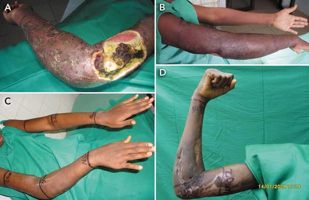
In June 2007, a woman who was 6-months pregnant with her first child and had a 7-month history of Buruli ulcer on her right upper limb (Box 1, A) was admitted to the Buruli ulcer treatment centre in Allada, Benin. She was otherwise in good health, and the fetal heart beat was normal. Routine laboratory examinations, including HIV serology tests, found no abnormalities. Swabs from the ulcer were positive for acid-fast bacilli by Ziehl–Neelsen stain, and for IS 2404 (DNA sequence specific for Mycobacterium ulcerans) by polymerase chain reaction testing, but no growth of M. ulcerans was obtained on culture. Histopathological analysis of punch biopsy specimens showed typical features of Buruli ulcer.
As streptomycin is contraindicated in pregnancy, we treated the patient with a combination of oral rifampicin (600 mg daily) and oral clarithromycin (500 mg twice daily) for 56 days, beginning 2 weeks after presentation. The treatment was well tolerated. We monitored the clinical response through serial photographs (Box 1) and measurements of the circumference of the affected and unaffected limbs at defined points (Box 2).
The patient gave birth to a healthy boy weighing 2.25 kg in September 2007, 2 weeks after completing antibiotic treatment. She underwent skin grafting a month later. The lesion healed without functional limitation (Box 1, D), and the patient was discharged in December 2007. At that time, the surface area affected by the lesion was reduced by 55%.
To our knowledge, this is the first report of successful treatment of Buruli ulcer using fully oral treament with rifampicin and clarithromycin alone. We hope our experience will contribute to future discussion and studies to find an oral treatment for this devastating disease.
1 Serial views of Buruli ulcer in a woman treated with rifampicin and clarithromycin
Acknowledgements
Fondation Luxembourgeoise Raoul Follereau, Luxembourg; Projet Burulico European Union (project reference number, INCO-CT-2005-051476); and the World Health Organization, Geneva.
References
- Portaels F, Johnson P, Meyers WM, editors. Buruli ulcer. Diagnosis of Mycobacterium ulcerans disease. A manual for health care providers. Geneva: World Health Organization, 2001: 92.
- World Health Organization. Provisional guidance on the role of specific antibiotics in the management of Mycobacterium ulcerans disease (Buruli ulcer). Geneva: WHO/CDS/CPE/GBUI.10, 2004. http://www.who.int/buruli/information/antibiotics/en/index.html (accessed 2004).
- Johnson PDR, Hayman JA, Quek TY, et al. Consensus recommendations for the diagnosis, treatment and control of Mycobacterium ulcerans infection (Bairnsdale or Buruli ulcer) in Victoria, Australia. Med J Aust 2007; 186: 64-68. 0_i1092093
- Portaels F, Traore H, De Ridder K, Meyers WM. In vitro susceptibility of Mycobacterium ulcerans to clarithromycin. Antimicrob Agents Chemother 1998; 42: 2070-2073. 0_i1092095
- Ji B, Chauffour A, Robert J, et al. Orally administered combined regimens for treatment of Mycobacterium ulcerans infection in mice. Antimicrob Agents Chemother 2007; 51: 3737-3739. 0_i1092099
Costs of Delivering the Hep B PAST Model of Care in Remote Australia: A Retrospective Pre–Post Study
Hoa Nguyen, Anh Le Tuan Nguyen, Paula Binks, Melita McKinnon, Emily Vintour-Cesar, Karen Wills, Nicola Stephens, Benjamin Cowie, Joshua S. Davis, Yuejen Zhao, Peter Nihill, Julie A. Campbell, Andrew J. Palmer, Jane Davies, Barbara de Graaff
Buruli Ulcer Acquired From Ringtail Possum Rescue
Daniel P. O'Brien, Naomi Clarke, Sara Bonnici, Mohammad A. Hussain
Q Fever Visceral Abscesses After Contact With Native Australian Marsupials
Samantha J. Laws, Enzo Binotto, James Stewart, Jennifer Ho, Trent Yarwood, Josh Hanson, Simon Smith
Hospital-Admitted Injection-Related Infections Among Incarcerated People Who Inject Drugs in Australia: A Retrospective Cohort Study
Andrew Palmer, Matthew Carter, Jeremy Yeo, Cecilia Shim, Jason Connor, Jeremy Hayllar, Gerald Holtmann, Naomi Moy, Elliott G. Playford, Naomi Runnegar, Paul J. Clark
The Live Attenuated Influenza Vaccine in Australia: An Additional Tool for Influenza Prevention
Cyra Patel, Alexis Pillsbury, Tran Nguyen, Xia Wang, Helen E. Quinn, Clayton K. Chiu, Allen C. Cheng, Katie L. Flanagan, Zhicheng Wang
Reflex Testing for Hepatitis D Infection: A Unique Opportunity to Reduce Hepatitis D-Related Chronic Liver Disease Deaths in Australia
Jessica Howell, Lauren Andersson, Miriam T. Levy, James O'Beirne, Leon Adams, Katharine Irvine, Avik Majumdar, Golo Ahlenstiel, Kathy Jackson, Krispin Hajkowicz, Joseph Doyle, Jane Davies, Sarah Cherian, Wayne Dimech, Alexander J. Thompson