




Volume 183 - Issue 1
Incidental finding of Dracunculus medinensis in Australia
Author: Tulsi Menon
Med J Aust 2005; 183 (1): 51-52. || doi: 10.5694/j.1326-5377.2005.tb06894.x
Published online: 20 June 2005
Published online: 20 June 2005
Tulsi Menon
Resident Medical Officer, Department of Orthopaedic Surgery, Royal Perth Hospital, 19 Morgan Road, Redcliffe, WA 6104
kaltulAToptusnet.com.au
To the Editor: I report an infection with Dracunculus medinensis diagnosed incidentally on x-ray. The patient was a Sudanese immigrant, who had sustained a left knee joint effusion after falling. During management at Royal Perth Hospital, a calcified lesion with a serpentine appearance was seen on x-ray within soft tissues near the left knee joint. Its characteristic appearance, combined with the patient’s background, led to a diagnosis of D. medinensis (known as guinea-worm), a nematode parasite which causes dracunculiasis.
The patient’s knee effusion was managed conservatively and improved within a few weeks. The effusion was secondary to the fall and believed to be unrelated to the calcified D. medinensis.
Transmission of dracunculiasis is through consumption of contaminated water. The guinea-worm larvae mature and migrate towards the skin surface over 1 year (during which the patient remains asymptomatic), with over 90% of the worms appearing from the lower limbs. When in contact with water, the exposed guinea-worm releases larvae, and the lifecycle is completed when people ingest contaminated water.1 If the guinea-worms die before maturation, they usually calcify.
The Global Dracunculiasis Eradication Campaign was established in 1981 with the aim of eliminating dracunculiasis through water sanitation efforts.1 With no vaccine or treatment, prevention is the only method of eliminating dracunculiasis. Since the campaign was established, the number of people affected by dracunculiasis has decreased by 98%. Currently, Sudan alone accounts for 73% of cases.1
Our patient migrated to Australia as a refugee from Sudan during the civil war. Her village in Sudan had only one source of water used for daily activities, including drinking. The patient knew many people with dracunculiasis, but did not know she had been infected.
When calcified guinea-worms are discovered during routine radiological examination, they usually do not need treatment. Many people are not aware they have been infected. Muller reported 89% of patients with calcified guinea-worms were asymptomatic.2
No known previous case of a radiologically diagnosed calcified guinea-worm has been reported in Australia. The consequences of war and famine, with a resultant increase in refugees and immigrants from affected nations, is likely to increase the number of incidental calcified guinea-worms found in non-endemic countries. This disease may have a significant impact if affected immigrants arrive during the incubation period (when asymptomatic) and the parasite emerges from the skin after immigration (rather than calcifying), similar to the patient described by Spring.3 Thus, it is important for health personnel to be aware of dracunculiasis, including its radiological manifestations.
Lateral view of left knee joint and distal left thigh
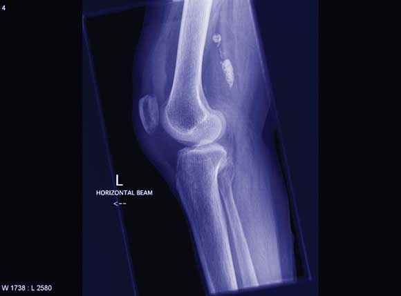
A calcified lesion, representing Dracunculus medinensis within the soft tissues, is visible posterior and lateral to the distal femur near the knee joint. The calcification has a serpentine appearance: the proximal part coiled in appearance, the middle having a string-like linear appearance, and the distal part having dense curvilinear opacity.
Acknowledgements
I thank Mr Alan Prosser, Dr Mike Ledger, Dr Vera Kinzel, and Dr Song for encouragement and assistance in preparing this letter.
References
- Greenaway C. Drancunculiasis (guinea worm disease). CMAJ 2004; 170: 495-500. CHDIFEDJ
- Muller R. Dracunculus and dracunculiasis. In: Dawes B, editor. Advances in parasitology. New York, NY: Academic Press, 1971; 73-140. i1085785
- Spring M, Spearman P. Dracunculiasis: report of an imported case in the United States. Clin Infec Dis 1997; 25: 749-750. i1085787
Costs of Delivering the Hep B PAST Model of Care in Remote Australia: A Retrospective Pre–Post Study
Hoa Nguyen, Anh Le Tuan Nguyen, Paula Binks, Melita McKinnon, Emily Vintour-Cesar, Karen Wills, Nicola Stephens, Benjamin Cowie, Joshua S. Davis, Yuejen Zhao, Peter Nihill, Julie A. Campbell, Andrew J. Palmer, Jane Davies, Barbara de Graaff
Buruli Ulcer Acquired From Ringtail Possum Rescue
Daniel P. O'Brien, Naomi Clarke, Sara Bonnici, Mohammad A. Hussain
Q Fever Visceral Abscesses After Contact With Native Australian Marsupials
Samantha J. Laws, Enzo Binotto, James Stewart, Jennifer Ho, Trent Yarwood, Josh Hanson, Simon Smith
Hospital-Admitted Injection-Related Infections Among Incarcerated People Who Inject Drugs in Australia: A Retrospective Cohort Study
Andrew Palmer, Matthew Carter, Jeremy Yeo, Cecilia Shim, Jason Connor, Jeremy Hayllar, Gerald Holtmann, Naomi Moy, Elliott G. Playford, Naomi Runnegar, Paul J. Clark
The Live Attenuated Influenza Vaccine in Australia: An Additional Tool for Influenza Prevention
Cyra Patel, Alexis Pillsbury, Tran Nguyen, Xia Wang, Helen E. Quinn, Clayton K. Chiu, Allen C. Cheng, Katie L. Flanagan, Zhicheng Wang
Reflex Testing for Hepatitis D Infection: A Unique Opportunity to Reduce Hepatitis D-Related Chronic Liver Disease Deaths in Australia
Jessica Howell, Lauren Andersson, Miriam T. Levy, James O'Beirne, Leon Adams, Katharine Irvine, Avik Majumdar, Golo Ahlenstiel, Kathy Jackson, Krispin Hajkowicz, Joseph Doyle, Jane Davies, Sarah Cherian, Wayne Dimech, Alexander J. Thompson