




Medical education
Lessons from practice
Volume 218 - Issue 9
Hypertensive retinopathy in the paediatric setting
Authors: Nandini Singh, Kanika Chaudhri and Caroline Catt
Med J Aust 2023; 218 (9): 392-393. || doi: 10.5694/mja2.51929
Published online: 15 May 2023
Published online: 15 May 2023
A 12-year-old girl presented to a general practitioner with a two-day history of progressively worsening blurry vision
Clinical record
A 12‐year‐old girl presented to a general practitioner with a two‐day history of progressively worsening blurry vision. The visual symptoms prompted the patient to be referred to the local optometrist. Blood pressure was not checked at this initial general practice visit. At the optometrist, fundoscopy revealed bilateral disc swelling, prompting urgent same‐day referral to our hospital's ophthalmology unit. There was no associated pain on extraocular eye movements, headaches, or diplopia. There was no past medical or relevant family history. However, her parents described abnormal behavioural patterns over the previous three months, including nocturnal enuresis, sleep walking, and wishing to take cold showers late at night.
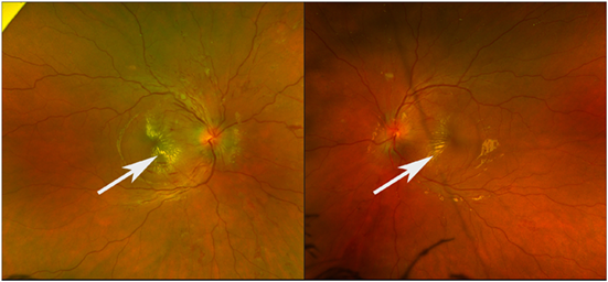
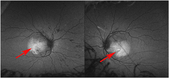
On examination, visual acuity was severely reduced to 6/76 and 6/38 in the right and left eyes respectively. Pupils were equal and reactive to light with no relative afferent pupillary defect. There was no ophthalmoplegia. Anterior segment examination was normal. Dilated fundus examination revealed bilateral optic disc swelling, scattered retinal nerve fibre layer and deep retinal haemorrhages, and exudative deposits at the macula in the form of classic macular stars (Box 1). Fundus autofluorescence revealed hyper‐autofluorescence in the region of the macular oedema (Box 2). Optical coherence tomography (OCT) through the fovea confirmed the presence of macular oedema and exudate (Supporting Information, figure 1).
The retinal signs prompted transfer to our emergency department for urgent systemic evaluation. Her blood pressure was severely elevated at 180/110mmHg. She was admitted to paediatric intensive care and required four antihypertensive agents to stabilise. Magnetic resonance imaging (MRI) scan of the brain and venogram were normal. A transthoracic echocardiogram revealed left ventricular hypertrophy with preserved systolic function. Biochemistry showed raised serum and urine catecholamine levels. On day 4 of admission, abdominal ultrasound revealed a large para‐aortic infrarenal mass, which was confirmed as a retroperitoneal paraganglioma on MRI. Renal vasculature was normal. A follow‐up positron emission tomography (PET) scan excluded any metastases.
The patient was diagnosed with malignant hypertension secondary to a catecholamine‐secreting paraganglioma, which was resected after medical stabilisation. Histopathology confirmed a 65×45×30mm paraganglioma. Immunohistochemistry was suggestive of a succinate dehydrogenase gene mutation. However, the family chose to defer genetic testing until the patient was older and able to participate in the decision making.
The ophthalmic status slowly improved. By day 10 of treatment, the visual acuity had increased to right 6/30 and left 6/12, and there was relative improvement in the macular oedema. By day 62, the visual acuity had improved to near normal at right 6/9.5 and left 6/7.6, and the OCT confirmed almost complete resolution of the macular oedema (Supporting Information, figure 2). Given the age of presentation, the size of the tumour, and that genetic testing was deferred, the patient was classified as high risk of developing other tumours. Follow‐up plans included annual blood pressure and ophthalmic testing, as well as MRI scans (base of skull to pelvis) every two years.
Discussion
Bilateral optic disc swelling should prompt an urgent workup for a broad set of differentials, including ocular, neurological, and other systemic causes. Optic disc swelling, in the presence of an exudative macular star, is referred to as neuroretinitis. The differentials for this include infectious (eg, viral infections, syphilis, tuberculosis, and Bartonella) and non‐infectious causes (eg, severe hypertension, diabetic papillopathy, and autoimmune disorders).
Unfortunately, it is relatively uncommon for vital signs to be routinely screened in a patient with an ophthalmic presentation, especially in a child. Yet, in this case, the visual symptoms and optic disc swelling prompted an early blood pressure check, leading to the diagnosis of malignant hypertension.
Malignant hypertension refers to severely raised blood pressure, with signs of end‐organ damage, such as grade 3 hypertensive retinopathy with disc swelling and/or cardiac, renal, and neurological sequelae.1 In adolescents with severe hypertension, secondary causes must be suspected, including renovascular disease, aldosteronism and phaeochromocytomas or paragangliomas.2 Physicians must consider these catecholamine‐secreting tumours in patients with sustained or paroxysmal hypertension. Given that the early symptomology of such tumours may be varied, diagnosis can be delayed, leading to end‐organ damage and metastases. Having a high index of suspicion is hence important.3
Treatment of a hypertensive crisis involves immediate control of blood pressure. As the systemic health is stabilised, the ocular complications slowly also improve. No specific local ophthalmic treatment is usually required. Macular oedema, followed by disc swelling, are usually the first to respond to treatment. Exudative deposits may resorb gradually, but not always completely. Hypertensive retinopathy can lead to other complications, such as ischaemic optic neuropathy or retinal vascular occlusions, which may lead to permanent vision loss.
In our patient, detection of the malignant hypertension led to the diagnosis of the underlying catecholamine‐secreting paraganglioma. A simple blood pressure check proved both sight‐ and lifesaving. Our patient was rapidly engaged with a multidisciplinary care team in our tertiary referral paediatric hospital and achieved good systemic and ophthalmic recovery.
This case should serve as a reminder to emergency specialists, primary care practitioners, physicians, and ophthalmic clinicians alike of the importance of checking blood pressure when assessing any neuro‐ophthalmic presentation, such as optic disc swelling.
Lessons from practice
- Bilateral optic disc swelling should prompt an urgent workup for ocular, neurological and systemic causes, including blood pressure.
- A blood pressure check can prove both sight‐ and lifesaving.
- Severe hypertension in the paediatric and adolescent population is often secondary to an underlying cause (unlike essential hypertension).
- The mainstay of treatment for hypertensive retinopathy is systemic control of the hypertension.
Competing interests
No relevant disclosures.
References
- Domek M, Gumprecht J, Lip GYH, Shantsila A. Malignant hypertension: does this still exist? J Hum Hypertens 2020; 34: 1‐4.
- Wijaya I, Siregar P. Hypertensive crises in the adolescent: evaluation of suspected renovascular hypertension. Acta Med Indones 2013; 45: 49‐54.
- Neumann HPH, Young WF, Eng C. Pheochromocytoma and paraganglioma. N Engl J Med 2019; 381: 552‐565.
Provenance: Not commissioned; externally peer reviewed.
Fatty Liver Disease in Australia: A Narrative Review on the Epidemiology, Natural History, Prognostication and Management in People With Metabolic Dysfunction
Karl Vaz, Daniel Clayton-Chubb, William W. Kemp, Stuart K. Roberts, Ammar Majeed
Sex Differences in Management and Outcomes of People With ST-Elevation Myocardial Infarction, New South Wales, 2011–2020: A Retrospective Cohort Study
Adam C. Livori, Jedidiah I. Morton, Dianna J. Magliano, Jonathan E. Shaw
Rethinking Rheumatic Fever Diagnosis: Progress, Pitfalls and the Path Towards a Definitive Test
Anna P. Ralph, Rachel Webb, Tom Parks, Nicole J. Moreland, Asha C. Bowen, Benjamin Jones, Emma Ndagire, Andrea Z. Beaton
Sex Differences in Management and Outcomes of People With ST-Elevation Myocardial Infarction, New South Wales, 2011–2020: A Retrospective Cohort Study
Samia Kazi, Simone Marschner, Clara K. Chow
Distribution of Cardiovascular Disease Risk Based on the Updated 2023 Guideline-Recommended Australian Cardiovascular Disease Risk Algorithm and Comparison With the 2012 Algorithm: An Observational Study
Nina Lazarevic, Meghana Bhat, Grace Joshy, Danielle C. Butler, Mark Woodward, Anushka Patel, Rod T. Jackson, Garry Jennings, Rosemary Wyber, Ellie Paige, Emily Banks
They Brought the Hospital to Me
Hugh N. Burrill