




Medical education
Lessons from practice
Volume 218 - Issue 1
An important case of atypical pneumonia
Authors: Arvind Yerramilli, Michelle Sam, Aadith Ashok and Eugene Athan
Med J Aust 2023; 218 (1): 16-17. || doi: 10.5694/mja2.51791
Published online: 16 January 2023
Published online: 16 January 2023
A 38-year-old woman presented to the emergency department with a 2-week history of fever, headache and mild dry cough
Clinical record
A 38‐year‐old woman presented to the emergency department with a 2‐week history of fever, headache and mild dry cough. She lived in an area that was undergoing a coronavirus disease 2019 (COVID‐19) outbreak at the time. A household contact had mild coryzal symptoms in the week prior but had recovered and was negative for severe acute respiratory syndrome coronavirus 2 (SARS‐CoV‐2). There were no other sick contacts and no travel history.
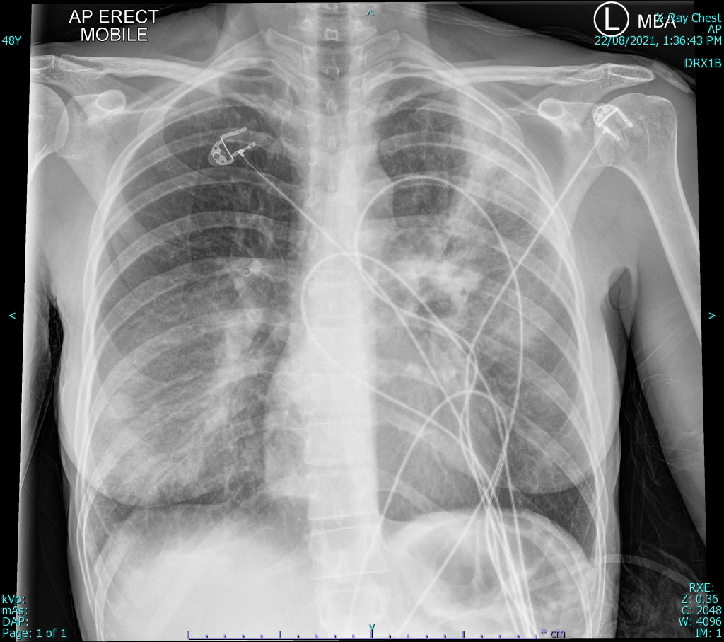
Blood film revealed a normal white cell count but a marked left shift and toxic changes. There was a mild transaminitis (Box 1). During assessment, she became profoundly hypoxic. Chest x‐ray demonstrated multilobar consolidation (Box 2). She was commenced on empirical ceftriaxone 2g every 24 hours and azithromycin 500mg every 24 hours and was transferred to the intensive care unit for high flow oxygen therapy. Despite a high suspicion for COVID‐19, the patient tested negative on two separate polymerase chain reaction (PCR) assays.
On further questioning, the family had purchased a cockatiel 3weeks prior. The antimicrobial regimen was switched to doxycycline monotherapy 100mg twice daily and the patient tested positive for Chlamydia psittaci via PCR of a nasopharyngeal specimen. The patient made a slow recovery, requiring ongoing high flow oxygen. A computed tomography pulmonary angiogram showed multiple small bilateral pulmonary embolisms and she was subsequently commenced on rivaroxaban 15mg twice daily for 3weeks with a plan for 20mg once daily thereafter and a 3‐month review. The patient made a gradual improvement and completed a 2‐week course of doxycycline with ongoing rivaroxaban and recovered.
Discussion
Psittacosis, also known as avian chlamydiosis, ornithosis or parrot fever, is a respiratory infection caused by the pathogen Chlamydia psittaci — an obligate, intracellular, gram‐negative bacterium.1 It classically presents as an atypical pneumonia syndrome with headache, fever and, less frequently, dry cough and dyspnoea.1,2 Diarrhoea, pharyngitis and altered mental state are other less common symptoms, and illness severity may be greater in both older and pregnant patients.2,3 Chest x‐ray is often discordant with clinical findings. Laboratory features include a normal neutrophil count with marked left shift and deranged liver function tests. Complications are varied and multisystem (Box 3), and differential diagnosis includes other causes of atypical pneumonia such as Chlamydia pneumoniae, Mycoplasma pneumoniae, Legionella spp, and viral and fungal pathogens.1,2 Although pulmonary embolism has not previously been directly linked to psittacosis, in the context of an acute deterioration and delayed response, pulmonary embolism was important to identify for our patient.
Sir Frank McFarlane Burnet was among the first to establish the zoonotic nature of psittacosis from Australian parrots in 1935.4 The respiratory infection is also listed as a Category B bioterrorism agent by the United States Centers for Disease Control and Prevention. Birds are the primary reservoir, with most human disease arising from the order Psittaciformes, which includes parakeets, parrots, cockatoos and cockatiels. Nevertheless, the pathogen has been found in over 30 different orders of both wild and domestic birds in Australia and rarely in mammals.2
Infection occurs via inhalation of bacteria in dried faeces shed by birds, often resulting from cage cleaning without protective equipment. Other routes such as lawn mowing, bird bites and mouth‐to‐beak contact have been described.3 Person‐to‐person transmission is rare, although there have been reports of nosocomial spread.3 National yearly notifications have been fewer than 50 in recent years, but there have been more substantial peaks in 2004 with more than 200 notifications in one year.4 Recently, there was a localised outbreak in the Victorian Alpine regions, but many of the patients affected had no direct contact with birds.5 Reliance on bird exposure may lead to delay in diagnosis, and in similar outbreak scenarios other clinical characteristics may be more reliable.
Culture is only done in specialised laboratories and requires Physical Containment Level 3 (PC3) isolation.2 Serological techniques include complement fixation based on the lipopolysaccharide antigen, and micro‐immunofluorescence based on species‐specific surface antigen. The former technique does not readily differentiate among chlamydial species and therefore testing for C. psittaci should be specifically requested.2 Due to poor sensitivity, both techniques require acute and convalescent testing to establish a diagnosis. A third specimen 8weeks later may be required if treated with antibiotics.3 PCR of respiratory specimens is the preferred diagnostic test and has the advantage of increased turnaround time, sensitivity and specificity.
Doxycycline is the antibiotic of choice based on intracellular activity, pharmacodynamics and clinical experience, although there have been no controlled trials.2 Macrolides are second line treatment, with similar efficacy and are preferred for children and pregnant patients. Case notification is required by testing laboratories, with outbreak management done by state health departments.3 Pets can be tested and treated by veterinarians. Education regarding cleaning of cages is paramount, and gloves, eye protection and P2/N95 respirators are recommended for preventing transmission. Wetting litter before cleaning might also reduce transmission risk.
Lessons from practice
- Psittacosis is a zoonotic infection, with a recent outbreak in the Victorian Alpine regions highlighting the importance of disease recognition.
- Presentation is of an atypical pneumonia syndrome, with respiratory symptoms often less prominent, although there can be multi‐organ involvement.
- Molecular testing with polymerase chain reaction (PCR) of respiratory specimens can establish a diagnosis, with the treatment of choice being doxycycline for at least 2weeks.
- Preventive measures such as the use of appropriate personal protective equipment when cage cleaning is important to reduce transmission risk.
Box 1 – Significant laboratory testing results
|
|
Result (reference interval) |
||||||||||||||
|
|
|||||||||||||||
|
White cell count (× 109 cells/L) |
6.8 (4.0–11.0) |
||||||||||||||
|
Neutrophil (× 109 cells/L) |
6.0 (2.0–8.0) |
||||||||||||||
|
Blood film |
Neutrophils show left shift and toxic changes |
||||||||||||||
|
AST (U/L) |
214 (<41) |
||||||||||||||
|
ALT (U/L) |
103 (<41) |
||||||||||||||
|
CK (U/L) |
544 (<161) |
||||||||||||||
|
CRP (mg/L) |
274 (<3.0) |
||||||||||||||
|
|
|||||||||||||||
|
AST = aspartate transaminase; ALT = alanine transaminase; CK = creatine kinase; CRP = C‐reactive protein. |
|||||||||||||||
Box 3 – Complications of psittacosis characterised by organ or systems affected
|
Organ/system |
Complications |
||||||||||||||
|
|
|||||||||||||||
|
Pulmonary |
Respiratory failure, acute respiratory distress syndrome, pleural effusion |
||||||||||||||
|
Cardiac |
Endocarditis, myocarditis, pericarditis, pericardial effusion |
||||||||||||||
|
Renal |
Proteinuria, oliguria, acute tubular necrosis, glomerulonephritis |
||||||||||||||
|
Haematological |
Cold agglutinins, haemophagocytic syndrome, thrombotic thrombocytopenic purpura, disseminated intravascular coagulation |
||||||||||||||
|
Hepatic |
Hepatitis, granulomas |
||||||||||||||
|
Neurological |
Encephalitis, meningitis, psychiatric symptoms, transverse myelitis, Guillain–Barré syndrome |
||||||||||||||
|
Skin |
Erythema nodosum, erythema multiforme, erythema marginatum |
||||||||||||||
|
|
|||||||||||||||
|
|
|||||||||||||||
Competing interests
No relevant disclosures.
References
- Yung AP, Grayson ML. Psittacosis — a review of 135 cases. Med J Aust 1988; 148: 228‐233.
- Stewardson AJ, Grayson ML. Psittacosis. Infect Dis Clin North Am 2010; 24: 7‐25.
- Department of Health and Aged Care. Psittacosis (ornithosis) — CDNA National Guidelines for Public Health Units; 2016. https://www.health.gov.au/resources/publications/psittacosis‐cdna‐national‐guidelines‐for‐public‐health‐units (viewed Feb 2022).
- Polkinghorne A, Weston KM, Branley J. Recent history of psittacosis in Australia: expanding our understanding of the epidemiology of this important globally distributed zoonotic disease. Intern Med J 2020; 50: 246‐249.
- Department of Health, State Government of Victoria. Health warning on increase in psittacosis in Victorian Alpine region [15 Dec 2020]. https://www.health.vic.gov.au/health‐advisories/health‐warning‐on‐increase‐in‐psittacosis‐in‐victorian‐alpine‐region (viewed Feb 2022).
Provenance: Not commissioned; externally peer reviewed.
Costs of Delivering the Hep B PAST Model of Care in Remote Australia: A Retrospective Pre–Post Study
Hoa Nguyen, Anh Le Tuan Nguyen, Paula Binks, Melita McKinnon, Emily Vintour-Cesar, Karen Wills, Nicola Stephens, Benjamin Cowie, Joshua S. Davis, Yuejen Zhao, Peter Nihill, Julie A. Campbell, Andrew J. Palmer, Jane Davies, Barbara de Graaff
Buruli Ulcer Acquired From Ringtail Possum Rescue
Daniel P. O'Brien, Naomi Clarke, Sara Bonnici, Mohammad A. Hussain
Q Fever Visceral Abscesses After Contact With Native Australian Marsupials
Samantha J. Laws, Enzo Binotto, James Stewart, Jennifer Ho, Trent Yarwood, Josh Hanson, Simon Smith
Hospital-Admitted Injection-Related Infections Among Incarcerated People Who Inject Drugs in Australia: A Retrospective Cohort Study
Andrew Palmer, Matthew Carter, Jeremy Yeo, Cecilia Shim, Jason Connor, Jeremy Hayllar, Gerald Holtmann, Naomi Moy, Elliott G. Playford, Naomi Runnegar, Paul J. Clark
The Live Attenuated Influenza Vaccine in Australia: An Additional Tool for Influenza Prevention
Cyra Patel, Alexis Pillsbury, Tran Nguyen, Xia Wang, Helen E. Quinn, Clayton K. Chiu, Allen C. Cheng, Katie L. Flanagan, Zhicheng Wang
Reflex Testing for Hepatitis D Infection: A Unique Opportunity to Reduce Hepatitis D-Related Chronic Liver Disease Deaths in Australia
Jessica Howell, Lauren Andersson, Miriam T. Levy, James O'Beirne, Leon Adams, Katharine Irvine, Avik Majumdar, Golo Ahlenstiel, Kathy Jackson, Krispin Hajkowicz, Joseph Doyle, Jane Davies, Sarah Cherian, Wayne Dimech, Alexander J. Thompson