




Medical education
Lessons from practice
Volume 208 - Issue 4
Sudden onset dyspnoea and recurrent stridor
Author: Clarissa Susanto
Med J Aust 2018; 208 (4): 159-160. || doi: 10.5694/mja16.01397
Published online: 5 March 2018
Published online: 5 March 2018
A 37-year-old woman admitted for elective reduction mammoplasty developed stridor after extubation
Clinical record
A 37-year-old woman admitted for elective reduction mammoplasty developed stridor after extubation. During this episode, she reported difficulty with inhalation and globus sensation. Vital measurements were within normal limits, with resting oxygen saturation measured by pulse oximetry (Spo2) of 99% on room air. There was no suggestion of difficult intubation in the anaesthetic report. Chest x-ray was unremarkable and full blood count revealed only mild neutrophilia. She was admitted to the high dependency unit for monitoring but required no additional support. Spirometry performed 24 hours later showed no airflow obstruction, although flow–volume curves revealed a flattened inspiratory loop. The stridor abated on its own over 48 hours.
The patient had a history of chronic episodic stridor dating back to childhood. It was often precipitated by exposure to fumes, exertion, respiratory tract infection or emotional stress. She had received a variety of diagnoses, including asthma, laryngeal dystonia and anxiety-related stridor, and had been trialled on nebulised adrenaline, prednisolone therapy and intralaryngeal botulinum toxin injection, without significant improvement. There were also occasions when intubation had been considered on presentation.
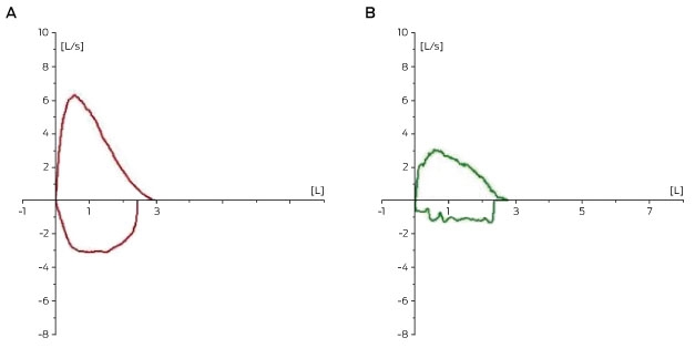
The patient had no features to suggest asthma. Previous computed tomography (CT) of the chest and neck, and neck ultrasound had been unremarkable. Repeat spirometry when she was asymptomatic was normal with no airflow obstruction, no bronchodilator reversibility and no flattening of the flow–volume curves (Box 1, A). Methacholine challenge test was negative.
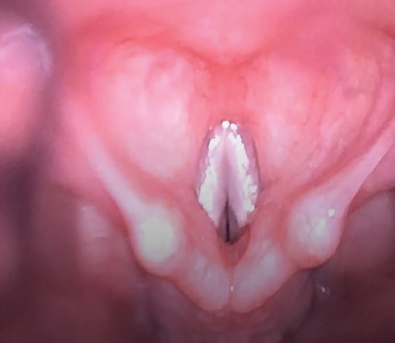
Bronchoscopy performed to exclude anatomical abnormality showed normal-looking vocal cords, no supra or subglottic stenosis and no tracheobronchomalacia; however, after the procedure, the patient developed abrupt onset of stridor with maintenance of oxygenation. The bronchoscope was re-introduced and paradoxical movement of the vocal cords was noted (Box 2). Repeat spirometry, performed while the patient was still symptomatic, revealed a variable flattening of the inspiratory curves (Box 1, B). A diagnosis of vocal cord dysfunction (VCD) was made.
VCD, also known as paradoxical vocal cord motion disorder, is a syndrome that describes inappropriate and unintentional vocal cord adduction during the respiratory cycle. This disorder can lead to episodic dyspnoea, wheezing, stridor and apparent upper-airway obstruction — symptoms that may be mistaken for anaphylaxis or asthma. Failure to recognise VCD has been associated with unnecessary treatments and hospitalisation.1
The estimated prevalence of this condition varies, ranging from 2.5% of patients presenting to asthma clinics to 22% of patients with recurrent emergency department presentation for dyspnoea.2,3 The literature suggests that VCD has a female preponderance, although it has also been seen in males,4 and it has been reported in both paediatric and adult populations.
The patient generally reports sudden onset dyspnoea; throat tightness; difficulty breathing in, rather than out; and cough. Stridor can be heard during inspiration, expiration or both, and it is generally heard loudest over the anterior neck. This episode may last minutes to days; in contrast to laryngospasm, which usually subsides over seconds to minutes. Occasionally, the patient can be left with dysphonia after the episode. In contrast to patients with asthma, there is a lack of symptomatic response to asthma therapy, and symptoms can sometimes be abated by distractions. Patients will usually have a normal SpO2 rate, although the presence of oxygen desaturations does not rule out VCD.
The pathophysiology of VCD is unclear, but autonomic imbalance, laryngeal sensory neuropathy and laryngeal hyper-responsiveness have been postulated as possible mechanisms.3 VCD is commonly precipitated by exercise, airborne irritants, gastroesophageal reflux disease or postnasal drip, and is associated with psychological conditions. Given the lack of prospective evaluation, the relationship between VCD and asthma is unclear, but the two conditions are known to coexist. A study by Newman and colleagues5 showed that 53 of the 95 patients fulfilling laryngoscopic criteria for VCD also had coexistent asthma. The medical literature records numerous cases in which VCD was misdiagnosed as asthma. Moreover, the diagnosis is further complicated by the fact that patients with VCD can have a positive methacholine challenge test, which should not be used to distinguish VCD from asthma.
Pulmonary function test with flow–volume curves and flexible laryngoscopy are the two most useful investigations to diagnose this condition. Typically, spirometry can look normal when the patient is asymptomatic (Box 1, A); however during symptoms the flow–volume curves may show variable flattening of the inspiratory curve but no airflow obstruction (Box 1, B). Flexible laryngoscopy during symptomatic periods is considered the gold standard for diagnosis —paradoxical complete adduction of the vocal cords during inspiration and occasionally during expiration is characteristic (Box 2). A novel imaging technique using dynamic volume CT scans has been used in research settings to diagnose VCD, but this technique is not yet standardised and is not currently widely adopted.6
The management of VCD in acute settings is centred on reassurance and supportive care until the episode resolves spontaneously. Breathing techniques such as forced sniffing, panting or pursed-lip breathing may sometimes help abort the symptoms. These techniques aim to relax the adducted vocal cords by activating the posterior cricoarytenoid muscle and widening the glottic aperture. Continuous positive airway pressure and inhalation of a helium–oxygen mixture have been reported to be useful in acute settings.3 Intralaryngeal botulinum toxin injection has been trialled with varying success.3 The mainstay of treatment for VCD is behavioural and speech therapy in addition to strategies that minimise the precipitating factors (eg, treatment of gastroesophageal reflux disease).Lessons from practice
Vocal cord dysfunction (VCD) is characterised by inappropriate adduction of the vocal cords during the breathing cycle, which can result, clinically, in sensation of dyspnoea and stridor.
VCD is an under-recognised condition that can be mistaken for asthma; misdiagnosis of this condition may lead to unnecessary treatments and significant morbidities.
Pulmonary function test with flow–volume loop is helpful in diagnosis; however, visualisation of the paradoxical vocal cord movement using flexible laryngoscopy is considered the gold standard in diagnosis.
Behavioural and speech therapy is generally considered the mainstay of treatment for this condition.
Competing interests
No relevant disclosures.
References
- Lawrence S. Laryngeal dyskinesia: an under-recognized condition. Emerg Med Australas 2007; 19: 96-104.
- Ciccolella DE, Brennan KJ, Borbely B, et al. Identification of vocal cord dysfunction (VCD) and other diagnoses in patients admitted to an inner city university hospital asthma center. Am J Respir Crit Care Med 1997; 155: A82.
- Kenn K, Balkissoon R. Vocal cord dysfunction: what do we know? Eur Respir J 2011; 37: 194-200.
- Morris MJ, Allan PF, Perkins PJ. Vocal cord dysfunction: aetiologies and treatment. Clin Pulm Med 2006; 13: 73-86.
- Newman KB, Mason UG, Schmaling KB. Clinical features of vocal cord dysfunction. Am J Respir Crit Care Med 1995; 152: 1382-1386.
- Holmes PW, Lau KK, Crossett M, et al. Diagnosis of vocal cord dysfunction with high resolution dynamic volume computerized tomography of the larynx. Respirology 2009; 14: 1106-1113.
Provenance: Not commissioned; externally peer reviewed.
Barriers and Facilitators to Timely Detection and Optimal Management of Chronic Wet Cough in Aboriginal Children Across Australia From the Perspectives of Carers of Aboriginal Children: A Qualitative Study
Gloria T. Y. Lau, Pam Laird, Robyn Aitken, Melanie Barwick, Liam Bedford, Emily R. Bowden, Jamie Everingham, Angela Fuery, Reece Griffin, Neha Jain, Mina Kinghorn, Renae Knapp, Gabrielle B. McCallum, Peter Morris, Richard Norman, Peter C. Richmond, Aarti Saiganesh, Slade Sibosado, Cameron Taylor, Maree Toombs, Roz Walker, Joan Wilson, Anne B. Chang, André Schultz
Barriers and Facilitators to Timely Detection and Optimal Management of Chronic Wet Cough in Aboriginal Children Across Australia From the Perspective of Health Care Providers: A Qualitative Study
Gloria T. Y. Lau, Pamela Laird, Robyn Aitken, Melanie Barwick, Liam Bedford, Emily R. Bowden, Corey Dalton, Jamie Everingham, Angela Fuery, Reece Griffin, Neha Jain, Mina Kinghorn, Renae Knapp, Gabrielle B. McCallum, Peter Morris, Richard Norman, Peter C. Richmond, Aarti Saiganesh, Slade Sibosado, Maree Toombs, Roz Walker, Joan Wilson, Anne B. Chang, André Schultz
Respiratory Syncytial Virus Disease Burden and Evidence Gaps in Australia Before Immunisation Introduction: A Retrospective Population-Based Study Using 2016–2019 Hospitalisation and 2023 Laboratory Surveillance Data
Chisato Imai-Boulter, Jean Li-Kim-Moy, Clayton Chiu, Sanjay Jayasinghe, Kristine Macartney
Not Gone but Often Forgotten: Action Needed to Address Tuberculosis in Aboriginal and Torres Strait Islander Communities
Nikkita Joshua, Emma L. Smith, Eunice (Cheeky) Love, Christopher P. Lowbridge, Nicole Tukana, Amy Bowden, Megan A. Campbell, Dawn Casey, James Ward
Uneven Ground: Survival Differences Among Victorian Lung Cancer Patients by Location of Residence (2011–2023): A Retrospective Cohort Study
Evangeline Samuel, Eldho Paul, Mike Lloyd, Sanuki Tissera, Craig Underhill, Sagun Parakh, Phillip Parente, Inger Olesen, Javier Torres, Katharine See, Gavin M. Wright, David Langton, Thomas John, Matthew Conron, James Bartlett, Nicola Atkin, Nikolajs Zeps, Susan V. Harden, Wasek Faisal, John R. Zalcberg, Rob G. Stirling
Treatment and Survival Outcomes for Indigenous and Non-Indigenous Australians Within the Victorian Lung Cancer Registry: A Retrospective Cross-Sectional Cohort Study
Melanie Wong, Mike Lloyd, Jessie Zeng, Sanuki Tissera, Kalinda E. Griffiths, Justine Clark, Jonathan Gillies, Lisa Briggs, Jacqueline Lesage, Tom Wood, Craig Underhill, Sagun Parakh, Louis B. Irving, Wasek Faisal, Rob Blum, Gary E. Richardson, Phillip Parente, Michelle Caldecott, Inger Olesen, Javier Torres, Evangeline Samuel, Christopher Lyne, Katharine See, David Langton, Thomas John, Gavin Wright, Matthew Conron, James Bartlett, Golsa Adabi, Maggie Moore, Susan Harden, Zoe K. McQuilten, John R. Zalcberg, Rob Stirling