




Volume 199 - Issue 7
Novel insights into the pathology of upper gut symptoms: new syndromes, new diseases
Authors: Nicholas J Talley and Marjorie M Walker
Med J Aust 2013; 199 (7): 440-441. || doi: 10.5694/mja13.11085
Published online: 7 October 2013
Published online: 7 October 2013
The importance of gut microflora in human health and immunity is just beginning to be realised.
The importance of gut microflora in human health and immunity is just beginning to be realised
Look and you will find it — what is unsought will go undetected — Sophocles, Oedipus Rex, 429 BC.
The failure to recognise the role of Helicobacter pylori in gastritis until the observations of Warren and Marshall were published1 was a dramatic illustration of human limitation. Despite looking at gastric biopsies, pathologists all over the world had failed to recognise the importance of these bacteria until what to look for became known — at which time (or, at least, after an initial period of scepticism) all became obvious.
A more recently recognised upper gut disease is eosinophilic oesophagitis (EoE).2 Until routine biopsy of the oesophagus became common practice, cases of food impaction or intermittent dysphagia were misattributed to other causes such as a Schatski ring or peptic ulcer disease. Oesophageal strictures have greatly decreased with improved control of reflux disease, but the incidence of EoE (associated with atopy) is increasing, as is the incidence of another atopic disease, asthma, and this is not explained by diagnostic bias.3 Atopy is a growing problem in Western society, attributed to gene–environment interaction.3 Eosinophilia is an abnormal finding in biopsies of the oesophageal squamous epithelium, and an underlying cause must be identified. EoE is diagnosed by clinicopathological correlation involving: symptoms of oesophageal dysfunction; eosinophil-predominant inflammation (> 15 eosinophils per high-power microscope field) isolated to the oesophagus which persists after a trial of therapy with proton pump inhibitors (PPIs); and excluding secondary causes of eosinophilia.2 A response to treatment (by dietary elimination or topical steroids) supports, but is not required to make the diagnosis.
Other causes of oesophageal eosinophilia include gastro-oesophageal reflux disease (GORD) and non-GORD PPI-responsive oesophageal eosinophilia (PPI-REE). While EoE per se does not respond to PPIs, PPI-REE will respond to these drugs and this condition (which some regard as a subset of EoE) is a separate entity within new guidelines.2 The choice of treatment for EoE depends on patient preference and clinician expertise, and includes topical steroids, dietary elimination and endoscopic treatment of complications (strictures). EoE is a refractory disease; symptoms recur and maintenance therapy is often necessary.2
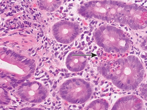
Another question under consideration is whether subtly abnormal histological appearances could similarly explain some functional gastrointestinal disorders (FGIDs), defined by chronic gut symptoms in the absence of known abnormalities. FGIDs affect at least one in three Australians from childhood to old age. Functional dyspepsia (FD) is a common syndrome presenting in primary care characterised by meal-related symptoms (such as early satiety, postprandial fullness and bloating, or recurrent epigastric pain and burning). FD overlaps with irritable bowel syndrome (IBS), and many patients also experience extra-intestinal problems (eg, asthma) at rates greater than would be expected by chance.4 Symptoms can be debilitating and, as the causes are obscure and biomarkers are unavailable, management is suboptimal. However eosinophilia has recently been described in duodenal biopsies taken from a subset of patients with early satiety symptoms in FD (Box).5 With the current upsurge in atopy and allergy, and of research into the importance of the microbiome and pathogens in the gut (which are known to give rise to postinfectious functional disorders), this observation is gaining credence as an abnormality responsible for some cases of FD.6 This may alter future management.
Coeliac disease is also in the news. There are likely more cases of this disease than are currently recognised, and new guidelines for diagnosis7 and disease definitions8 will ensure a greater capture of cases. Lymphocytic duodenosis (normal duodenal architecture and > 25 intraepithelial lymphocytes per 100 enterocytes) on biopsy is now recognised as an early manifestation of coeliac disease. However, lymphocytic duodenosis occurs in 5% of the population, and other causes (eg, H. pylori, non-steroidal anti-inflammatory drugs, small intestinal bacterial overgrowth and autoimmune disorders) must be considered if this condition is diagnosed at biopsy and coeliac disease is excluded.7 Furthermore, it is important to emphasise that serological tests and biopsies must be performed while the patient is eating a diet containing gluten, as a gluten-free diet can obscure the diagnosis.
The recently described associated entity of non-coeliac gluten sensitivity (gluten sensitivity symptoms without biopsy evidence of coeliac disease) may explain some cases of IBS, and the diagnosis can only be considered when coeliac disease is excluded by appropriate testing.7
Coeliac disease, EoE and IBS are subject to genetic predisposition. In coeliac disease, expression of the disease is reliant on having the HLA-DQ2 and/or HLA-DQ8 genes and exposure to gluten,8 and most patients with EoE have a background of an atopic diathesis.2 The expression of disease thus depends on multiple factors, including genetic background and environmental exposure, which likely includes the gut microbiome. The microbiome can influence disease expression in coeliac disease — those with classical gastrointestinal symptoms and those with extra-intestinal manifestations show marked differences in the intestinal microbiota, suggesting the same disease can be expressed differently depending on the microbiome.9
The microbiome influences the gut immune system. The previously held “hygiene hypothesis” presumed that exposure to microbes in early life prevents later development of an atopic diathesis. However, this is being overtaken by the concept of “the disappearing microbiome” which postulates that lifestyle changes (eg, antibiotics, caesarean section births, lower rates of breastfeeding) has changed the original microbiota of the gut, which is thought to affect the equilibrium and contribute to disease risk, as seen by the rapid rise in obesity, asthma and gastro-oesophageal reflux.10
As our knowledge expands, we are beginning to realise that many gastrointestinal diseases may have their origins in a complex interplay of factors both within and beyond our control. Our genetic background, environmental influence and the balance between “good” and “bad” gut bacteria in the microbiome may help explain many of the newly recognised gut disorders.
Competing interests
Nick Talley has received research grants from the National Health and Medical Research Council, the United States Institutes of Health and industry, including Forest Pharmaceuticals, Ironwood Pharmaceuticals, Janssen Pharmaceuticals and Takeda Pharmaceutical Company. He has been a consultant including for Danone, Abbott Laboratories and Prometheus Laboratories.
References
- Marshall BJ, Warren JR. Unidentified curved bacilli in the stomach of patients with gastritis and peptic ulceration. Lancet 1984; 1: 1311-1315. 0_i1139922
- Dellon ES, Gonsalves N, Hirano I, et al. American College of Gastroenterology. ACG clinical guideline: evidence based approach to the diagnosis and management of esophageal eosinophilia and eosinophilic esophagitis (EoE). Am J Gastroenterol 2013; 108: 679-692. 0_i1139924
- Custovic A, Marinho S, Simpson A. Gene–environment interactions in the development of asthma and atopy. Expert Rev Respir Med 2012; 6: 301-308. 0_i1139926
- Powell N, Huntley B, Beech T, et al. Upper gastrointestinal symptoms and asthma: a manifestation of allergy? Gut 2008; 57: 1026-1027. 0_i1139928
- Talley NJ, Walker MM, Aro P, et al. Non-ulcer dyspepsia and duodenal eosinophilia: an adult endoscopic population-based case-control study. Clin Gastroenterol Hepatol 2007; 5: 1175-1183. 0_i1139930
- Moayyedi P. Dyspepsia. Curr Opin Gastroenterol 2012; 28: 602-607. 0_i1139932
- Rubio-Tapia A, Hill ID, Kelly CP, et al; American College of Gastroenterology. ACG clinical guidelines: diagnosis and management of celiac disease. Am J Gastroenterol 2013; 108: 656-676. 0_i1139934
- Ludvigsson JF, Leffler DA, Bai JC, et al. The Oslo definitions for coeliac disease and related terms. Gut 2013; 62: 43-52. 0_i1139936
- Wacklin P, Kaukinen K, Tuovinen E, et al. The duodenal microbiota composition of adult celiac disease patients is associated with the clinical manifestation of the disease. Inflamm Bowel Dis 2013; 19: 934-941. 0_i1139938
- Blaser MJ, Falkow S. What are the consequences of the disappearing human microbiota? Nat Rev Microbiol 2009; 7: 887-894. 0_i1139941
Provenance: Commissioned; externally peer reviewed.
Early-Onset Colorectal Cancer With Liver-Only Metastases: A Retrospective Cohort Study Integrating Prospectively Collected Real-World Clinical and Molecular Data From an Australian National Database (2009–2024) to Guide Treatment Planning
Savio G. Barreto, Christos S. Karapetis, Shahid Ullah, Matthew Burge, Susan Caird, Angus Campbell, Azim Jalali, Ross Jennens, Muhammad A. Khattak, Belinda Lee, Stephanie H. Lim, Shehara Mendis, Louise Nott, Timothy J. Price, Jeremy D. Shapiro, Jeanne Tie, Javier Torres, Colin Williams, Rachel Wong, Vanessa Wong, Peter Gibbs
Australian Pathways for Specialist Pain Management and Early Palliative Care for People With Pancreatic Cancer: Developed Using a Community Consensus Approach
Jennifer Philip, Melanie R. Lovell, Kylee Bellingham, Gail Garvey, Gregory B. Crawford, Nicole M. Rankin, Kara Burns, Isabel Young, Vivienne Milch, Dorothy Keefe, Katrina Anderson, James Lawson, Meinir Krishnasamy
Cause‐specific mortality among Queensland people with cirrhosis, by cirrhosis aetiology and decompensation status, 2007–22: a retrospective cohort study
Vikas Bhasker, Jessica R Fong, Paul J Clark, Gunter F Hartel, Richard Skoien, James O’Beirne, Elizabeth E Powell, Patricia C Valery
Clinical practice guidelines for hepatocellular carcinoma surveillance for people at high risk in Australia: summary of recommendations
Jacob George, Nicole L Allard, Stuart K Roberts, Leon A Adams, Jane Davies, Behzad Hajarizadeh, Jennifer H MacLachlan, Suzanne E Mahady, Rosalie Altus, Catherine Brown, David C Fry, Belinda Greenwood‐Smith, Natali Smud, Patricia C Valery, Nafisa Yussf, Kate Broun, Denise Campbell, Karen Canfell, Chelsea Carle Harrison, Victoria Freeman, Paul Grogan, Catherine Holliday, Suzanne Hughes, Anna Kelly, Cathelijne Kemenade, Claire Latumahina, Amanda McAtamney, Megan Varlow, Joachim Worthington, Susan Yuill, Eleonora Feletto
Assessment of metabolic dysfunction‐associated fatty liver disease in primary care: a consensus statement summary
Leon A Adams, William W Kemp, Kate R Muller, Elizabeth E Powell, Stuart K Roberts, Luis Calzadilla Bertot, Stephanie Best, Gary Deed, Jon D Emery, Samantha L Hocking, Graham R Jones, John S Lubel, Sinead Sheils, Stephen M Twigg, Gerald F Watts, Jacob George
Treatment outcomes for people with hepatitis C referred to tertiary care in Victoria, 2021–22: a retrospective observational study
Elly Layton, Nicole Matthews, Brendan Quinn, Nasra Higgins, Gabrielle Lindeman, Mielle Abbott, Jennifer MacLachlan, Elizabeth Birbilis, Margaret E Hellard, Joseph Doyle, Benjamin C Cowie, Mark Stoové