




Volume 188 - Issue 11
A case of periportal fibrosis in a Sudanese refugee
Authors: James Daveson and Graeme Macdonald
Med J Aust 2008; 188 (11): 677-678. || doi: 10.5694/j.1326-5377.2008.tb01834.x
Published online: 2 June 2008
Published online: 2 June 2008
To the Editor: A 37-year-old male Sudanese refugee presented with lethargy, nausea, abdominal discomfort and bloating. He had chronic hepatitis B and a 2-year history of hazardous levels of alcohol consumption (90 g/day). On examination, there were no features of chronic liver disease.
His liver enzyme levels were elevated (alkaline phosphatase, 189 U/L [reference range (RR), 40–110 U/L], γ-glutamyltransferase, 456 U/L [RR, < 50 U/L], alanine aminotransferase, 51 U/L [RR, < 45 U/L], and aspartate aminotransferase, 53 U/L [RR, < 40 U/L]), but synthetic function was preserved and serum bilirubin level was normal. Hepatitis B virus DNA was 1.3 × 10³ IU/mL, consistent with a low-level viraemia, while HBeAg and anti-HBeAb were both non-reactive. His platelet count was reduced (115 × 109/L [RR, 140–400 × 109/L]), suggesting portal hypertension. The remainder of his chronic liver disease screen was unremarkable. Endoscopy revealed four grade 1 oesophageal varices, mild portal hypertensive gastritis, and patchy erosive duodenitis.
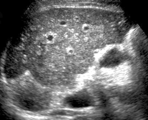
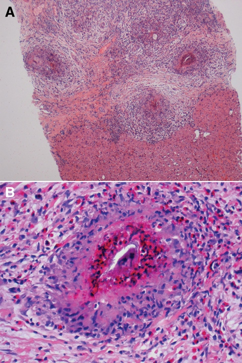
The irregular liver and periportal fibrosis seen on ultrasound (Box 1) raised the possibility of cirrhosis. Subsequently, a biopsy of the liver showed preserved liver architecture, with periportal fibrosis and active schistosomiasis (Box 2). A diagnosis of Schistosoma mansoni infection was made, based on the histological appearance of the ova.
S. mansoni is the leading cause of chronic liver disease and portal hypertension in sub-Saharan Africa.1,2 Adult worms reside in mesenteric vessels, but their migrating eggs lodge in hepatic presinusoidal radicals, resulting in inflammation and granuloma formation. The inflammatory reaction eventually leads to occlusion of portal veins and secondary portal hypertension.3 Hepatocellular function usually remains normal.1
Although the “gold standard” for diagnosis of S. mansoni infection is microscopic examination of faeces, this test may be negative (as it was in this case). Serological screening is recommended, but these assays cross-react with other helminthic infections and are unable to distinguish active infections from previous exposure.1
Praziquantel should be offered to previously untreated patients with positive serology results; after a single dose, 70%–100% of patients cease to excrete eggs.1 In patients who have left S. mansoni-endemic areas, an oral dose of 60 mg/kg split in two and given several hours apart should ensure cure.1
Our patient was treated with praziquantel, with ongoing follow-up for hepatitis B and portal hypertension. In retrospect, the patient’s history and the sonographic appearances were consistent with schistosomiasis. This clinical scenario is of increasing relevance, with a growing number of people from Africa now living in Australia.
1 Liver ultrasound
 | |||||||||||||||
|
Ultrasound shows an irregular liver with marked periportal fibrosis. There is no intra- or extrahepatic biliary tree dilatation. The portal vein flow is antegrade. No focal hepatic lesion is seen. | |||||||||||||||
2 Liver biopsy specimen
References
- Gryseels B, Polman K, Clerinx J, Kestens L. Human schistosomiasis. Lancet 2006; 368: 1106-1118.
- Doumenge JP, Mott KE. Global distribution of schistosomiasis: CEGET/WHO atlas. World Health Stat Q 1984; 37: 186-199. 0_i1091855
- Cheever AW, Hoffmann KF, Wynn TA. Immunopathology of schistosomiasis mansoni in mice and men. Immunol Today 2000; 21: 465-466. 0_i1091858
Early-Onset Colorectal Cancer With Liver-Only Metastases: A Retrospective Cohort Study Integrating Prospectively Collected Real-World Clinical and Molecular Data From an Australian National Database (2009–2024) to Guide Treatment Planning
Savio G. Barreto, Christos S. Karapetis, Shahid Ullah, Matthew Burge, Susan Caird, Angus Campbell, Azim Jalali, Ross Jennens, Muhammad A. Khattak, Belinda Lee, Stephanie H. Lim, Shehara Mendis, Louise Nott, Timothy J. Price, Jeremy D. Shapiro, Jeanne Tie, Javier Torres, Colin Williams, Rachel Wong, Vanessa Wong, Peter Gibbs
Australian Pathways for Specialist Pain Management and Early Palliative Care for People With Pancreatic Cancer: Developed Using a Community Consensus Approach
Jennifer Philip, Melanie R. Lovell, Kylee Bellingham, Gail Garvey, Gregory B. Crawford, Nicole M. Rankin, Kara Burns, Isabel Young, Vivienne Milch, Dorothy Keefe, Katrina Anderson, James Lawson, Meinir Krishnasamy
Cause‐specific mortality among Queensland people with cirrhosis, by cirrhosis aetiology and decompensation status, 2007–22: a retrospective cohort study
Vikas Bhasker, Jessica R Fong, Paul J Clark, Gunter F Hartel, Richard Skoien, James O’Beirne, Elizabeth E Powell, Patricia C Valery
Clinical practice guidelines for hepatocellular carcinoma surveillance for people at high risk in Australia: summary of recommendations
Jacob George, Nicole L Allard, Stuart K Roberts, Leon A Adams, Jane Davies, Behzad Hajarizadeh, Jennifer H MacLachlan, Suzanne E Mahady, Rosalie Altus, Catherine Brown, David C Fry, Belinda Greenwood‐Smith, Natali Smud, Patricia C Valery, Nafisa Yussf, Kate Broun, Denise Campbell, Karen Canfell, Chelsea Carle Harrison, Victoria Freeman, Paul Grogan, Catherine Holliday, Suzanne Hughes, Anna Kelly, Cathelijne Kemenade, Claire Latumahina, Amanda McAtamney, Megan Varlow, Joachim Worthington, Susan Yuill, Eleonora Feletto
Assessment of metabolic dysfunction‐associated fatty liver disease in primary care: a consensus statement summary
Leon A Adams, William W Kemp, Kate R Muller, Elizabeth E Powell, Stuart K Roberts, Luis Calzadilla Bertot, Stephanie Best, Gary Deed, Jon D Emery, Samantha L Hocking, Graham R Jones, John S Lubel, Sinead Sheils, Stephen M Twigg, Gerald F Watts, Jacob George
Treatment outcomes for people with hepatitis C referred to tertiary care in Victoria, 2021–22: a retrospective observational study
Elly Layton, Nicole Matthews, Brendan Quinn, Nasra Higgins, Gabrielle Lindeman, Mielle Abbott, Jennifer MacLachlan, Elizabeth Birbilis, Margaret E Hellard, Joseph Doyle, Benjamin C Cowie, Mark Stoové