




Volume 169 - Issue 4
Spina bifida
Author: Debra S Kennedy
Med J Aust 1998; 169 (4): 182-183.
Published online: 17 August 1998
Published online: 17 August 1998
Spina bifida
We can reduce the incidence of this congenital defect with folate fortification of food
MJA 1998; 169: 182-183
|
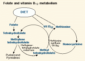
Each year about 80 babies in Australia are born with spina bifida, a congenital malformation. Spina bifida, including meningocele and meningomyelocele, is one of the spectrum of birth defects known as neural tube defects (NTDs). These defects result from failure of the neural tube to close normally during the first five weeks of embryonic life. The severity of the defect depends on where it occurs and whether nervous tissue or only meninges are involved. At least 80% of people with spina bifida have an Arnold-Chiari malformation and associated hydrocephalus, often requiring a ventricular shunt. Other chronic problems include impaired mobility, bladder and bowel dysfunction, and scoliosis, all of which contribute to high medical and psychosocial morbidity. The incidence of NTDs varies considerably according to geography and ethnicity, ranging from 0.8 to more than 10 per 1000 live births. The incidence in Australia is currently 1.1 per 1000 live births.1 Recurrence risks, which are dependent on the background incidence, reflect this variation. In Australia the recurrence risk for a couple with a previous NTD-affected pregnancy is 3%-4%, and the risk for any first-degree relatives is 1%.2 About 10% of NTDs are caused by chromosomal anomalies, predominantly trisomy 18 and triploidy, or by single-gene disorders. Maternal factors, including diabetes mellitus and exposure to the anticonvulsants valproic acid and carbamazepine, also predispose to NTDs.3 However, most NTDs are isolated malformations and are thought to be multifactorial in origin, caused by an interaction between a genetic predisposition and environmental factors.4 The B group vitamins folic acid (folate) and vitamin B12 appear to be involved in NTDs. These vitamins share metabolic pathways involved in DNA synthesis (Figure), so that a deficiency or metabolic derangement affecting either vitamin may result in impaired cell proliferation and an increased risk of congenital defects. The underlying biology is complex -- several studies have shown that women with NTD-affected pregnancies do not have folate and vitamin B12 levels in the defined deficiency ranges, although some have abnormalities such as hyperhomocysteinaemia, which suggest subtle metabolic defects.5,6
One important genetic influence may be a mutation in the gene for 5,10-methylenetetrahydrofolate reductase, an enzyme involved in folate metabolism (Figure). About 12% of the Caucasian population is homozygous for a mutation which produces a heat-labile form of this enzyme with reduced activity and higher folate requirements.7 This causes higher serum homocysteine levels, and is associated with a greater risk of NTD-affected pregnancies.8 NTDs can be prevented by reducing the incidence in all pregnancies (primary prevention), or by termination of affected pregnancies (secondary prevention). In Australia the rate of spina bifida in live births declined gradually from 7.1 per 10 000 births in 1987 to 2.9 per 10 000 births in 1994. During the same period the number of induced abortions carried out before 20 weeks' gestation for spina bifida increased, although notification of these therapeutic abortions was incomplete.1 These figures indicate that secondary prevention of NTDs is occurring throughout Australia, largely because of improvements in antenatal diagnosis. Antenatal diagnosis of NTDs usually involves maternal serum alpha-fetoprotein (MSAFP) estimation at 15-17 weeks' gestation (raised in NTD-affected pregnancies), followed by targeted ultrasound scanning at 18-19 weeks' gestation.9 The combined sensitivity of these two investigations is greater than 90%. Women at increased risk of an NTD-affected pregnancy may be offered first-trimester vaginal ultrasound and amniotic fluid alpha-fetoprotein and acetylcholinesterase estimation. Alpha-fetoprotein is raised, and acetylcholinesterase detectable, in NTD-affected pregnancies.10 Primary prevention of NTDs is preferable to secondary prevention. Although the mechanisms are not entirely understood, it is well established that periconceptional supplementation with folic acid plays a role in reducing by more than 70% the risk of both the first occurrence11 and the recurrence of NTDs.12 Therefore, current recommendations are that women planning a pregnancy should supplement their diets with 0.5 mg (500 µg) of folic acid daily, starting from at least one month before conception and continuing for the first 12 weeks of pregnancy. Women at increased risk of an NTD-affected pregnancy, including those with a previous NTD-affected pregnancy or a close family history of NTDs, should supplement their diet with 5 mg of folic acid daily.13,14 Despite apparent acceptance of the need for folic acid and the realisation that many Western women do not consume sufficient dietary folate, there has been relatively poor compliance with regimens of periconceptional folic acid supplementation.15,16 One of the main reasons is that at least half of all pregnancies are unplanned. Another reason is that some people cannot afford a folate-rich diet and others are unaware of what constitutes such a diet. There are also indications that obstetricians and general practitioners do not adequately educate and counsel their female patients of reproductive age about the need for periconceptional folic acid supplementation.17 Although education of women and their physicians to ensure consumption of folate-rich foods (such as green leafy vegetables) must continue, the relatively simple and inexpensive strategy of fortification of cereals, breads, pasta and milk would be more efficacious, as it does not involve specific planning or significant alteration of a woman's normal lifestyle or behaviour. This strategy is being applied in the United States -- in January 1998 the Food and Drug Administration, after considerable debate, made fortification of cereals with folic acid mandatory. In Australia, unfortunately, the National Health and Medical Research Council's Food Fortification Panel recommended only voluntary fortification, pending future review.18 Australian companies have not acted; the only folate-fortified breakfast cereals available in Australia are marketed by an American company. Let us hope that local companies will develop the vision to play a role in public health, and adopt voluntary food fortification policies. Debra S Kennedy | |
References |
|
-
Readers may print a single copy for personal use. No further reproduction or distribution of the articles should proceed without the permission of the publisher. For permission, contact the Australasian Medical Publishing Company
Journalists are welcome to write news stories based on what they read here, but should acknowledge their source as "an article published on the Internet by The Medical Journal of Australia <http://www.mja.com.au>".
<URL: http://www.mja.com.au/>