




Volume 207 - Issue 8
Clinical practice guidelines for the diagnosis and management of melanoma: melanomas that lack classical clinical features
Authors: Victoria J Mar, Alex J Chamberlain, John W Kelly, William K Murray and John F Thompson
Med J Aust 2017; 207 (8): 348-350. || doi: 10.5694/mja17.00123
Published online: 9 October 2017
Published online: 9 October 2017
New guidelines provide greater emphasis on atypical presentations of melanoma
Abstract
Introduction: A Cancer Council Australia multidisciplinary working group is currently revising and updating the 2008 evidence-based clinical practice guidelines for the management of cutaneous melanoma. While there have been many recent improvements in treatment options for metastatic melanoma, early diagnosis remains critical to reducing mortality from the disease. Improved awareness of the atypical presentations of this common malignancy is required to achieve this. A chapter of the new guidelines was therefore developed to aid recognition of atypical melanomas.
Main recommendations: Because thick, life-threatening melanomas may lack the more classical ABCD (asymmetry, border irregularity, colour variegation, diameter > 6 mm) features of melanoma, a thorough history of the lesion with regard to change in morphology and growth over time is essential. Any lesion that is changing in morphology or growing over a period of more than one month should be excised or referred for prompt expert opinion.
Changes in management as a result of the guidelines: These guidelines provide greater emphasis on improved recognition of the atypical presentations of melanoma, in particular nodular, desmoplastic and acral lentiginous subtypes, with particular awareness of hypomelanotic and amelanotic lesions.
There is evidence that the rate of early detection of superficial spreading melanomas in Australia has improved, with a corresponding reduction in both the median tumour thickness and in melanoma mortality from this subtype.1 However, a number of studies in Australia and other countries have shown an increasing or stable incidence rate of thick melanomas.2-7 Nodular melanoma (NM), desmoplastic melanoma (DM) and acral lentiginous melanoma (ALM) are often diagnosed when they are much thicker lesions compared with superficial spreading melanoma (SSM).3,4,6,8-10 This is in part due to their atypical clinical features. Improved diagnostic accuracy of these subtypes can significantly reduce mortality from melanoma.
The 2008 evidence-based clinical practice guidelines for the management of cutaneous melanoma (http://www.cancer.org.au/content/pdf/HealthProfessionals/ClinicalGuidelines/ClinicalPracticeGuidelines-ManagementofMelanoma.pdf) are currently being revised and updated in a staged process by a multidisciplinary working group under the auspices of Cancer Council Australia. This article summarises the atypical clinical features of melanoma. The new guidelines chapter containing more detailed descriptions of the clinical presentations of distinct melanoma subtypes, as well as other completed chapters of the revised guidelines, can be accessed online at http://wiki.cancer.org.au/australia/Guidelines:Melanoma.
Method
The study question “How do atypical melanomas present?” was determined by the multidisciplinary Melanoma Guidelines Working Party. To collect all relevant evidence, a literature search was performed in 2015 by the Cancer Council Australia Clinical Guidelines Network using PubMed and Embase, with the key words “atypical melanoma”, “typical melanoma”, “nodular melanoma”, “desmoplastic melanoma”, “amelanotic melanoma”, “amelanosis”, “acral lentiginous melanoma”, “subungal melanoma”, “scalp melanoma” and “clinical features”. Given the nature of the study question, a broad range of study designs including case–control, cohort and comparative studies were included in the search criteria. Information from the search was collated and summarised, and key findings that directly addressed the study question formed the main evidence summary, with level of evidence determined using the National Health and Medical Research Council evidence hierarchy (https://www.nhmrc.gov.au/_files_nhmrc/file/guidelines/developers/nhmrc_levels_grades_evidence_120423.pdf). Practice points were included based on expert consensus.
Recommendations
Melanomas may not conform to the usual ABCD (asymmetry, border irregularity, colour variegation, diameter > 6 mm) criteria. They are sometimes symmetrical, dome shaped and skin coloured (Box 1 and Box 2). A useful extension of the ABCD criteria involves adding EFG (elevated, firm and growing) criteria — any lesion that is elevated, firm and growing over a period of more than one month should raise suspicion for melanoma.11
Lack of pigment is significantly associated with poorer diagnostic accuracy.12 Up to 20% of all melanomas are only partially pigmented (hypomelanotic), with completely amelanotic melanomas much less common.13,14 Nodular, desmoplastic and acral lentiginous subtypes are more commonly hypomelanotic (over 40% of cases) compared with superficial spreading and lentigo maligna subtypes (about 10–25% of cases).13,15,16 Spitzoid melanomas are also commonly non-pigmented.17 Hypomelanotic melanomas may mimic basal cell carcinomas clinically, with a slightly shiny surface and atypical vessels on dermoscopy (Box 1). Other dermoscopic clues include scar-like depigmentation, an inverse network, irregular blue-grey dots, a blue-white veil, and milky pink areas14,18 or shiny white streaks (on polarised dermoscopy only; Box 2). While dermoscopic sensitivity is about 90% for pigmented lesions, it is much lower for predominantly amelanotic lesions.
Although many NMs are seemingly non-pigmented, closer inspection reveals faint pigmentation in some and focal pigmentation in others. Dermoscopy shows melanin pigment in 90% of NMs, although 27% in one large series were lightly or focally pigmented and 9.6% were completely amelanotic.19 Compared with non-nodular subtypes, dermoscopic features such as blue-white veil, homogeneous blue areas, black areas, milky pink areas, atypical vessels and symmetrical (rather than asymmetrical) pigment patterns are more commonly identified.19
Dermoscopy is less useful in diagnosing DM unless features of an associated radial growth phase melanoma are present. It may be misdiagnosed clinically as a dermatofibroma, scar or non-melanoma skin cancer. Recurrence at the site of a previous biopsy diagnosed as benign on histopathology (eg, as dermatofibroma, neurofibroma, scar) is not an uncommon presentation of DM, as the histopathology can be difficult in some cases, particularly with a partial biopsy. Review of previous pathology may be helpful when there is clinical suspicion.
Over 30% of ALMs are hypomelanotic16 (Box 3 and Box 4). Occasionally, ALMs are verrucous and may mimic a plantar wart or macerated tinea infection. If an ALM were to be incorrectly diagnosed as a wart and inadvertently pared down, it would not show the typical pinpoint vessels of a wart (Box 4). Dermoscopy is helpful when the parallel ridge pattern of pigment can be identified. Hypomelanotic subungual melanoma may present as nail dystrophy and be readily mistaken for nail trauma or infection (Box 5).
Tumour thickness is not necessarily related to diagnostic delay.2,20-22 While some melanomas grow slowly over a number of years, others will become thick and life- threatening over weeks or months. More rapid growth has been associated with nodular and desmoplastic subtypes as well as amelanosis.23-26 These subtypes are more common on chronically sun-damaged skin, typically on the head and neck and predominantly in older men.10
Perhaps the most helpful clinical feature of biologically significant melanomas is that they are changing, regardless of their other clinical features. If these changes have been accurately recognised by the patient or if there is photographic evidence to demonstrate stability or change, this may be very helpful in determining the index of suspicion. Radial growth phase melanomas change in size, shape or colour, and vertical growth phase melanomas evolve with elevation and ulceration and may bleed. A history of the duration of a lesion and any change in its appearance is a minimum requirement for the assessment of any potential skin cancer.
A summary of the evidence relating to atypical clinical presentations of melanomas and the impact on diagnostic accuracy is shown in Box 6.
Practice points
-
Melanomas are generally distinguished from benign lesions by their history of change, and thick melanomas often do not conform to the ABCD rule but usually meet the EFG criteria. Therefore, careful history taking is important, and any lesion that continues to grow or change in size, shape, colour or elevation over a period of more than one month should have a biopsy taken and be assessed histologically or referred for expert opinion.
-
Suspicious raised lesions should be excised rather than monitored.
Box 1 – Amelanotic desmoplastic melanoma on the scalp (5.8 mm thick)
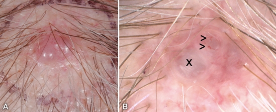
A: Shiny, symmetrical, dome-shaped scalp nodule. B: Polarised dermoscopy (magnification, × 10) demonstrates atypical polymorphic vessels (>), milky pink areas (x) and lacks the typical in-focus arborising vessels of a basal cell carcinoma.
Box 2 – Superficial spreading melanoma (0.5 mm thick) on the posterior leg
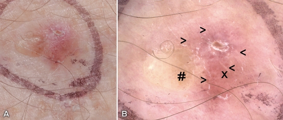
A: New and growing shiny pink plaque with focal hyperkeratosis, easily mistaken for an inflamed seborrhoeic keratosis. B: Polarised dermoscopy (magnification, × 10) demonstrates an atypical vascular pattern with both dotted vessels and red globules (>) along with focal hyperkeratosis, shiny white streaks (crystalline structures) (<), milky pink structureless areas (x) and trace light brown pigment (#). Pathology revealed a 0.5 mm thick superficial spreading melanoma with 2 mitoses/mm2.
Box 3 – Acral lentiginous melanoma (3.1 mm thick)
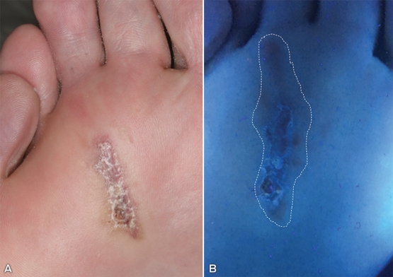
A: Large linear hyperkeratotic and focally eroded plantar plaque with faint pigment at the rim and inferior pole (easily mistaken for verruca or chronic scar). B: Subtle pigmentation is more obvious under Wood lamp examination (white dotted line).
Box 4 – Ulcerated hypomelanotic acral lentiginous melanoma (4.8 mm thick)
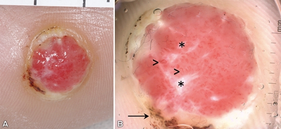
A: Well circumscribed pink/red nodule, which could be mistaken for a verruca; however, it does not have the typical pinpoint vessels. B: Instead, polarised dermoscopy (magnification, × 10) reveals atypical vessels (>), shiny white streaks (*) and focal pigmentation (arrow).
Box 5 – Ulcerated subungal melanoma (6 mm thick) previously mistaken for nail trauma
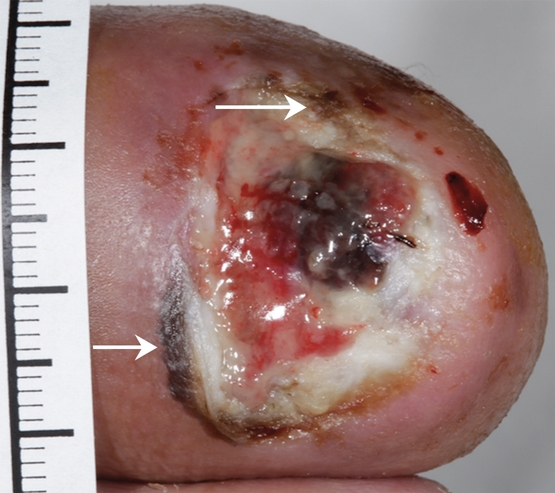
Melanin pigment is evident at the proximal and lateral nail folds (Hutchinson sign shown by arrows) and there is pigment in the nail bed with haemorrhage.
Box 6 – Evidence summary for atypical presentations of melanoma
|
Evidence |
NHMRC level* |
||||||||||||||
|
|
|||||||||||||||
|
Nodular, acral lentiginous and desmoplastic subtypes more commonly present as thick lesions; improved diagnostic accuracy of these is therefore critical3,4,6-8,10 |
III-2, III-3, IV |
||||||||||||||
|
Nodular melanomas are associated with more rapid vertical growth compared with superficial spreading melanomas23-26 |
III-3, IV |
||||||||||||||
|
Up to 20% of all melanomas are amelanotic or only partially pigmented, with this being more common among nodular, acral lentiginous and desmoplastic subtypes10,13,16 |
IV |
||||||||||||||
|
Amelanosis/hypomelanosis is significantly associated with poorer diagnostic accuracy12,14 |
III-2, III-3 |
||||||||||||||
|
|
|||||||||||||||
|
* For details of the National Health and Medical Research Council (NHMRC) evidence hierarchy, see https://www.nhmrc.gov.au/_files_nhmrc/file/guidelines/developers/nhmrc_levels_grades_evidence_120423.pdf. |
|||||||||||||||
Competing interests
No relevant disclosures.
Acknowledgements
We thank Laura Wuellner, Jutta von Dincklage and Jackie Buck from the Cancer Council Australia Clinical Guidelines Network for their assistance in this work. Development of the new Clinical Practice Guidelines for the Diagnosis and Management of Melanoma was funded by Cancer Council Australia and Melanoma Institute Australia, with additional support from the Skin Cancer College Australasia.
References
- Smithson SL, Pan Y, Mar V. Differing trends in thickness and survival between nodular and non-nodular primary cutaneous melanoma in Victoria, Australia. Med J Aust 2015; 203: 20.
- Baade PD, English DR, Youl PH, et al. The relationship between melanoma thickness and time to diagnosis in a large population-based study. Arch Dermatol 2006; 142: 1422-1427.
- Criscione VD, Weinstock MA. Melanoma thickness trends in the United States, 1988-2006. J Invest Dermatol 2010; 130: 793-797.
- Demierre MF, Chung C, Miller DR, Geller AC. Early detection of thick melanomas in the United States: beware of the nodular subtype. Arch Dermatol 2005; 141: 745-750.
- Lipsker DM, Hedelin G, Heid E, et al. Striking increase of thin melanomas contrasts with stable incidence of thick melanomas. Arch Dermatol 1999; 135: 1451-1456.
- Mar V, Roberts H, Wolfe R, et al. Nodular melanoma: a distinct clinical entity and the largest contributor to melanoma deaths in Victoria, Australia. J Am Acad Dermatol 2013; 68: 568-575.
- Tejera-Vaquerizo A, Mendiola-Fernandez M, Fernandez-Orland A, Herrera-Ceballos E. Thick melanoma: the problem continues. J Eur Acad Dermatol Venereol 2008; 22: 575-579.
- Baumert J, Schmidt M, Giehl KA, et al. Time trends in tumour thickness vary in subgroups: analysis of 6475 patients by age, tumour site and melanoma subtype. Melanoma Res 2009; 19: 24-30.
- Bergenmar M, Ringborg U, Mansson Brahme E, Brandberg Y. Nodular histogenetic type – the most significant factor for thick melanoma: implications for prevention. Melanoma Res 1998; 8: 403-411.
- Chamberlain AJ, Fritschi L, Giles GG, et al. Nodular type and older age as the most significant associations of thick melanoma in Victoria, Australia. Arch Dermatol 2002; 138: 609-614.
- Kelly JW, Chamberlain AJ, Staples MP, McAvoy B. Nodular melanoma. No longer as simple as ABC. Aust Fam Physician 2003; 32: 706-709.
- Lin MJ, Mar V, McLean C, et al. Diagnostic accuracy of malignant melanoma according to subtype. Australas J Dermatol 2014; 55: 35-42.
- Liu WD, Dowling JP, Murray WK, et al. Amelanotic primary cutaneous melanoma – clinical associations and dynamic evolution. Australas J Dermatol 2006; 47(Suppl 1): A1.
- Menzies SW, Kreusch J, Byth K, et al. Dermoscopic evaluation of amelanotic and hypomelanotic melanoma. Arch Dermatol 2008; 144: 1120-1127.
- Chamberlain AJ, Fritschi L, Kelly JW. Nodular melanoma: patients’ perceptions of presenting features and implications for earlier detection. J Am Acad Dermatol 2003; 48: 694-701.
- Phan A, Dalle S, Touzet S, et al. Dermoscopic features of acral lentiginous melanoma in a large series of 110 cases in a white population. Br J Dermatol 2010; 162: 765-771.
- McCormack CJ, Conyers RK, Scolyer RA, et al. Atypical Spitzoid neoplasms: a review of potential markers of biological behavior including sentinel node biopsy. Melanoma Res 2014; 24: 437-447.
- Pizzichetta MA, Talamini R, Stanganelli I, et al. Amelanotic/hypomelanotic melanoma: clinical and dermoscopic features. Br J Dermatol 2004; 150: 1117-1124.
- Menzies SW, Moloney FJ, Byth K, et al. Dermoscopic evaluation of nodular melanoma. JAMA Dermatol 2013; 149: 699-709.
- Betti R, Martino P, Vergani R, et al. Nodular melanomas: analysis of the casistic and relationship with thick melanomas and diagnostic delay. J Dermatol 2008; 35: 643-650.
- Richard MA, Grob JJ, Avril MF, et al. Delays in diagnosis and melanoma prognosis (II): the role of doctors. Int J Cancer 2000; 89: 280-285.
- Richard MA, Grob JJ, Avril MF, et al. Delays in diagnosis and melanoma prognosis (I): the role of patients. Int J Cancer 2000; 89: 271-279.
- Lin MJ, Mar V, McLean C, Kelly JW. An objective measure of growth rate using partial biopsy specimens of melanomas that were initially misdiagnosed. J Am Acad Dermatol 2014; 71: 691-697.
- Liu W, Dowling JP, Murray WK, et al. Rate of growth in melanomas: characteristics and associations of rapidly growing melanomas. Arch Dermatol 2006; 142: 1551-1558.
- Martorell-Calatayud A, Nagore E, Botella-Estrada R, et al. Defining fast-growing melanomas: reappraisal of epidemiological, clinical, and histological features. Melanoma Res 2011; 21: 131-138.
- Tejera-Vaquerizo A, Barrera-Vigo MV, Lopez-Navarro N, Herrera-Ceballos E. Growth rate as a prognostic factor in localized invasive cutaneous melanoma. J Eur Acad Dermatol Venereol 2010; 24: 147-154.
Linked content
-
MJA InSight: Practical guide to atypical melanomas
-
MJA Podcast: Dr Victoria Mar
Provenance: Not commissioned; externally peer reviewed.
Diagnosing Acute Kava Dermopathy: A Case Report of a Characteristic Cutaneous Eruption
Ali Abid, Nicholas Allen, Abeer Hagelamin, Christopher Henderson, Artiene Tatian
Localised Herpes Simplex Following Midline Laparotomy
Jessica S. Bulluss, Paul Chee, Matthew J. Verheyden
No silver lining with health misinformation: argyria caused by intentional silver consumption
Luke Collins, Logesh Palanikumar, Stephen Bacchi
Lingual Raynaud phenomenon
Michael Taggart, Mina John
Melasma in the male: a less well recognised entity
Tim Aung, Rowland Noakes
Age group‐specific changes in keratinocyte cancer treatment rates in Australia, 2012–2021: a retrospective cohort study based on MBS claims data
Catherine M Olsen, Nirmala Pandeya, Rachel E Neale, David C Whiteman