




Medical education
Lessons from practice
Volume 214 - Issue 7
More than meets the eye: a missed traumatic laser in situ keratomileusis flap dislocation
Authors: Shivesh Varma, Yi Fan Tang and Salim Okera
Med J Aust 2021; 214 (7): 305-305.e1. || doi: 10.5694/mja2.50985
Published online: 19 April 2021
Published online: 19 April 2021
A 61-year-old man was referred to our ophthalmology service 3 days after a tree branch injury to the left eye
Clinical record
A 61‐year‐old man was referred to our ophthalmology service 3 days after a tree branch injury to the left eye. He had a history of laser in situ keratomileusis (LASIK) refractive surgery 17 years earlier. He had attended his local emergency department on the day of the injury with pain and reduced vision. He was diagnosed with a corneal abrasion and treated with topical chloramphenicol drops; he then presented to our service as he failed to improve.
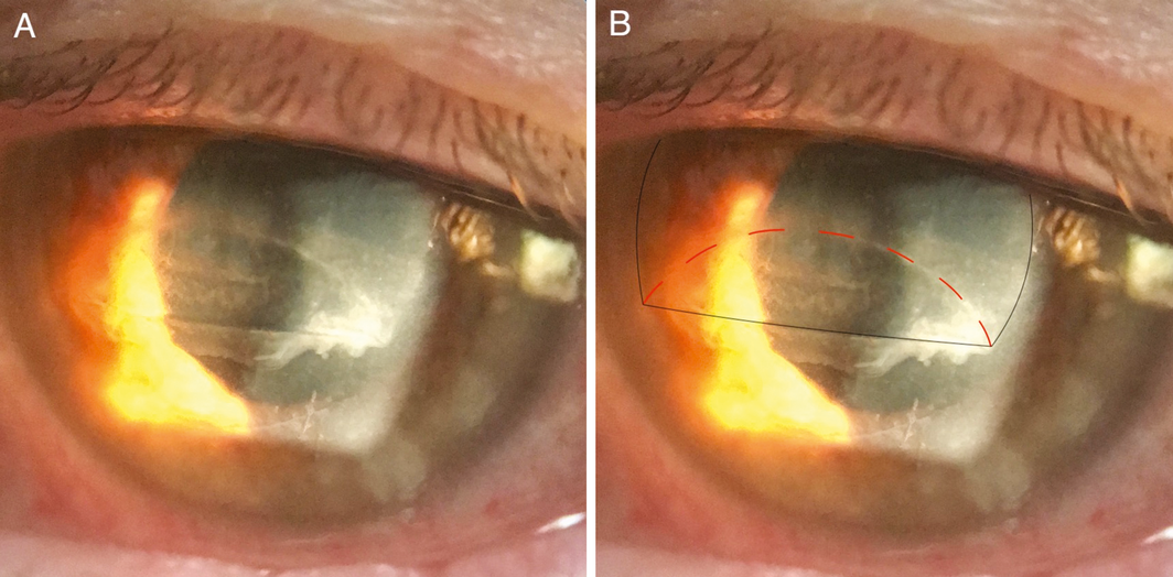
Visual acuity was 6/6 in the right eye and 6/30 in the left eye. The inferior third of the left eye LASIK flap was dislocated and folded underneath the superior flap (Box, A). Image B in the Box highlights the normal position of the superior flap edge and the new horizontal fold in the flap (black line) and the reflected position of the dislocated inferior flap (red dashed line). The adjacent cornea was hazy and oedematous, and there were fine grey sheets of epithelial regrowth over the exposed stroma inferiorly. The patient was taken to the operating theatre the next day, and the LASIK flap was lifted, debrided, repositioned, and a bandage contact lens was placed for 1 week. After 4 weeks of topical steroid and antibiotic drops, the flap had healed in its normal position and visual acuity returned to 6/6.
Discussion
Laser refractive surgery is an increasingly popular group of procedures that correct refractive error, allowing 99.5% of patients to achieve spectacle independence, with 98.8% of those treated being satisfied with their outcome.1 LASIK is a form of refractive surgery in which a circular flap of corneal tissue about 120 µ in thickness and 8 mm in diameter is cut and reflected on a superior hinge, allowing access to the underlying corneal stroma for excimer laser reshaping to correct refractive error. The flap is replaced and heals by the formation of weak scar tissue at its edges. This makes the LASIK flap vulnerable to traumatic dislocation, which has been reported following a variety of mechanisms of blunt and sharp ocular trauma including gardening, sports, finger poking, and motor vehicle accidents.2,3 Traumatic flap dislocation is a lifelong risk following surgery, and has been reported up to 14 years after LASIK.2 Moreover, trauma to the flap may result in formation of subtle flap striae without dislocation, which threatens vision and may also require surgery.4 Traumatic flap dislocations or striae require prompt repair in the operating theatre.2,4 Visual outcomes following surgical repair of dislocated flaps are positive, with 55% of patients achieving their pre‐injury uncorrected visual acuity as in our case, and a further 33% losing only one line of Snellen visual acuity.3
This case highlights the importance of enquiring about a history of previous laser refractive surgery in cases of ocular trauma, as urgent surgical management may be required to prevent flap‐related complications and to restore vision.
Lessons from practice
- Always ask about a history of refractive laser surgery, as patients who have had laser eye surgery may not immediately volunteer this history at the time of presenting with ocular trauma.
- Laser in situ keratomileusis (LASIK) flap dislocation can be the result of minor trauma and can mimic a corneal abrasion.
- Risk of traumatic LASIK flap dislocation is lifelong.
- Suspected LASIK flap dislocation warrants urgent ophthalmic referral.
Box – Patient’s left eye showing (A) the dislocated laser in situ keratomileusis flap which mimics a simple corneal abrasion, and (B) the normal position of the superior flap edge and the new horizontal fold in the flap (black line) and the reflected position of the dislocated inferior flap (red dashed line)
Competing interests
No relevant disclosures.
References
- Kim T, del Barrio JLA, Wilkins M, et al. Refractive Surgery. Lancet 2019; 393: 2085–2098.
- Tsai TH, Peng KL, Lin CJ. Traumatic corneal flap displacement after laser in situ keratomileusis (LASIK). Int Med Case Rep J 2017; 10: 143–148.
- Landau D, Levy J, Solomon A, et al. Traumatic corneal flap dislocation one to six years after LASIK in nine eyes with a favourable outcome. J Refract Surg 2006; 22: 884–889.
- Ursea R, Feng MT. Traumatic flap striae 6 years after LASIK: case report and literature review. J Refract Surg 2010; 26: 899–905.
Provenance: Not commissioned; externally peer reviewed.
A Diagnostic Headache
Thomas Glynn, Michelle T. Leech, Stacy K. Goergen, Emily Lin, Nadeem Toodayan, Ralph Junckerstorff
Neisseria meningitidis: an uncommon cause of conjunctivitis
Chloe Story, Tina Marinelli, Simeon Crawford, Amrita Ronnachit
Dismantling barriers to research and clinical care for individuals with a vision impairment
Eden G Robertson, Kate Hetherington, Meredith Prain, Julia Hall, Leighton Boyd AM, Rosemary Boyd OAM, Emily Shepard, Hollie Feller, Sally Karandrews, Fleur O'Hare, Kanae Yamamoto, Matthew P Simunovic, Robyn V Jamieson, Alan Ma, Lauren Ayton AM, Anai Gonzalez‐Cordero
The New South Wales Pharmacy Trial for herpes zoster: on the nose?
Christian P Pappas, Timothy R Holmes, Minas T Coroneo
Skin fragility disorder misdiagnosed as child abuse: a cautionary tale
Heather G Mack, Fred K Chen, John R Grigg
Health economic aspects of inherited retinal diseases: looking for cost‐effective treatments
Benjamin Kamien, Rachael Heath Jeffery, Fred K Chen