




Medical education
Lessons from practice
Volume 213 - Issue 3
Necrotising enterocolitis caused by Clostridium perfringens: a life‐threatening manifestation of a common foodborne infection
Authors: Harry N Walker, Kwee‐Chin Liew, Vicki Adams, Sarah Larcombe, Sonal S Nagra, Glenn Guest and Eugene Athan
Med J Aust 2020; 213 (3): 114-115.e1. || doi: 10.5694/mja2.50568
Published online: 27 April 2020
Published online: 27 April 2020
Clinical record
A 40‐year‐old woman of Karen ethnicity presented with 5 days of generalised abdominal pain. The pain was worsening and associated with vomiting in the 12 hours preceding presentation. She had been constipated for 5 weeks. There was no diarrhoea, no blood or mucous in the stool and no haematemesis. There were no sick family, friends or colleagues and no recent travel. She had an omnivorous diet, which had not changed recently, and worked casually at a vegetable farm.
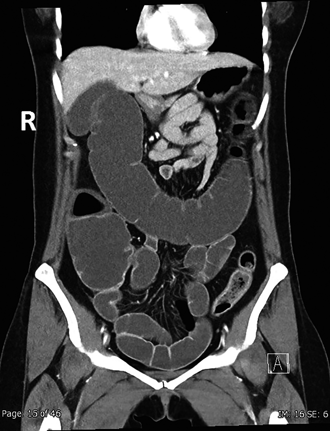
In the emergency department, vital signs were normal and the abdomen was mildly distended and tender. Initial blood tests showed a neutrophil count of 9.6 × 10⁹/L (reference range [RR], 2.0–8.0 × 10⁹/L), serum bicarbonate 22 mmol/L (RR, 22–32 mmol/L), normal renal function, and C‐reactive protein below 2.9 mg/L (RR, < 3.0 mg/L). Computed tomography of the abdomen and pelvis was consistent with colitis of the descending colon and ileus (Box 1). She was admitted for observation under the general surgical team.
Five hours after admission, her condition rapidly deteriorated. Blood pressure was 80/40 mmHg, heart rate 129 beats per minute in sinus rhythm, and there was severe abdominal tenderness with generalised guarding. Repeat tests showed serum creatinine 175 μmol/L (RR, 60–110 μmol/L), bicarbonate below 10 mmol/L (RR, 22–32 mmol/L), blood pH 6.97 (RR, 7.35–7.45), and lactate 14.8 mmol/L (RR, < 1.5 mmol/L).
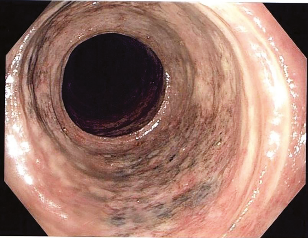
At emergency surgery, colonoscopy and ileoscopy revealed mucosal inflammation affecting the distal 20 cm of terminal ileum, caecum, transverse and sigmoid colon, with patches of frank mucosal necrosis (Box 2). The bowel was grossly dilated, with small patches of full thickness caecal necrosis but no perforation. Subtotal colectomy, terminal ileectomy, and formation of an end ileostomy were performed, with resultant resolution of circulatory shock.
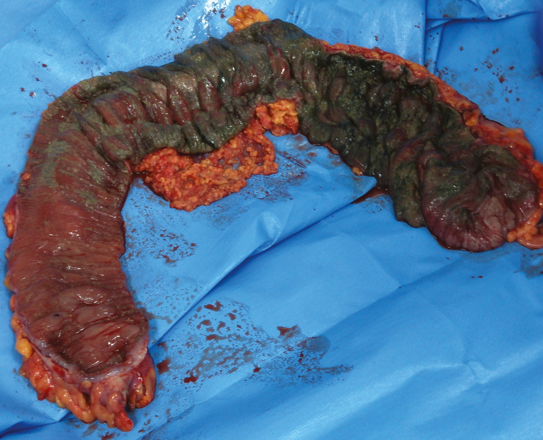
Pathological examination demonstrated severe acute pancolitis and extensive mucosal necrosis (Box 3). Clostridium perfringens infection was suspected, due to a striking similarity to published cases.1,2C. perfringens was isolated from biopsy specimens of the necrotic colonic mucosa using selective culture media. The isolate expressed C. perfringens enterotoxin and α‐toxin, defined as toxinotype F.3
Postoperative management included vancomycin both orally and per rectum. Three months after the operation, she was pain‐free, with a normal appetite and functional state. Reversal of ileostomy is planned in coming months.
Discussion
C. perfringens is a gram‐positive bacillus that forms hardy spores, is ubiquitous in environmental soil and water, and can be part of normal bowel flora. Toxigenic strains commonly cause both foodborne and sporadic cases of acute, self‐limiting diarrhoea. The typical foodborne strain F produces C. perfringens enterotoxin and α‐toxin and was found in our patient.3 The organism exhibits the shortest known doubling time of any cell when grown at 42°C in cooked minced beef.4 Exposure to large inocula of toxigenic organisms may arise when meat is kept lukewarm before consumption. Enteric infections are characterised by adherence of organisms to small bowel mucosa before concurrent sporulation and release of toxin. This typically induces self‐limiting diarrhoea 10–12 hours after eating.
Necrotising enterocolitis is a manifestation of C. perfringens enteric infection that is rare in high income countries. Some reported cases associate the condition with constipation, either pre‐existing due to medication side effects or induced by the high protein content of ingested contaminated meat. Constipation has been proposed to impair the usual expulsion of C. perfringens bacteria and spores, leading to mucosal necrosis and shock as opposed to the usual syndrome of transient diarrhoea. Mortality in case series is greater than 50%.1,2
Despite thorough assessment, the cause of our patient's constipation and means of exposure remain unclear. She most likely contracted the infection hours before the onset of her pain, 5 days before presentation. Exposure could have occurred during food preparation at home, meals with social groups, or via soil at her workplace.
While rare in Australia, a type of C. perfringens necrotising enteritis was endemic throughout the 20th century in the Papua New Guinea highlands, caused by β‐toxin‐producing strains. Called “pigbel” in Tok Pisin, the disease is closely associated with traditional pig feasts. In the 1960s and 1970s, pigbel accounted for almost a quarter of paediatric deaths in highlands hospitals. Implementation of a β‐toxoid vaccine in 1979 resulted in an eightfold reduction in incidence and an even greater reduction in disease‐specific mortality.5
C. perfringens infection is an important differential diagnosis in cases of acute severe enteritis or colitis, particularly if accompanied by circulatory shock. Prompt operative intervention is necessary in such situations. In Papua New Guinea, toxoid vaccination has proven very successful. In Australia, food safety practices likely play the greatest role in controlling disease burden.
Lessons from practice
- Toxigenic Clostridium perfringens type F commonly causes acute diarrhoea, with illness typically commencing hours after meat consumption.
- Cases are usually self‐limiting, and require symptomatic management only.
- Very rarely, toxigenic C. perfringens strains can cause fulminant bowel necrosis requiring emergency bowel resection, which may be associated with pre‐existing constipation.
- In the highlands of Papua New Guinea, necrotising enteritis has been a common cause of paediatric morbidity and mortality, where it is caused by a locally endemic strain of toxigenic C. perfringens.
Box 1 – Coronal computed tomography image of the abdomen and pelvis with portal venous phase contrast, demonstrating mural thickening of the descending colon in the left lower quadrant and fluid‐filled distension of the transverse colon, ascending colon, and ileum
Competing interests
No relevant disclosures.
Acknowledgements
We thank staff at Australian Clinical Labs: Ancy Joseph, Owen Harris and Eddie Chan for their work coordinating and analysing microbiological specimens.
References
- Bos J, Smithee L, McClane B, et al. Fatal necrotizing colitis following a foodborne outbreak of enterotoxigenic Clostridium perfringens type A infection. Clin Infect Dis 2005; 40: e78–e83.
- Sobel J, Mixter CG, Kolhe P, et al. Necrotizing enterocolitis associated with Clostridium perfringens type A in previously healthy North American adults. J Am Coll Surg 2005; 201: 48–56.
- Rood JI, Adams V, Lacey J, et al. Expansion of the Clostridium perfringens toxin‐based typing scheme. Anaerobe 2018; 53: 5–10.
- Labbe RG, Huang TH. Generation times and modeling of enterotoxin‐positive and enterotoxin‐negative strains of Clostridium perfringens in laboratory media and ground beef. J Food Prot 1995; 58: 1303–1306.
- Lawrence GW, Lehmann D, Anian G, et al. Impact of active immunisation against enteritis necroticans in Papua New Guinea. Lancet 1990; 336: 1165–1167.
Provenance: Not commissioned; externally peer reviewed.
Costs of Delivering the Hep B PAST Model of Care in Remote Australia: A Retrospective Pre–Post Study
Hoa Nguyen, Anh Le Tuan Nguyen, Paula Binks, Melita McKinnon, Emily Vintour-Cesar, Karen Wills, Nicola Stephens, Benjamin Cowie, Joshua S. Davis, Yuejen Zhao, Peter Nihill, Julie A. Campbell, Andrew J. Palmer, Jane Davies, Barbara de Graaff
Buruli Ulcer Acquired From Ringtail Possum Rescue
Daniel P. O'Brien, Naomi Clarke, Sara Bonnici, Mohammad A. Hussain
Q Fever Visceral Abscesses After Contact With Native Australian Marsupials
Samantha J. Laws, Enzo Binotto, James Stewart, Jennifer Ho, Trent Yarwood, Josh Hanson, Simon Smith
Hospital-Admitted Injection-Related Infections Among Incarcerated People Who Inject Drugs in Australia: A Retrospective Cohort Study
Andrew Palmer, Matthew Carter, Jeremy Yeo, Cecilia Shim, Jason Connor, Jeremy Hayllar, Gerald Holtmann, Naomi Moy, Elliott G. Playford, Naomi Runnegar, Paul J. Clark
The Live Attenuated Influenza Vaccine in Australia: An Additional Tool for Influenza Prevention
Cyra Patel, Alexis Pillsbury, Tran Nguyen, Xia Wang, Helen E. Quinn, Clayton K. Chiu, Allen C. Cheng, Katie L. Flanagan, Zhicheng Wang
Reflex Testing for Hepatitis D Infection: A Unique Opportunity to Reduce Hepatitis D-Related Chronic Liver Disease Deaths in Australia
Jessica Howell, Lauren Andersson, Miriam T. Levy, James O'Beirne, Leon Adams, Katharine Irvine, Avik Majumdar, Golo Ahlenstiel, Kathy Jackson, Krispin Hajkowicz, Joseph Doyle, Jane Davies, Sarah Cherian, Wayne Dimech, Alexander J. Thompson