




Medical education
Lessons from practice
Volume 212 - Issue 6
Cutaneous gnathostomiasis in Vietnam
Authors: Minh Cuong Duong, Phuc VD Le, Oanh NK Pham and Hong Quang Huynh
Med J Aust 2020; 212 (6): 252-253.e1. || doi: 10.5694/mja2.50514
Published online: 6 April 2020
Published online: 6 April 2020
A 33-year-old male tour guide presented at our clinic with rash and swelling on the anterior upper arms
Clinical record
A 33‐year‐old male tour guide presented at our clinic with rash and swelling on the anterior upper arms. He was not alcohol or drug dependent and had no relevant medical and family history. He never walked barefoot. His symptoms developed 3–4 weeks after he had last eaten grilled frog. His physical examination was unremarkable other than the rash. He developed raised, erythematous and serpiginous skin lesions accompanied by pruritus, mild pain, redness and hot feeling on the middle‐third forearms for 10 days (Box 1). The rash spread distally and caused formication. He was well and had no fever or other specific symptoms.
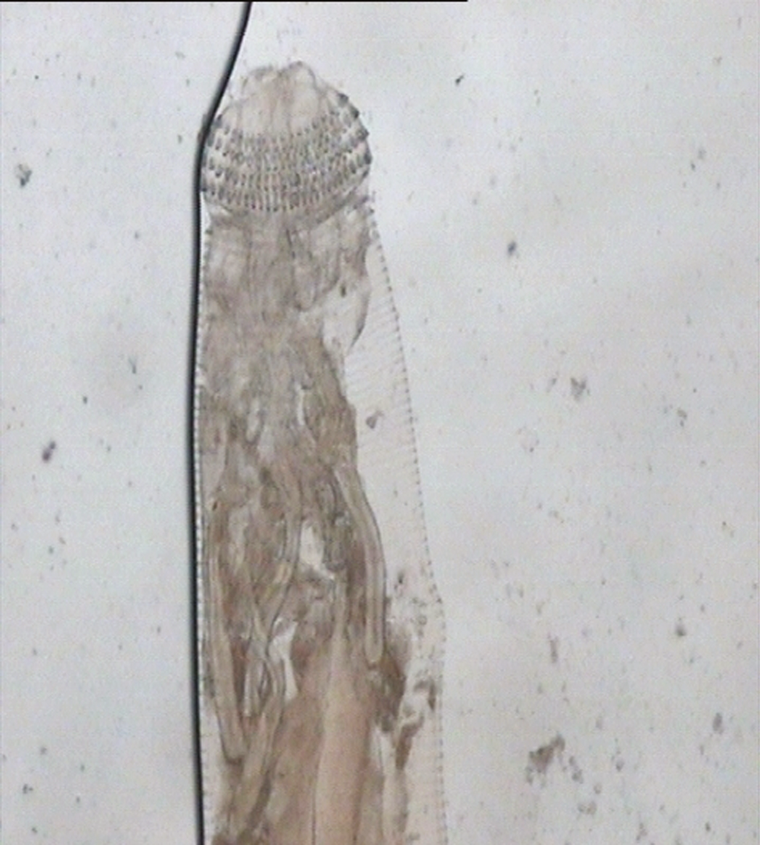
Differential diagnosis included gnathostomiasis, cutaneous larva migrans, toxocariasis and larva currens (Strongyloides). Stool microscopy, complete blood count and enzyme‐linked immunosorbent assay (ELISA) were carried out at our clinic to detect antibodies against Toxocara spp., Gnathostoma spp. and Strongyloides stercoralis. His stool sample was normal. His peripheral leucocyte count was 8830/mm3 (reference interval, 4000–10 000/mm3), and his absolute eosinophil count was elevated at 940/mm3 (reference interval, 0–500/mm3). Only Gnathostoma serological results were positive. Skin biopsy showed third‐stage Gnathostoma spinigerum larvae (Box 2). Polymerase chain reaction (PCR) testing reconfirmed the parasite species. Details regarding the use of PCR in identifying Gnathostoma spp. are discussed elsewhere.1
He was treated with a single dose of 200 μg ivermectin per kilogram of body weight.2 Skin lesions responded well to treatment and normal eosinophil levels were reached after 2 weeks. Follow‐up every 3 months until 1 year after treatment found no relapse. He was advised to avoid raw or undercooked food.
Discussion
Raised, serpiginous skin lesions in a systemically well patient may suggest an infection caused by helminth species. Our patient's rash could have been a symptom of cutaneous larva migrans caused by the migration of zoonotic nematode larvae (eg, Ancylostoma ceylanicum) through the skin.3 Such a rash usually occurs in the lower extremities, especially the feet, and is associated with walking barefoot. Although our patient lived in Vietnam, where cutaneous larva migrans is endemic, his symptoms and habits helped exclude this diagnosis. Larva currens caused by transdermal migration of Strongyloides larvae may induce serpiginous cutaneous lesions. The rashes are usually linear and recurrent with an intervening symptom‐free period.3 Our patient's Strongyloides serology results were negative, and his rashes did not have the typical characteristics of migrating Strongyloides larvae. Nodular migratory panniculitis, which is usually located at the trunk or upper limbs, is the typical symptom of cutaneous gnathostomiasis.4 These lesions are ill defined and can be pruritic, painful or erythematous.2 The appearance of our patient's rash suggested cutaneous gnathostomiasis. His Gnathostoma serological test result was positive, and Toxocara serology was negative.
Gnathostoma serological testing includes immunoblot and ELISA.2,5 ELISA, which is easier to perform and has good sensitivity (98%) and specificity (88%), should be used.5 However, it can only detect IgG and cross‐reactivity with other parasitic infections, so its specificity is not high.4 Diagnosing the causative agent based on clinical symptoms is not always reliable — especially in countries such as Vietnam, where multiple helminth species are endemic — because different species can cause overlapping cutaneous manifestations.3 The typical cutaneous manifestations of parasitic infections are also uncommon. Only 0.5–3.4% of patients present with the typical symptoms of zoonotic nematode larvae, Strongyloides larvae (1.9–8.3%) and Gnathostoma spp. infections (0.5–3.8%) (unpublished data, Institute of Malariology Parasitology and Entomology Quy Nhon annual report, January 2018). Moreover, the chances of detecting the larva are low.2 Therefore, antihelminthic therapy can be instituted if the patient has epidemiological history (eg, eating raw or undercooked fish, eels, frogs or chickens in endemic areas4) and develops clinical presentation plus suggestive serological testing and either blood or tissue eosinophilia, although the definitive diagnosis of the causative agent remains unknown.2 Our patient fully met all the criteria. Third‐stage G. spinigerum larvae were molecularly and microscopically detected in his skin sample. He was definitely diagnosed with cutaneous gnathostomiasis caused by G. spinigerum.
If left untreated, gnathostomiasis may last up to 12 years.2 Visceral gnathostomiasis, including ocular gnathostomiasis, can also develop with only cutaneous impairment.2 Although surgical removal of larvae is the best treatment for cutaneous gnathostomiasis, this is difficult when larvae are in inaccessible areas or cannot be located.2 Complete removal of larvae also depends on the clinician's surgical skills and experiences. Therefore, our patient was subsequently treated with ivermectin and fully recovered. Current antihelminthic therapy includes oral albendazole or ivermectin (Box 3).2 Relapses can be treated using the same antihelminthic therapy.2 Since relapses are not rare, patients should be symptom‐free 12 months after treatment to indicate sufficient evidence of cure.4 However, an optimal follow‐up interval remains unknown. Our patient was monitored 3‐monthly up to 12 months and had no relapse.
Given the increased number of travellers worldwide, cutaneous gnathostomiasis is not restricted to endemic countries where diagnostic error is common due to atypical skin lesions and low probability of obtaining the parasite. Non‐endemic countries such as Australia have also experienced diagnosis and treatment difficulties.5 Travellers and physicians need to be aware of gnathostomiasis.
Lessons from practice
- Cutaneous gnathostomiasis is an emerging imported disease in developed countries; diagnostic error is common in endemic countries because skin lesions are non‐specific, and few clinicians are familiar with this condition.
- Based on the migration route, a skin biopsy can be performed to detect third‐stage Gnathostoma spinigerum larvae, and surgical removal of larvae can probably be undertaken even though the success of these procedures depends on the clinician's skills and experiences.
- Follow‐up every 3 months until 1 year after treatment completion may be suggested to ensure there is no relapse, provided that a follow‐up 2 weeks after treatment shows good clinical response and normal eosinophil levels.
- Gnathostomiasis can be diagnosed using serological tests including ELISA and immunoblot. However, compared with immunoblot, ELISA is more commonly used as it is easier to perform even in low resource settings, with an acceptable sensitivity of 98% and a specificity of 88%. ◆
Box 1 – Movable skin lesions on the middle third of the patient's right and left arms
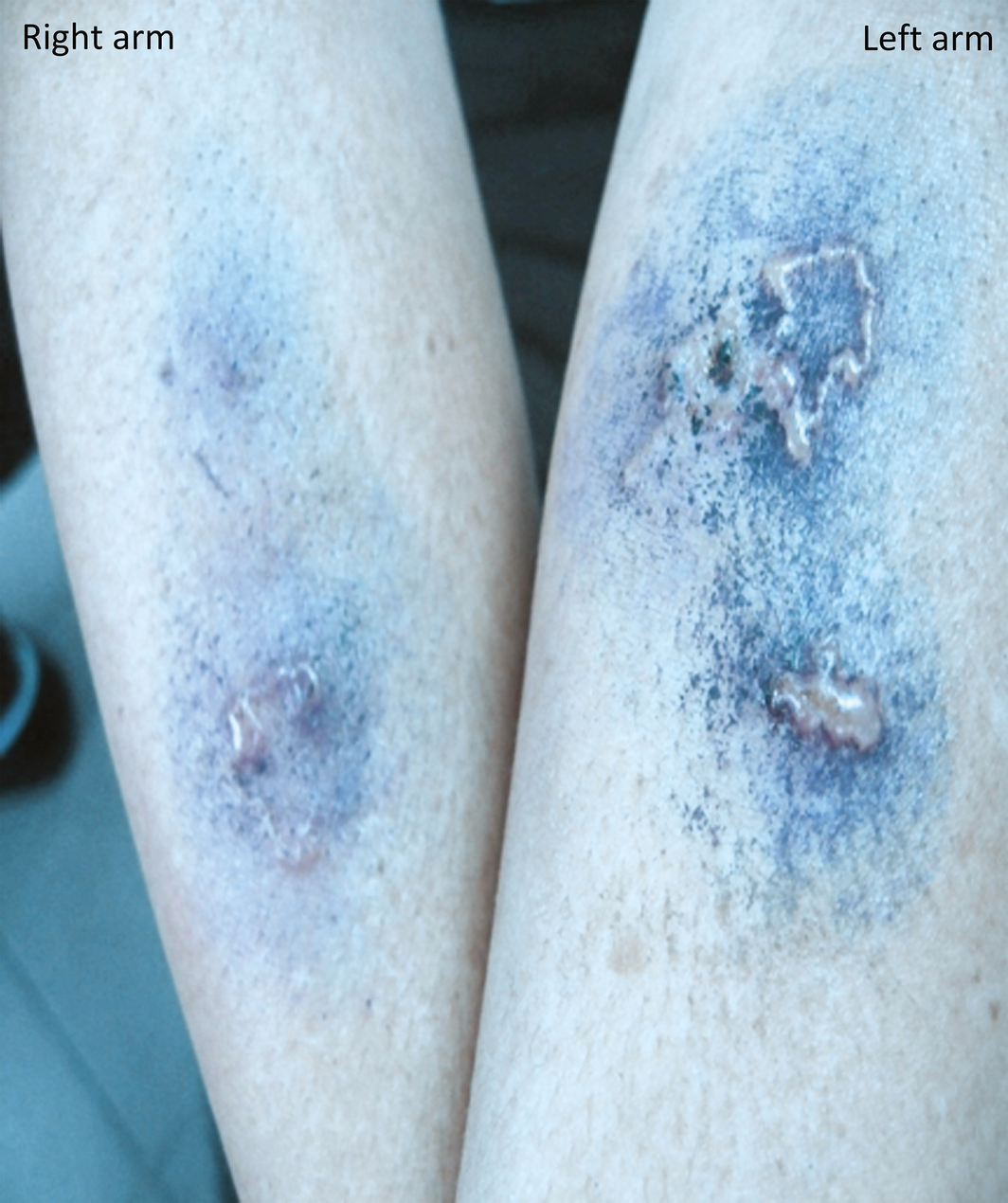
Note: Blue colour is due to the application of methylene blue.
Box 3 – Antihelminthic therapy for gnathostomiasis
Initial infection
- Oral albendazole (400 mg twice daily for 21 days) or ivermectin (0.2 mg/kg single dose or may be repeated after 7 days)
- Antihistamine if mild symptoms occur (ie, transient redness and/or itching in the area previously affected) and no evidence of new migration and eosinophilia
- Same antihelminthic drug and dose as treatment for initial infection if there is evidence of new migration
- Simultaneous use of albendazole and ivermectin may be considered when migration occurs in the head and neck region
Competing interests
No relevant disclosures.
References
- Boonroumkaew P, Sanpool O, Rodpai R, et al. Molecular identification and genetic diversity of Gnathostoma spinigerum larvae in freshwater fishes in southern Lao PDR, Cambodia, and Myanmar. Parasitol Res 2019; 118: 1465–1472.
- Bravo F, Gontijo B. Gnathostomiasis: an emerging infectious disease relevant to all dermatologists. Anais Brasileiros Dermatologia 2018; 93: 172–180.
- Hamilton WL, Agranoff D. Imported gnathostomiasis manifesting as cutaneous larva migrans and Loffler's syndrome. BMJ Case Rep 2018; 2018: bcr2017223132.
- Herman JS, Chiodini PL. Gnathostomiasis, another emerging imported disease. Clin Microbiol Rev 2009; 22: 484–492.
- Jeremiah CJ, Harangozo CS, Fuller AJ. Gnathostomiasis in remote northern Western Australia: the first confirmed cases acquired in Australia. Med J Aust 2011; 195: 42–44. https://www.mja.com.au/journal/2011/195/1/gnathostomiasis-remote-northern-western-australia-first-confirmed-cases-acquired.
Provenance: Not commissioned; externally peer reviewed.
Costs of Delivering the Hep B PAST Model of Care in Remote Australia: A Retrospective Pre–Post Study
Hoa Nguyen, Anh Le Tuan Nguyen, Paula Binks, Melita McKinnon, Emily Vintour-Cesar, Karen Wills, Nicola Stephens, Benjamin Cowie, Joshua S. Davis, Yuejen Zhao, Peter Nihill, Julie A. Campbell, Andrew J. Palmer, Jane Davies, Barbara de Graaff
Buruli Ulcer Acquired From Ringtail Possum Rescue
Daniel P. O'Brien, Naomi Clarke, Sara Bonnici, Mohammad A. Hussain
Q Fever Visceral Abscesses After Contact With Native Australian Marsupials
Samantha J. Laws, Enzo Binotto, James Stewart, Jennifer Ho, Trent Yarwood, Josh Hanson, Simon Smith
Hospital-Admitted Injection-Related Infections Among Incarcerated People Who Inject Drugs in Australia: A Retrospective Cohort Study
Andrew Palmer, Matthew Carter, Jeremy Yeo, Cecilia Shim, Jason Connor, Jeremy Hayllar, Gerald Holtmann, Naomi Moy, Elliott G. Playford, Naomi Runnegar, Paul J. Clark
The Live Attenuated Influenza Vaccine in Australia: An Additional Tool for Influenza Prevention
Cyra Patel, Alexis Pillsbury, Tran Nguyen, Xia Wang, Helen E. Quinn, Clayton K. Chiu, Allen C. Cheng, Katie L. Flanagan, Zhicheng Wang
Reflex Testing for Hepatitis D Infection: A Unique Opportunity to Reduce Hepatitis D-Related Chronic Liver Disease Deaths in Australia
Jessica Howell, Lauren Andersson, Miriam T. Levy, James O'Beirne, Leon Adams, Katharine Irvine, Avik Majumdar, Golo Ahlenstiel, Kathy Jackson, Krispin Hajkowicz, Joseph Doyle, Jane Davies, Sarah Cherian, Wayne Dimech, Alexander J. Thompson