




Volume 207 - Issue 3
Optimising assessment of kidney function when managing localised renal masses
Authors: Robert J Ellis, Andre Joshi, Keng L Ng, Ross S Francis, Glenda C Gobe and Simon T Wood
Med J Aust 2017; 207 (3): 127-133. || doi: 10.5694/mja17.00161
Published online: 7 August 2017
Published online: 7 August 2017
Strategies to improve the detection and management of patients with an increased risk of post-operative CKD
Summary
- Increased early and incidental detection, improved surgical techniques and technological advancement mean that the management of renal mass lesions is constantly evolving.
- The treatment of choice for renal mass lesions has historically been radical nephrectomy.
- Partial nephrectomy is now recommended for localised renal masses, owing to favourable renal functional outcomes.
- Ablative renal surgery confers a significant risk of chronic kidney disease.
- There are few studies assessing long term outcomes of nephrectomy on renal outcomes, and virtually no studies assessing long term outcomes for less invasive therapies such as ablation.
- Unless a renal mass is clearly benign on imaging, management decisions will be made with an assumption of malignancy. The content of this review applies to both benign and malignant renal mass lesions.
- We advocate for improved strategies for kidney function assessment and risk stratification, early targeted referral, and regular screening for chronic kidney disease for all patients after surgery.
Suspicious renal mass lesions are considered malignant until proven otherwise. The most common diagnosis for intra-renal masses is renal cell carcinoma (RCC).1 Gold standard management of malignant renal mass lesions is surgical resection, either through radical (total) or partial (nephron-sparing) nephrectomy. Although curative in the vast majority of localised tumours, there are risks associated with nephrectomy, particularly relating to post-operative decline in kidney function and chronic kidney disease (CKD) onset and progression. CKD is defined as abnormalities in kidney structure or function with health implications, generally defined as estimated glomerular filtration rate (eGFR) reduction < 60 mL/min/1.73 m2 and/or evidence of kidney damage (typically manifested as albuminuria) for a period = 3 months.2 CKD increases the risk of both cardiovascular disease and all-cause mortality. In many patients after tumour nephrectomy, CKD may be prevented through use of alternative management strategies.
This review uses the most up-to-date relevant original research, systematic reviews and meta-analyses, identified via PubMed and Google Scholar searches, to evaluate current clinical practice guidelines and standard of care for localised renal masses. We identify potential strategies to improve the detection and management of patients with an increased risk of post-operative CKD. The review focuses on benign and malignant lesions limited to the kidney; however, advice regarding kidney function may still be applicable for advanced tumours.
Epidemiology
Worldwide, kidney cancer is the ninth most common cancer in men and the fourteenth most common in women.1 It is the third most common genitourinary malignancy, associated with the highest genitourinary cancer mortality rate, and the 16th most common cause of cancer death worldwide.1,3 Incidence is higher and increasing in many developed compared with developing countries, and this is significantly more pronounced in men than women. RCC incidence rates range from 22 per 100 000 in Czech men to < 1 per 100 000 in some African countries. In Australia, the respective incidence rates for men and women are 10.4 and 5.1 per 100 000.1 Australian projections predict there will be 19 280 new cases of kidney cancer from 2016 to 2020.4 Although Australia currently lacks formalised population data collection for kidney cancer management, a Victorian population study reported that in 2009, 67.3% and 15.4% of patients with diagnosed kidney cancer were managed with radical and partial nephrectomy respectively.5 A retrospective study of 488 nephrectomy patients from Victoria further identified that 61.3% and 26.9% of radical and partial nephrectomy patients experience an eGFR below 60 mL/min/1.73 m2 at a minimum of 6 months post-operatively.6 A rough comparison of these figures indicates that about 45.4% of patients diagnosed with kidney cancer will experience a post-operative eGFR below 60 mL/min/1.73 m2 which, if assumed to be representative of the Australian population, encompasses an estimated 8753 of the projected number of patients with kidney cancer from 2016 to 2020.
Renal tumours
RCCs originate from the renal tubular epithelium and encompass 90% of intra-renal neoplasms.1 There are several distinct histological subtypes,7 the most common of which are clear cell, papillary and chromophobe RCCs, constituting 70%, 10–15% and 5% of RCC diagnoses respectively.1 Best practice prognostication is through tumour–node–metastasis staging (Box 1) and the World Health Organization/International Society of Urological Pathology grading system.8,9 Localised tumours (T1/2–N0–M0) have a favourable prognosis compared with advanced disease (T3/4–N1–M1).8,9 Grade is prognostic for clear cell and papillary RCC, but not for chromophobe and most other less common RCC subtypes.8 Benign tumours of renal cell origin include papillary adenomas and oncocytomas. Benign and malignant primary tumours of non-renal cell origin may be of metanephric, nephroblastic, mesenchymal (eg, benign angiomyolipoma), neuroendocrine, hematopoietic and lymphoid (eg, lymphoma) or germ cell origin, or distant metastases.7
Current standard of care
There are no specific Australian guidelines for managing renal masses. Best practice follows international consensus, for example, the European Association of Urology (EAU) or American Urological Association guidelines.10,11
Clinical evaluation
About 6–10% of patients present with the classic triad of flank pain, haematuria and a palpable mass, which is associated with advanced disease.10 In current practice, the majority of lesions are identified incidentally during unrelated or routine investigation.12 As a result, small renal masses (SRMs) account for up to 66% of renal masses.12 These lesions, defined as = 4 cm in maximum dimension, are usually asymptomatic.12,13 Physical examination and history consequently play a limited role in SRM diagnosis.
Laboratory investigations
A patient’s kidney function is a key factor in determining management options. Routine examination of serum creatinine level, eGFR and urinalysis is recommended.10 For central masses with suspicion of collecting system invasion, urine cytology and potential endoscopic investigation are useful for staging, and to diagnose urothelial tumours which may have poorer outcomes.10 Abnormal liver function test results and haemoglobin, serum calcium, alkaline phosphatase or lactate dehydrogenase levels may indicate paraneoplastic syndromes (eg, anaemia), which are less common presentations associated with worse outcomes.10,14,15
Imaging
After the identification of a renal mass lesion, imaging studies are used to characterise and stage the tumour, and provide anatomical information relevant to management decisions (Box 2). Abdominal ultrasound is useful for initial screening but provides limited information outside of the location and nature of the lesion. Computed tomography (CT) and magnetic resonance imaging (MRI) have more clinical utility for operative planning and staging. MRI is indicated in patients with intravenous iodinated contrast allergies or for whom cumulative radiation exposure due to serial imaging is of concern.16
Imaging assessment of tumours can distinguish between clearly benign (simple cysts and angiomyolipomas) and clearly malignant masses with some certainty. About 5–8% of lesions remain indeterminate on CT scan,17 and are considered malignant until proven otherwise. Renal masses can be divided into cystic and solid, and by their enhancement pattern (Box 3); 85% of enhancing lesions are malignant.18 Cystic lesions are classified and managed according to the Bosniak classification system (Box 4).19,20 Solid lesions are more likely malignant if they are large and/or have poorly defined borders, significant tissue heterogeneity, focal calcification or central necrosis.18 Likelihood of malignancy is proportional to tumour size; about 46% of tumours < 1 cm are benign, compared with 6.3% of tumours > 7 cm.21 For further details on imaging characteristics for various renal tumours and sensitivity/specificity compared by imaging modality, see reviews by Pallwein-Prettner and colleagues18 and Sankineni and colleagues.22
Imaging for tumour staging is indicated in all patients with renal masses unless malignancy has been ruled out (Box 5).10 It can be accomplished effectively with abdominal cross-sectional imaging (CT and/or MRI) to assess involvement of the renal vein or inferior vena cava, local lymphadenopathy, or contiguous extension or metastasis to the adrenal gland or other abdominal organs.23 A chest CT scan or radiograph is also indicated for staging. A bone scan may be further indicated if bony metastases are suspected (pathological fracture, bone pain, lumbosacral radiculopathy, or elevated levels of serum calcium or alkaline phosphatase). Future directions for tumour staging may include an increased role for functional imaging techniques such as positron emission tomography using novel radiotracers (eg, prostate-specific membrane antigen).24
Anatomical information acquired from CT or MRI scans facilitates operative planning. Vascular anatomy, presence of a normal contralateral kidney, feasibility of nephron-sparing surgery, and functional nephron mass are particularly relevant. Triple-phase CT provides the most accurate information about differential enhancement characteristics and collecting system viability. If asymmetric kidney function is suspected, based on imaging or abnormal kidney function test results, these investigations can be supplemented with a dimercaptosuccinic acid or mercaptoacetyltriglycine tracer renogram to characterise the relative contributions of both kidneys to GFR. Tumour size and anatomical location are important when choosing the management strategy. Standardised nephrometry scoring may assist; for example, the RENAL Nephrometry Scoring System (http://www.nephrometry.com) quantifies risk based on these metrics.25
Renal mass biopsy
A proportion of SRMs may be benign or have a low malignant potential, and conservative management could be advocated. In a study of 2770 cases of surgically resected unilateral non-metastatic renal tumours, 13% of all lesions and 23% of lesions < 4 cm were benign.21 Renal mass biopsy may assist in the management decision-making process for indeterminate tumours; however, it is not uniformly adopted into clinical practice. As high-level evidence is lacking, its use is largely up to the discretion of the treating clinician.10 EAU guidelines do not recommend biopsy for localised tumours suspected for malignancy in patients with long life expectancy.10 Biopsy may be indicated in advanced cases when choosing targeted therapy, or if there is an unusual anatomical distribution which points towards a tumour of non-renal origin that may benefit from non-standard management.11 In Queensland, biopsies are performed on only 6.8% of patients; most of these are for advanced disease (Joshi, Jordan and Wood [Princess Alexandra Hospital and University of Queensland], unpublished data presented at the Urological Society of Australia and New Zealand Northern Section Meeting, 14–15 Oct 2016, Brisbane). Despite a high positive predictive value, there are concerns regarding accuracy, low negative predictive value, relatively high non-diagnostic rate, difficulties in interpreting histological subtypes, the occurrence of hybrid lesions, and significant tumour heterogeneity.26,27 Seeding of malignant cells along the biopsy tract is a rare complication, with only eight cases reported.28
Surgical management
International consensus for management of localised renal masses is partial nephrectomy where technically feasible.23,24 Clinical factors, tumour location and size, as well as patient and surgeon preference, determine if a lesion is amenable to partial or radical nephrectomy. Technical difficulty arises with large endophytic (inwardly growing) or locally invasive tumours, or tumours with a central location or proximal to hilar structures. Technical difficulty is associated with a high nephrometry score. EAU guidelines recommend partial nephrectomy with an open, laparoscopic or robot-assisted approach, based on the surgeon’s expertise and skills.29,30
Active surveillance
Patients with clinical stage T1a SRMs may be suitable for active surveillance (AS), especially elderly and comorbid patients. Such patients can be observed with serial imaging (every 6–12 months) to determine tumour growth rate and to delay intervention until tumours show growth or clinical progression. In these cases, alternative treatment modalities may be considered. AS has obvious benefits regarding preservation of kidney function and does not expose patients to the risks of surgery; however, it may carry mildly increased oncological risk.
In a systematic review of 18 retrospective studies (880 patients, 936 SRMs), the tumour growth rate was low and progression to metastatic disease was reported in only 18 lesions.31 The calculated linear (n = 251) and volumetric (n = 284) growth rates were 0.31 ± 0.38 cm/year and 6.3 ± 27.4 cm3/year.31 The Delayed Intervention and Surveillance for Small Renal Masses Registry, a prospective observational study comparing AS with primary intervention, has shown that in an appropriately selected AS cohort, delayed intervention (resection after surveillance, indicated if the growth rate was > 0.5 cm/year, size was > 4 cm or haematuria was present) was not inferior to primary intervention (resection at diagnosis).13
Ablation
There is minimal high-level evidence for the efficacy of radiotherapy, robotic radiosurgery, radiofrequency ablation or cryoablation.10 These therapies may be offered to older patients with comorbidities who have limited life expectancy.10 An Australian single-centre retrospective study of 168 patients managed with percutaneous radiofrequency ablation reported 98% and 87% disease-free survival for 3 and 5 years respectively.32 A European retrospective study of 808 patients managed with laparoscopic-assisted cryoablation at eight separate centres reported 90% and 80% disease-free survival for 5 and 10 years respectively.33
Evaluating kidney function
Current best practice provides little guidance for screening patients at risk of adverse renal functional outcomes. Given that existing CKD is associated with long term post-nephrectomy decline of eGFR,34 clinicians should consider major CKD risk factors when assessing patients suspected of having renal masses.
Chronic kidney disease risk
CKD is most commonly diagnosed by an eGFR < 60 mL/min/1.73 m2 and/or albuminuria, persisting for = 3 months.2 In Australia, at least one in ten adults aged = 18 years has CKD, and a third of adults have one or more of the following major risk factors that can increase the risk of CKD by 20–40%: obesity; hypertension; diabetes mellitus; cigarette smoking; established cardiovascular disease; age > 60 years; Aboriginal, Torres Strait Islander, Maori, or Pacific Islander heritage; family history of stage 5 CKD; hereditary kidney disease; and severe socio-economic disadvantage.35
The presence of one or more of these risk factors (with the exception of age > 60 years in isolation) is an indication for CKD screening in the general population.35 We recommend assessing these risk factors when approaching management decisions. In patients with complicated, chronic or systemic diseases, we recommend a case-by-case assessment with thorough evaluation of the literature regarding CKD risk conferred by the relevant comorbidity. Examples of relevant conditions in this category include systemic lupus erythematosus (lupus nephritis) and human immunodeficiency virus infection (HIV-associated nephropathy).
Kidney function after nephrectomy
Kidney function decline and CKD risk after nephrectomy have been associated with a variety of factors, many of which are also risk factors for CKD in the general population. In addition to the factors previously listed, hyperuricaemia and tumour size = 4 cm have been associated with worse renal functional outcomes after tumour nephrectomy, with radical nephrectomy having a greater detrimental effect than partial nephrectomy.6,36-41
There are few studies assessing return to pre-operative levels of kidney function; however, in a study of 571 patients who underwent radical nephrectomy, Zabor and colleagues found that 44% and 58% of patients with an eGFR = 60 and < 60 mL/min/1.73 m2 respectively, returned to baseline levels of functioning after 24 months.42 Notwithstanding, pre-operative eGFR is inversely proportional to the long term risk of CKD, as identified in an American retrospective cohort study of 4299 patients who underwent radical nephrectomy with a median follow-up of 9.4 years.34 Risk of CKD progression in patients with a pre-operative eGFR > 60 mL/min/1.73 m2 was lower than in patients with pre-operative eGFR < 60 mL/min/1.73 m2.34 This disparity is likely related to the fact that patients with pre-operative CKD generally have a greater absolute risk of CKD progression than those with normal kidney function.43 Post-operative CKD in patients after tumour nephrectomy is associated with increased cardiovascular and all-cause mortality risk.34,44
Pre-operative assessment
Pre-operative investigations should include serum creatinine measurements (at multiple time points if possible) and eGFR calculated using the CKD Epidemiology Collaboration equation (https://www.qxmd.com/calculate/calculator_251/egfr-using-ckd-epi).34,45 Electrolyte or acid–base disturbances should be ruled out. Assessing urate levels may be useful given their association with post-operative CKD.37 Although not currently standard of care, the urinary albumin–creatinine ratio (ACR) should be obtained for each patient (first void if practical) to stage for albuminuria which, along with eGFR, is used to diagnose and stratify CKD (Box 6).2
Management decisions should be made concurrently with a thorough assessment of urological, oncological and nephrological factors, in consultation with evidence-based guidelines. We recommend the Kidney Health Australia — Caring for Australasians with Renal Impairment guideline for CKD screening,35 and the Kidney Disease: Improving Global Outcomes guideline for acute kidney injury (AKI) management.46
Referral
In the presence of several CKD risk factors or indications of abnormal kidney function, it may be pertinent to refer a prospective patient to a nephrologist for specialist assessment pre-operatively (Box 7).45
Some patients may require post-operative referral. This could be because of AKI (assessed by post-operative changes in creatinine levels or urine output)46 or pathological diagnosis of subclinical kidney disease, based on radical nephrectomy specimens with abnormalities in non-neoplastic renal parenchyma not adjacent to the tumour.47-49
Peri-operative and intra-operative factors
Appropriate peri-operative management is vital for preventing post-operative decline in renal function. Ensuring adequate pre-operative nutritional status and hydration for patients, avoiding the use of nephrotoxic drugs, and appropriate intra-operative monitoring of blood pressure may reduce kidney damage sustained in both radical and partial nephrectomy procedures. Anaesthetists are advised against the use of vasodilating anaesthetic agents to minimise the risk of hypotension intra-operatively. Partial nephrectomy performed without hilar clamping may assist favourable renal outcomes compared with procedures that use hilar clamping; although associated with increased blood loss, the remaining ipsilateral nephrons are not subject to ischaemic injury.50 Although some studies report minimal impact of ischaemia duration on kidney function, given current levels of evidence, it is difficult to draw robust conclusions.51
Post-operative care and follow-up
After surgery, kidney function is expected to decrease initially (especially after radical nephrectomy) and return to baseline within a period of days to weeks. A new baseline eGFR is usually established within 3 months; however, equations which estimate GFR may only be accurate after an extended period in excess of 12 post-operative months.52 Before discharge, creatinine levels should be monitored. Serum creatinine persistently elevated to two or more times the pre-operative value may indicate clinically significant AKI.46 Nephrotoxic drugs should be avoided for all patients after nephrectomy. Caution should be applied regarding the use of non-steroidal anti-inflammatory drug use, especially in patients managed for hypertension.
There is limited evidence regarding appropriate follow-up periods for patients after tumour nephrectomy. Guidance is provided in EAU guidelines based only on tumour stage, and follow-up periods are determined based on urological oncological risk.10 Owing to the increased CKD risk conferred by nephrectomy surgery, general practitioners of patients who have undergone nephrectomy are encouraged to perform annual and opportunistic CKD screening.
Serum creatinine concentration and eGFR should be documented at each surgical follow-up clinic, and it is advisable that urinary ACR be assessed and documented at least annually. Blood pressure and fasting lipid and glucose levels should also be assessed regularly. Adequate blood pressure and glycaemic control are paramount to reducing CKD risk.35,53 Notable declines in kidney function or the presence of albuminuria should be treated with caution and re-evaluated within 3 months (Box 6). Persistent albuminuria (ACR = 30 mg/mmol), eGFR decline < 45 mL/min/1.73 m2 or an eGFR reduction of > 5 mL/min/1.73 m2 within 6 months (if new baseline eGFR is < 60 mL/min/1.73 m2) are indications for referral to a nephrologist (Box 8).34,35,53 Weight reduction and smoking cessation should be encouraged in all patients as applicable.
Conclusion
We have evaluated current management approaches for localised renal masses and have identified that many improvements can be made in the assessment of kidney function and CKD risk. We highlight the importance of pre-operative urinary ACR in conjunction with evaluating eGFR, and the assessment of relevant CKD risk factors in determining management strategy, follow-up periods and early referral to a nephrologist. We suggest strategies for improving CKD risk assessment during follow-up, through regular evaluation of eGFR, urinary ACR, blood pressure, and fasting glucose and lipid levels. We also advocate for GP involvement in regular CKD screening, and for opportunistic screening coinciding with scheduled follow-up clinic visits. Patients who have undergone nephrectomy are at considerably higher risk of CKD than the general population, and this carries increased risk of cardiovascular and all-cause mortality. Management plans should reflect this, with strategies in place for primary, secondary and tertiary prevention of adverse outcomes.
Box 1 – Renal tumour staging9
|
Stage |
Characteristics |
||||||||||||||
|
|
|||||||||||||||
|
Tx |
|
Primary tumour cannot be assessed |
|||||||||||||
|
T0 |
|
No evidence of primary tumour |
|||||||||||||
|
T1 |
a |
Tumour = 4 cm in greatest dimension, limited to kidney |
|||||||||||||
|
|
b |
Tumour > 4 cm but = 7 cm in greatest dimension, limited to kidney |
|||||||||||||
|
T2 |
a |
Tumour > 7 cm but = 10 cm in greatest dimension, limited to kidney |
|||||||||||||
|
|
b |
Tumour > 10 cm, limited to kidney |
|||||||||||||
|
T3 |
a |
Tumour extends into the renal vein or its segmental veins, or tumour invades pelvicalyceal system but not beyond Gerota’s fascia |
|||||||||||||
|
|
b |
Tumour grossly extends into the vena cava below the diaphragm |
|||||||||||||
|
|
c |
Tumour grossly extends into the vena cava above the diaphragm or invades the wall of the vena cava |
|||||||||||||
|
T4 |
|
Tumour invades beyond Gerota’s fascia (including contiguous extension into the ipsilateral adrenal gland) |
|||||||||||||
|
|
|||||||||||||||
|
|
|||||||||||||||
Box 2 – Clinical utility of common imaging modalities16,18,20
|
Modality |
Indications |
Clinical utility |
Limitations |
||||||||||||
|
|
|||||||||||||||
|
Ultrasound |
Non-specific local/systemic symptoms/signs pointing to malignancy |
Screening for renal masses |
Poor detection of very small tumours (< 5 mm) |
||||||||||||
|
Computed tomography |
Renal mass identified on ultrasound or signs/symptoms suggestive of malignancy |
Rule out most benign lesions |
Some masses remain indeterminate (particularly oncocytomas and fat-free angiomyolipomas) |
||||||||||||
|
Magnetic resonance imaging |
If serial imaging is necessary or for patients in whom intravenous contrast is contraindicated |
Rule out most benign lesions |
May not be cost-effective |
||||||||||||
|
Positron emission tomography |
Renal mass identified and metastases suspected |
Staging of metastatic disease and screening for recurrence |
Sensitivity and specificity dependent on radiotracer used |
||||||||||||
|
|
|||||||||||||||
|
|
|||||||||||||||
Box 3 – Enhancing and non-enhancing renal masses
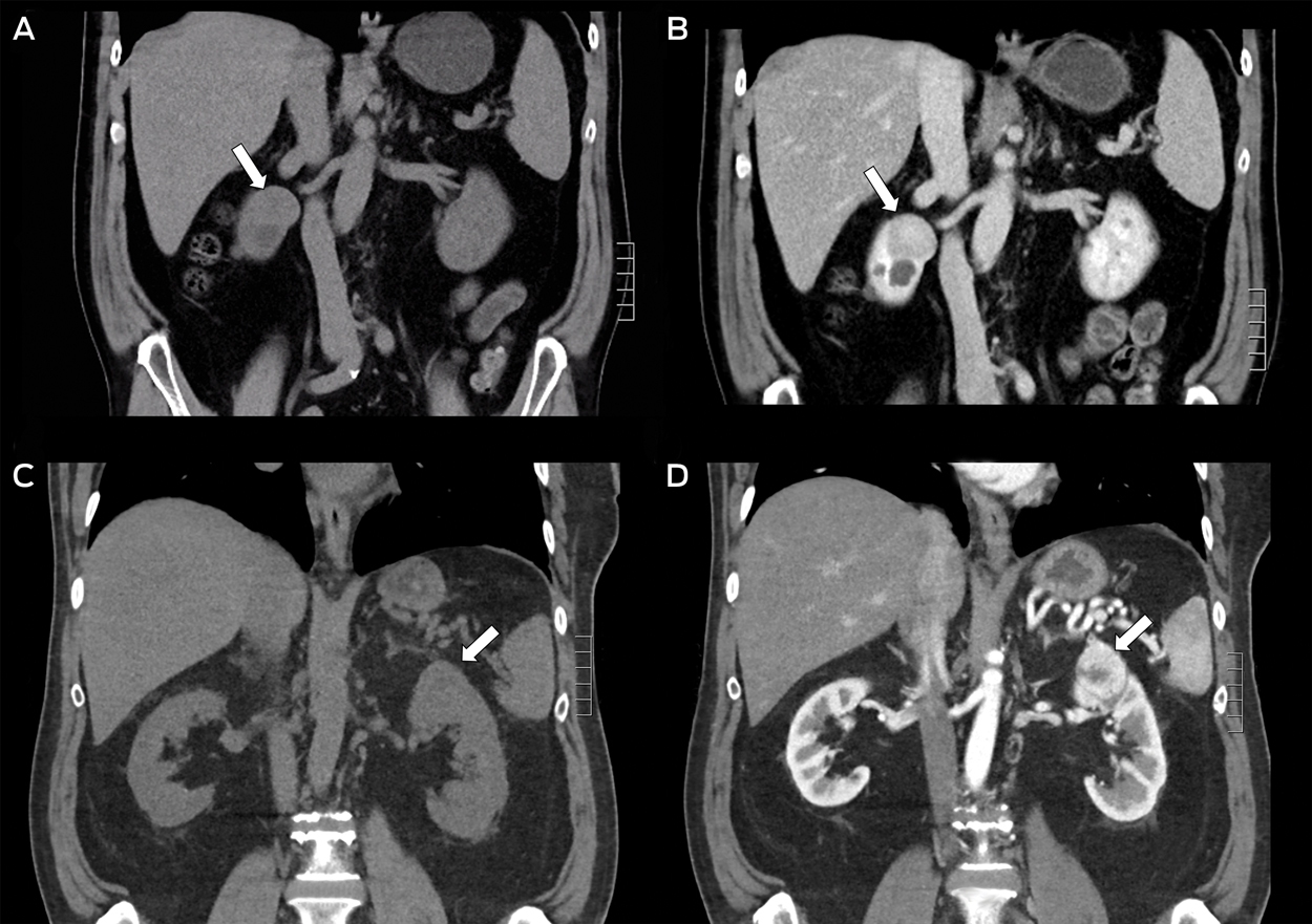
Coronal sections of computed tomography scans comparing two cases of incidentally identified small renal masses. Case 1. A: Non-contrast study. There is an indiscriminate upper polar mass in the right kidney with a mid-lower polar hypodense region. B: Portal-venous phase study with contrast. The upper polar lesion does not enhance significantly compared with the surrounding normal renal parenchyma. This lesion was diagnosed as a benign renal oncocytoma, 30 mm at maximum dimension. The hypodense region below the tumour is a simple cortical cyst; note the complete lack of enhancement. Case 2. C: Non-contrast study. There is an indiscriminate, bulging upper polar lesion in the left kidney. D: Arterial phase study with contrast. The upper polar lesion enhances distinctly, with reference to the surrounding renal parenchyma, in a heterogeneous pattern. This lesion was diagnosed as a clear cell renal cell carcinoma, 40 mm at maximum dimension. Enhancement is likely due to hypervascularity, which is typical of this subtype.
Box 4 – Bosniak classification system of cystic renal masses19,20
|
Category |
Features |
Risk |
Management |
||||||||||||
|
|
|||||||||||||||
|
I |
Simple benign cyst |
No risk |
No further treatment |
||||||||||||
|
II |
Benign cyst |
No risk |
No further treatment |
||||||||||||
|
IIF |
Minimally complex cysts |
About 5% malignant |
Regular repeat imaging (6-monthly) |
||||||||||||
|
III |
Indeterminate cystic masses |
> 50% malignant |
Surgery or active surveillance |
||||||||||||
|
IV |
Clearly malignant, containing enhancing soft tissue components; eg, solid mass with a large cystic or a necrotic component |
100% malignant |
Surgery |
||||||||||||
|
|
|||||||||||||||
|
|
|||||||||||||||
Box 5 – Decision-making algorithm for surgical management of renal masses
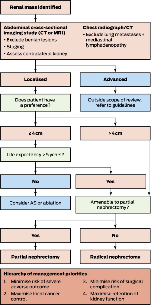
AS = active surveillance. CT = computed tomography. MRI = magnetic resonance imaging.
Box 6 – Risk of chronic kidney disease progression based on albumin–creatinine ratio (ACR) and estimated glomerular filtration rate (eGFR)2,35,53
|
Variable |
ACR (mg/mmol) |
||||||||||||||
|
|
|||||||||||||||
|
Sex |
|
|
|
||||||||||||
|
Male |
< 2.5 |
2.5–25.0 |
> 25.0 |
||||||||||||
|
Female |
< 3.5 |
3.5–35.0 |
> 35.0 |
||||||||||||
|
eGFR (mL/min/1.73 m2) |
Risk level
|
||||||||||||||
|
= 60 |
na |
Low |
High |
||||||||||||
|
45–59 |
Low |
Moderate |
High |
||||||||||||
|
30–45 |
Moderate |
Moderate |
High |
||||||||||||
|
< 30 |
High |
High |
High |
||||||||||||
|
|
|||||||||||||||
|
|
|||||||||||||||
Box 7 – Screening tool for referral to a nephrologist*47
|
Pre-operative findings |
Score |
||||||||||||||
|
|
|||||||||||||||
|
Albumin–creatinine ratio (mg/mmol) |
|
||||||||||||||
|
< 3 |
0 |
||||||||||||||
|
3–30 |
1 |
||||||||||||||
|
> 30 |
3 |
||||||||||||||
|
Diabetes mellitus |
2 |
||||||||||||||
|
Hypertension |
1 |
||||||||||||||
|
eGFR (mL/min/1.73 m2) |
|
||||||||||||||
|
= 60 |
0 |
||||||||||||||
|
< 60† |
1 |
||||||||||||||
|
< 45† |
2 |
||||||||||||||
|
< 30† |
2 |
||||||||||||||
|
Hereditary kidney cancer syndrome‡ |
3 |
||||||||||||||
|
|
|||||||||||||||
|
eGFR = estimated glomerular filtration rate. * Referral is recommended if the score is = 3, or = 1 if the patient is of Aboriginal, Torres Strait Islander, Maori or Pacific Islander heritage. This algorithm is not exhaustive and discretion should be applied in complex cases. † Cumulative criteria. ‡ Referral of patients with hereditary kidney cancer is recommended because of the likelihood of recurrence and subsequent renal ablation. |
|||||||||||||||
Box 8 – Suggestions for assessing kidney function34,35,53
Pre-operative assessment- Radiological assessment:
- supplement with nuclear medicine scan for differential kidney function assessment, if indicated
- Blood pressure and fasting glucose
- Evaluate CKD risk factors*
- Serum creatinine
- eGFR*
- Urinary ACR*
- Serum creatinine:
- refer to a nephrologist if two or more times the pre-operative value
- eGFR (unlikely to be a stable value)
- Urinary ACR:
- evaluate before discharge; if = 30 mg/mmol, re-assess within 3 months, or refer to nephrologist if accompanied by significantly elevated serum creatinine
- Non-neoplastic parenchyma:
- refer to a nephrologist if kidney disease is identified
- Serum creatinine and eGFR; refer to a nephrologist if:
- eGFR < 45 ml/min/1.73 m2; or
- eGFR has declined > 5 mL/min/1.73 m2 within 6 months (with a new baseline < 60 ml/min/1.73 m2); or
- eGFR has declined > 10 mL/min/1.73 m2 within 6 months (with a new baseline < 90 mL/min/1.73 m2)
- for other indications, see Kidney Health Australia guidelines53
- Urinary ACR (assess at least annually):
- re-assess within 3 months if = 30 mg/mmol
- refer to a nephrologist if persistently elevated = 30 mg/mmol
- Blood pressure and glucose (CKD risk)
- Fasting lipids and weight (cardiovascular risk)
Competing interests
No relevant disclosures.
Acknowledgements
Robert Ellis was supported by an Australian Government Research Training Scholarship.
References
- Znaor A, Lortet-Tieulent J, Laversanne M, et al. International variations and trends in renal cell carcinoma incidence and mortality. Eur Urol 2015; 67: 519-530.
- Kidney Disease: Improving Global Outcomes CKD Workgroup. KDIGO 2012 clinical practice guideline for the evaluation and management of chronic kidney disease. Kidney Int Suppl 2013; 3: 1-150.
- Kim SP, Murad MH, Thompson RH, et al. Comparative effectiveness for survival and renal function of partial and radical nephrectomy for localized renal tumors: a systematic review and meta-analysis. J Urol 2012; 188: 51-57.
- Australian Institute of Health and Welfare. Cancer Incidence Projections: Australia, 2011-2020 (AIHW Cat. No. CAN 62; Cancer Series No. 66). Canberra: AIHW; 2012.
- Ta AD, Bolton DM, Dimech MK, et al. Contemporary management of renal cell carcinoma (RCC) in Victoria: implications for longer term outcomes and costs. BJU Int 2013; 112 Suppl 2: 36-43.
- Satasivam P, Reeves F, Rao K, et al. Patients with medical risk factors for chronic kidney disease are at increased risk of renal impairment despite the use of nephron-sparing surgery. BJU Int 2015; 116: 590-595.
- Srigley JR, Delahunt B, Eble JN, et al. The International Society of Urological Pathology (ISUP) Vancouver classification of renal neoplasia. Am J Surg Pathol 2013; 37: 1469-1489.
- Samaratunga H, Gianduzzo T, Delahunt B. The ISUP system of staging, grading and classification of renal cell neoplasia. J Kidney Cancer VHL 2014; 1: 26-39.
- Amin MB, Edge S, Greene F, et al. AJCC Cancer Staging Manual. 8th ed. New York: Springer, 2017.
- Ljungberg B, Bensalah K, Canfield S, et al. EAU guidelines on renal cell carcinoma: 2014 update. Eur Urol 2015; 67: 913-924.
- Campbell SC, Novick AC, Belldegrun A, et al. Guideline for management of the clinical T1 renal mass. J Urol 2009; 182: 1271-1279.
- Sanchez-Martin FM, Millan-Rodriguez F, Urdaneta-Pignalosa G, et al. Small renal masses: incidental diagnosis, clinical symptoms, and prognostic factors. Adv Urol 2008: 310694.
- Pierorazio PM, Johnson MH, Ball MW, et al. Five-year analysis of a multi-institutional prospective clinical trial of delayed intervention and surveillance for small renal masses: the DISSRM registry. Eur Urol 2015; 68: 408-415.
- Kim HL, Belldegrun AS, Freitas DG, et al. Paraneoplastic signs and symptoms of renal cell carcinoma: implications for prognosis. J Urol 2003; 170: 1742-1746.
- Yap NY, Ng KL, Ong TA, et al. Clinical prognostic factors and survival outcome in renal cell carcinoma patients — a Malaysian single centre perspective. Asian Pac J Cancer Prev 2013; 14: 7497-7500.
- Giannarini G, Petralia G, Thoeny HC. Potential and limitations of diffusion-weighted magnetic resonance imaging in kidney, prostate, and bladder cancer including pelvic lymph node staging: a critical analysis of the literature. Eur Urol 2012; 61: 326-340.
- Fowler C, Reznek R. The indeterminate renal mass. Imaging 2001; 13: 27-43.
- Pallwein-Prettner L, Flöry D, Rotter CR, et al. Assessment and characterisation of common renal masses with CT and MRI. Insights Imaging 2011; 2: 543-556.
- Bosniak MA. The Bosniak renal cyst classification: 25 years later. Radiology 2012; 262: 781-785.
- Warren KS, McFarlane J. The Bosniak classification of renal cystic masses. BJU Int 2005; 95: 939-942.
- Frank I, Blute ML, Cheville JC, et al. Solid renal tumors: an analysis of pathological features related to tumor size. J Urol 2003; 170: 2217-2220.
- Sankineni S, Brown A, Cieciera M, et al. Imaging of renal cell carcinoma. Urol Oncol 2016; 34: 147-155.
- Mueller-Lisse UG, Mueller-Lisse UL. Imaging of advanced renal cell carcinoma. World J Urol 2010; 28: 253-261.
- Rhee H, Blazak J, Tham CM, et al. Pilot study: use of gallium-68 PSMA PET for detection of metastatic lesions in patients with renal tumour. EJNMMI Res 2016; 6: 76.
- Okhunov Z, Rais-Bahrami S, George AK, et al. The comparison of three renal tumor scoring systems: C-Index, P.A.D.U.A., and R.E.N.A.L. nephrometry scores. J Endourol 2011; 25: 1921-1924.
- Patel HD, Johnson MH, Pierorazio PM, et al. Diagnostic accuracy and risks of biopsy in the diagnosis of a renal mass suspicious for localized renal cell carcinoma: systematic review of the literature. J Urol 2016; 195: 1340-1347.
- Marconi L, Dabestani S, Lam TB, et al. Systematic review and meta-analysis of diagnostic accuracy of percutaneous renal tumour biopsy. Eur Urol 2016; 69: 660-673.
- Andersen MF, Norus TP. Tumor seeding with renal cell carcinoma after renal biopsy. Urol Case Rep 2016; 9: 43-44.
- Masson-Lecomte A, Bensalah K, Seringe E, et al. A prospective comparison of surgical and pathological outcomes obtained after robot-assisted or pure laparoscopic partial nephrectomy in moderate to complex renal tumours: results from a French multicentre collaborative study. BJU Int 2013; 111: 256-263.
- Choi JE, You JH, Kim DK, et al. Comparison of perioperative outcomes between robotic and laparoscopic partial nephrectomy: a systematic review and meta-analysis. Eur Urol 2015; 67: 891-901.
- Smaldone MC, Kutikov A, Egleston BL, et al. Small renal masses progressing to metastases under active surveillance: a systematic review and pooled analysis. Cancer 2012; 118: 997-1006.
- Su MZ, Memon F, Lau HM, et al. Safety, efficacy and predictors of local recurrence after percutaneous radiofrequency ablation of biopsy-proven renal cell carcinoma. Int Urol Nephrol 2016; 48: 1609-1616.
- Nielsen TK, Lagerveld BW, Keeley F, et al. Oncological outcomes and complication rates after laparoscopic-assisted cryoablation: a European Registry for Renal Cryoablation (EuRECA) multi-institutional study. BJU Int 2017; 119: 390-395.
- Lane BR, Demirjian S, Derweesh IH, et al. Survival and functional stability in chronic kidney disease due to surgical removal of nephrons: importance of the new baseline glomerular filtration rate. Eur Urol 2015; 68: 996-1003.
- Johnson DW, Atai E, Chan M, et al. KHA-CARI guideline: early chronic kidney disease: detection, prevention and management. Nephrology 2013; 18: 340-350.
- Jeon HG, Jeong IG, Lee JW, et al. Prognostic factors for chronic kidney disease after curative surgery in patients with small renal tumors. Urology 2009; 74: 1064-1068.
- Jeon HG, Choo SH, Jeong BC, et al. Uric acid levels correlate with baseline renal function and high levels are a potent risk factor for postoperative chronic kidney disease in patients with renal cell carcinoma. J Urol 2013; 189: 1249-1254.
- Torricelli FC, Danilovic A, Marchini GS, et al. Can we predict which patients will evolve to chronic kidney disease after nephrectomy for cortical renal tumors? Int Braz J Urol 2012; 38: 637-643; discussion 644.
- Malcolm JB, Bagrodia A, Derweesh IH, et al. Comparison of rates and risk factors for developing chronic renal insufficiency, proteinuria and metabolic acidosis after radical or partial nephrectomy. BJU Int 2009; 104: 476-481.
- Klarenbach S, Moore RB, Chapman DW, et al. Adverse renal outcomes in subjects undergoing nephrectomy for renal tumors: a population-based analysis. Eur Urol 2011; 59: 333-339.
- Jeon HG, Choo SH, Sung HH, et al. Small tumour size is associated with new-onset chronic kidney disease after radical nephrectomy in patients with renal cell carcinoma. Eur J Cancer 2014; 50: 64-69.
- Zabor EC, Furberg H, Mashni J, et al. Factors associated with recovery of renal function following radical nephrectomy for kidney neoplasms. Clin J Am Soc Nephrol 2016; 11: 101-107.
- Tangri N, Stevens LA, Griffith J, et al. A predictive model for progression of chronic kidney disease to kidney failure. JAMA 2011; 305: 1553-1559.
- Weight CJ, Larson BT, Fergany AF, et al. Nephrectomy induced chronic renal insufficiency is associated with increased risk of cardiovascular death and death from any cause in patients with localized cT1b renal masses. J Urol 2010; 183: 1317-1323.
- Levey AS, Stevens LA, Schmid CH, et al. A new equation to estimate glomerular filtration rate. Ann Intern Med 2009; 150: 604-612.
- Kidney Disease: Improving Global Outcomes AKI Workgroup. KDIGO clinical practice guideline for acute kidney injury. Kidney Int Suppl 2012; 2: 1-138.
- Hu SL, Chang A, Perazella MA, et al. The nephrologist's tumor: basic biology and management of renal cell carcinoma. J Am Soc Nephrol 2016; 27: 2227-2237.
- Henriksen KJ, Meehan SM, Chang A. Nonneoplastic kidney diseases in adult tumor nephrectomy and nephroureterectomy specimens: common, harmful, yet underappreciated. Arch Pathol Lab Med 2009; 133: 1012-1025.
- Cho A, Lee JE, Kwon GY, et al. Post-operative acute kidney injury in patients with renal cell carcinoma is a potent risk factor for new-onset chronic kidney disease after radical nephrectomy. Nephrol Dial Transplant 2011; 26: 3496-3501.
- Trehan A. Comparison of off-clamp partial nephrectomy and on-clamp partial nephrectomy: a systematic review and meta-analysis. Urol Int 2014; 93: 125-134.
- Mir MC, Pavan N, Parekh DJ. Current paradigm for ischemia in kidney surgery. J Urol 2016; 195: 1655-1663.
- Chung JS, Son NH, Byun SS, et al. Trends in renal function after radical nephrectomy: a multicentre analysis. BJU Int 2014; 113: 408-415.
- Kidney Health Australia. Chronic kidney disease (CKD) management in general practice. 3rd ed. Melbourne: KHA, 2015. http://kidney.org.au/health-professionals/prevent/chronic-kidney-disease-management-handbook (accessed June 2017).
Linked content
-
MJA InSight: GP follow-up all important for renal mass
-
MJA Podcast: Mr Robert Ellis
Provenance: Not commissioned; externally peer reviewed.
The first Australian evidence‐based guidelines on male infertility
Darren J Katz, Liza O’Donnell, Robert I McLachlan, Tim J Moss, Clare V Boothroyd, Veena Jayadev, Sarah R Catford
The epidemiology of haemodialysis catheter infections in Australia, 2016–20: a prospective cohort study
Benjamin Lazarus, Kevan R Polkinghorne, Martin P Gallagher, Jayson Catiwa, Nicholas A Gray, Sarah Coggan, Kathryn R Higgins, Girish Talaulikar, Stephen P McDonald AM, Sradha Kotwal
Am I on the list? Clinician‐reported factors for kidney transplantation non‐waitlisting among Aboriginal and Torres Strait Islander people with kidney failure: a cross‐sectional study
Stephen P McDonald AM, Katie Cundale, Christopher E Davies, Kelli Karrikarringka Owen (Kaurna, Nharangga, and Ngarrindjeri), Kerry Dole, Feruza Kholmurodova, Matilda D’Antoine (Paakantyi), Jaquelyne T Hughes (Wagadagam)
Hyperkalaemic cardiac arrest due to cream of tartar ingestion
Daniel Yee Lee Ng, Laksmi Govindasamy, Andrew Hughes, Hwee Min Lee
Dialysis care in Australia: a call for reform and innovation
Dharshana Sabanayagam, Angela Makris, Frederika Sciberras, Nicole J Scholes‐Robertson, Germaine Wong
Closing the gap in kidney disease: validating the reporting of Aboriginal and/or Torres Strait Islander identification in a clinical quality registry using linked data
Heather J Baldwin, Nicole De La Mata, Grant Sara, Faye McMillan, Brett Biles, Jianyun Wu, Paul Lawton, Stephen McDonald, Angela C Webster