




Volume 194 - Issue 8
Diagnosis and management of iron deficiency anaemia: a clinical update
Author: Alan E Dugdale
Med J Aust 2011; 194 (8): 429. || doi: 10.5694/j.1326-5377.2011.tb03046.x
Published online: 18 April 2011
Published online: 18 April 2011
To the Editor: The levels of ferritin and folate in the blood are regarded as the “gold standards” for measuring deficiencies in iron and folate, but they are complex and expensive tests. The red cell distribution width (RDW-CV%) measures variability in red blood cell (RBC) size, helps in early detection of deficiencies and is available in all automated analyses of RBCs. It is seldom listed on haematology reports, though it can be printed out at no extra cost. A raised RDW-CV% should prompt the treating doctor to consider underlying latent deficiencies and to order specific tests.
Pasricha and colleagues outlined the most efficient ways of diagnosing and managing iron deficiency anaemia.1 However, they did not mention RDW-CV%.
Haemoanalysers measure the haemoglobin (Hb) content and size of individual RBCs. The average RBC size is the mean cell volume (MCV), and the variation in sizes is calculated as a standard deviation (SD). The haemoanalyser converts the MCV and SD to a coefficient of variation (CV) using the formula:

Normal RBCs vary slightly in size, so the normal value of the RDW-CV% is 10%–15%. Greater variability gives a higher RDW-CV%.
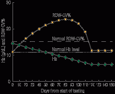
When a person becomes iron deficient, he or she forms small RBCs. The circulation will then contain a mixture of older normal RBCs and increasing numbers of newer small cells, until all the normal cells reach the end of their 120-day lifespan. It therefore takes several weeks for the Hb level and MCV to drop low enough to diagnostic levels for iron deficiency. However, the mixture of normal and small RBCs rapidly increases the variability in the size of circulating RBCs, so the RDW-CV% reaches pathological levels before other RBC measurements do.2 A raised RDW-CV% is the first haematological sign of iron deficiency.
My computer model demonstrating the mechanism and timing of changes in Hb level and RDW-CV% showed a large and early rise in the initial stages of iron deficiency before anaemia (Box).3 In the anaemia of thalassaemia, the RDW-CV% is usually normal. In combination with other parameters, the RDW-CV% helps to classify anaemias. The sensitivity and specificity for diagnoses is about 80%,4 so the RDW-CV% result should be used as a screening tool to alert the clinician to a possible disorder that can be investigated using standard tests. I recommend the RDW-CV% be included in all routine blood reports.
References
- Pasricha SS, Flecknoe-Brown SC, Allen KJ, et al. Diagnosis and management of iron deficiency anaemia: a clinical update. Med J Aust 2010; 193: 525-532.
- Uchida T. Change in red cell distribution width with iron deficiency. Clin Lab Haematol 1989; 11: 117-121. 0_i1095851
- Dugdale AE. Predicting iron and folate deficiency anaemias from standard blood testing: the mechanism and implications for clinical medicine and public health in developing countries. Theor Biol Med Model 2006; 3: 34. 0_i1095853
- Demir A, Yarali N, Fisgin T, et al. Most reliable indices in differentiation between thalassemia trait and iron deficiency anemia. Pediatr Int 2002; 44: 612-616. 0_i1095855
Splenic injury in severe cases of the zoonoses Q fever and rickettsial infection: diagnostic challenges
Ashleigh Drury, Philippa Harrison, Aiveen Bannan
Health and economic benefits of improving pre‐hospital identification of stroke in Australian women: a modelling study
Thomas Gadsden, Lei Si, Emily R Atkins, Cheryl Carcel, Xia Wang, Stephen Jan, Mark Woodward, Laura E Downey
Updating the diagnosis and management of elevated serum ferritin levels in the era of routine ferritin testing of blood donors by Australian Red Cross Lifeblood
Gary D Zhang, James Chen, Daniel M Johnstone, Martin B Delatycki, Katie Allen, John K Olynyk
Early cardiovascular collapse after envenoming by snakes in Australia, 2005–2020: an observational study (ASP‐31)
Geoffrey K Isbister, Katherine Z Isoardi, Angela L Chiew, Shane Jenkins, Nicholas A Buckley
Updated recommendations for warfarin reversal in the setting of four‐factor prothrombin complex concentrate
Danielle Robinson, James McFadyen, Eileen Merriman, Chee Wee Tan, Ross Baker, Huyen Tran
Updating the diagnosis and management of iron deficiency in the era of routine ferritin testing of blood donors by Australian Red Cross Lifeblood
Gary D Zhang, Daniel Johnstone, Michael F Leahy, John K Olynyk