




Volume 198 - Issue 4
Medicine on the walls of the art gallery
Authors: George M Weisz and William R Albury
Med J Aust 2013; 198 (4): 229-230. || doi: 10.5694/mja12.11263
Published online: 4 March 2013
Published online: 4 March 2013
Distinguishing medical conditions from artists’ stylistic embellishments in old master paintings can contribute to our historical understanding of diseases like arthritis.
Distinguishing medical conditions from artists’ stylistic embellishments can contribute to our historical understanding of diseases like arthritis
When visiting art galleries, one often sees abnormal anatomical features depicted in paintings and sculptures. In the recent Brisbane exhibition of works from the Prado museum in Spain, viewers could find instances of medical conditions in the works of several artists — for example, dwarfism (Velasquez) and club-foot (Ribera), as well as mandibular abnormalities in Hapsburg family members (Titian).1 Here, we report that we have observed what we argue to be pathological changes, albeit subtle ones, depicted in the hands of two portrait subjects in the Art Gallery of New South Wales.
Portrait 1: Cosimo I de’ Medici in armour
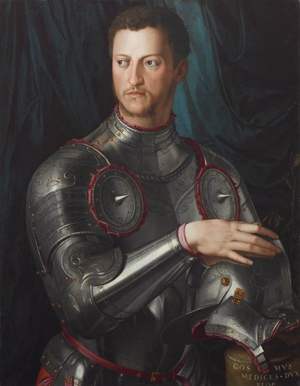
Cosimo I de’ Medici (1519–1574), Duke of Florence from 1537 and Grand Duke of Tuscany from 1569, was painted in about 1545 by his court artist Agnolo Bronzino (1503–1572), a master of the late Renaissance mannerist style.2,3 This painting is an official portrait, of which many copies were made by Bronzino’s workshop. Cosimo, young and determined, in armour, like a warrior, is shown with his right hand situated prominently in the foreground, resting on his helmet.
Agnolo Bronzino (1503–1572). Cosimo I de’ Medici in armour, c. 1545. Oil on poplar, 86.0 × 67.0 cm. Art Gallery of New South Wales. Art Gallery of NSW Foundation Purchase 1996. Photo © Art Gallery of NSW. (Reproduced by permission of the Art Gallery of NSW.)
The fingers of the right hand in this portrait present certain deformities that are also seen in the left hand of three other portraits of the duke, one by Bronzino and two by his older contemporary Jacopo Pontormo (1494–1557).4 The metacarpophalangeal (MP) joints are depressed and dorsiflexed, the proximal interphalangeal (PIP) joints of the four fingers are swollen and flexed, and the distal interphalangeal joints appear to be somewhat extended. This is not a natural position for the fingers, and it would be very painful for someone with normal hands to hold the finger joints in this position voluntarily for any length of time (as would be required when sitting for a portrait).
This particular representation of the joints of the fingers is not found in Bronzino’s or Pontormo’s portraits of other individuals, so we conclude that it is not a stylistic preference on any of these artists’ part but the representation of a condition existing in the subject.
By contrast, although one can also see that the two middle fingers of Cosimo’s hand in Bronzino’s portrait are pressed together as they would be in syndactyly, this feature is so commonly found in the paintings of the period — both by Bronzino (for example, in his 1543 portrait of Cosimo’s wife Eleonora of Toledo [1522–1562], National Gallery in Prague, http://www.wga.hu/html/b/bronzino/1/eleonorb.html) and by many other artists — that we must regard it as a matter of artistic style rather than pathological abnormality.5
Throughout the three centuries of the Medici family’s predominance in Florence (from the mid 15th to the mid 18th centuries), many of its members suffered joint ailments. The evidence suggests a complex hereditary condition that affected both the senior line of the family and also the cadet line from which Cosimo was descended.4,6,7
Cosimo was reported to have suffered from disabling joint problems throughout his middle and later years, and further information was gained about these problems when his pathology was recently investigated in the exhumed skeleton. X-rays showed no evidence of gout, which was the premodern explanation of Medici joint pathologies, but they identified a number of arthritic and other pathological changes in the spine and leg bones. The hand had a localised erosion of the joint only at the PIP joint level, which indicates a specific pathology and not accidental damage due to the ravages of time.4 This erosion corresponds to the swollen PIP joints in Cosimo’s portrait.
Portrait 2: Portrait of an officer
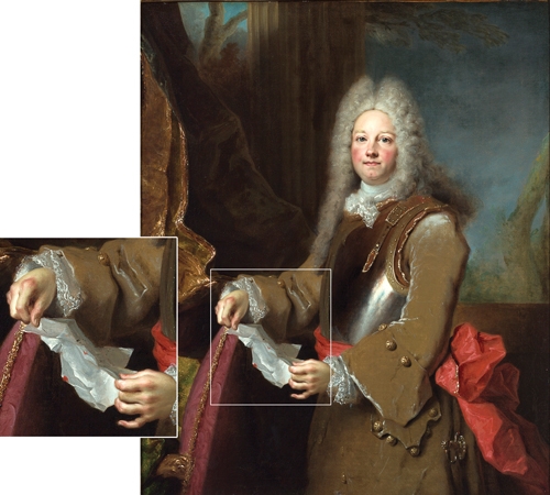
Portrait of an officer, by the French rococo artist Nicolas de Largillière (1656–1746), probably completed by 1715, shows a young man of around 20 years of age dressed in military attire. The figure wears an elegant lace cravat and cuffs, a high powdered wig and an armoured breastplate. The work was initially considered to be a portrait of John Churchill, 1st Duke of Marlborough (1650–1722); however, this suggestion has not been confirmed and the identity of the figure remains unknown.8
On visual inspection, two anatomical anomalies are obvious — red cheeks and swollen knuckles. We can draw no conclusion concerning the red cheeks, which are common in Largillière’s portraits of both men and women. They may indicate an ongoing pathological condition such as lupus or a temporary one such as fever. Equally, they may have been caused by an overheated room in which the subject posed for his portrait or by the use of cosmetics (which was fashionable among upper class men and women at the time). Or they may be the result of the artist’s stylistic embellishment of the sitter’s complexion.
More significant, however, are the swollen knuckles — namely, the MP joints in both hands — not subluxated, but inflamed and reddish. Largillière is considered by art historians to have had a “genius for depicting hands”,9 so the accuracy of his representation on this point is highly probable. His other works show men’s hands both with normal joints and with swellings in other joints apart from the MP joints,8 from which we conclude that the swollen MP joints in Portrait of an officer are not a regular stylistic feature of Largillière’s male portraits but a naturalistic representation.
Any diagnosis proposed on the basis of the limited information available would have to be highly tentative. However, if one treats the redness of the officer’s face as a non-pathological feature, as we have argued, then a possible diagnosis in the context of swollen but undeformed fingers could be rheumatic fever or juvenile rheumatoid arthritis.
Discussion
We argue that these two portraits in the Art Gallery of NSW, from different epochs of European art, reliably depict certain rheumatic diseases in young men.
When investigating apparent pathological conditions in old master paintings, one must always be conscious of the fact that artists’ representations are not clinical photographs. The artist may exaggerate or tone down different aspects of the sitter’s anatomical features, or even depict wholly imaginary features that the person does not have.10,11 As we have done here, it is always necessary to look for evidence that will assist the investigator in distinguishing between features that are likely to be naturalistic representations of what the artist saw, and those that are likely to be stylistic embellishments added for aesthetic effect.
This requires investigation of such factors as the medical history of the period concerned, the characteristic features of the artist’s style, the biography of the sitter (if his or her identity is known) and the history of the artwork itself (especially the circumstances and purpose of its origin).
Deciphering the content of artworks is a specialised discipline, which art historians have cultivated since the 16th century. However, we believe this decoding system would not be complete without taking account of the medical conditions that were at times represented, intentionally or inadvertently, in the people shown in paintings or statues.
When relevant information is available and caution is exercised in the research, the results of this analysis can add much to our historical understanding of the occurrence and appearance of diseases in earlier times.
Competing interests
No relevant disclosures.
References
- Museo Nacional del Prado. Portrait of Spain: masterpieces from the Prado [exhibition catalogue]. Brisbane: Queensland Art Gallery, 2012. 0_CHDECGGA
- Brock M. Bronzino. Paris: Flammarion, 2002: 52-54. 0_CHDEAFFC
- Langedijk K. The portraits of the Medici: 15th–18th centuries. Florence: Studio per Edizioni Scelte, 1981: 79-120. 0_i1115653
- Weisz GM, Albury WR, Lippi D, Matucci-Cerinic M. Who was Pontormo's halberdier? The evidence from pathology. Rheumatol Int 2012; 32: 1915-1920. 0_i1115655
- Herman PJ. A systematic study of the pictorial hand from Botticelli to Rembrandt. In: Appelboom T, editor. Art, history and antiquity of rheumatic diseases. Brussels: Elsevier, 1987: 38-40. 0_i1115657
- Weisz GM, Matucci-Cerinic M, Lippi D, Albury WR. The ossification diathesis in the Medici family: DISH and other features. Rheumatol Int 2011; 31: 1649-1652. 0_i1115659
- Weisz GM, Matucci-Cerinic M, Albury WR, Lippi D. The illnesses of Carlo di Ferdinando I de’ Medici: a second opinion. Clin Exp Rheumatol 2012; 30: -6-11. 0_i1115661
- Rosenfeld MN. Largillièrre and the eighteenth-century portrait [exhibition catalogue]. Montreal: Montreal Museum of Fine Arts, 1982. 0_i1115663
- Bolton R, editor. The collectors: old master paintings [exhibition catalogue]. London: Sphinx Fine Art, 2009: 166. 0_i1115665
- Rothschild B. Style versus substance in artistic depiction. Rheumatology 2005; 44: 1464-1465. 0_i1115667
- Appelboom T. Style versus substance in artistic depiction. Rheumatology 2005; 44: 1465. 0_i1115670
Provenance: Not commissioned; externally peer reviewed.
Prisoner of war pathology in Changi, 1942–1945
Kate Ariotti, Elizabeth Roberts‐Pedersen
Should medical eponyms continue to be used in everyday practice?
Leya Nedumannil, Diana Lewis
100 years on: the first use of insulin in Australia
Sophie Templer
Reflections on the life and career of Professor Dame Valerie Beral AC DBE FRS FRCOG FMedSci (1943–2022)
Karen Canfell, Bette Liu, Emily Banks
The Honourable Dr Donald James Grimes AO
Michael Kirby, Patti Warn
From wipeout to drill out: a history of exostosis management and Australian surfing
Alon Taylor, Hannah North, Narinder P Singh, Paul A Fagan