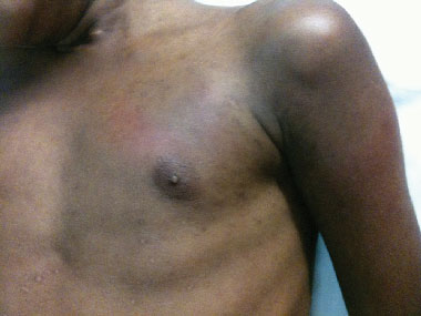
A 10-year-old boy from Papua New Guinea with multidrug-resistant tuberculosis and multibacillary leprosy developed acute glomerulonephritis while being treated as an inpatient at Thursday Island Hospital in the Torres Strait, Queensland. This is the first such case to be reported in Australia, where these diseases are uncommon and the combination is extremely rare, and it outlines important learning points regarding the aetiology of renal disease among patients with tuberculosis and leprosy. (MJA 2011; 195: 150-152)
A 10-year-old boy from a remote village in Western Province, Papua New Guinea (PNG), presented to Saibai Island Primary Health Centre in the northern Torres Strait, Queensland, with a 4-year history of intermittent malaise, fevers, night sweats, recurrent skin sores and a cough productive of green sputum. He had received treatment for leprosy for 1 month the previous year at Daru Hospital (Western Province, PNG). There was a strong family history of leprosy and tuberculosis among both first- and second-degree relatives.
On initial examination, the patient appeared cachectic. There was evidence of recent impetigo on both lower limbs and depigmented areas on his upper and lower limbs. He had thickened ulnar and posterior tibial nerves bilaterally, with normal sensation and motor function on repeated clinical assessments. His lungfields were clear to auscultation, but he had an ejection systolic murmur; he was also found to have hepatomegaly and enlarged inguinal, anterior and posterior cervical lymph nodes. He was transferred to Thursday Island Hospital for inpatient management.
Slit skin smears were performed; phenotypic analysis of the right earlobe smear was positive for Mycobacterium leprae, confirming the diagnosis of multibacillary leprosy.1 An initial chest x-ray showed left upper lobe changes consistent with pulmonary tuberculosis; subsequent sputum samples and an aspirate of an anterior cervical lymph node cultured Mycobacterium tuberculosis resistant to rifampicin, isoniazid, streptomycin and ethionamide, in keeping with a diagnosis of disseminated multidrug-resistant tuberculosis (MDR-TB).2 HIV and hepatitis serological tests were negative. On admission, his serum creatinine level was 30 μmol/L (reference range [RR], 46–81 μmol/L).
The patient was given intravenous amikacin and oral moxifloxacin, isoniazid, pyrazinamide, ethambutol, pyridoxine and rifampicin; cycloserine, aminosalicylic acid (mesalazine), dapsone and clofazimine were later added.
One week after admission, the patient developed acute glomerulonephritis, which manifested as fluid retention (pulmonary oedema, peripheral oedema and ascites), hypertension (maximum blood pressure, 160/112 mmHg), haematuria, proteinuria (up to 9700 mg/L [RR, < 100 mg/L]) and impaired kidney function (serum creatinine level peaked at 69 μmol/L). He was treated with frusemide, nifedipine and prednisolone. There was serological evidence of recent infection with Streptococcus pyogenes (elevated antistreptolysin O and anti-DNAse B titres) and hypocomplementaemia (decreased complement component 3 [C3] concentration with normal complement component 4 [C4] concentration). The nephritic illness resolved slowly over the next few weeks.
During this period, the patient developed painful, erythematous nodules over his upper torso and limbs (Box 1). His mother reported that he had experienced several such episodes in the past. The lesions measured 5–10 mm in diameter and were tender to palpation; in association with the cardiac murmur and elevated streptococcal serology there was some initial concern about the possibility of acute rheumatic fever and the patient was commenced on penicillin prophylaxis.
However, expert consensus was that the lesions more likely represented erythema nodosum leprosum (ENL) — an immune complex-mediated inflammatory reaction that can occur in both acute and chronic relapsing forms among leprosy patients with a high mycobacterial load. Treatment options for this condition have historically included simple analgesics, steroids, non-steroidal anti-inflammatory drugs, clofazimine and thalidomide. A recent Cochrane review found a paucity of evidence for most treatments of ENL, and only a modest benefit from clofazimine and thalidomide.3
As the patient was already taking clofazimine, thalidomide was not considered to be a practical or necessary option in his case (given its significant side effects, the need for monitoring and the patient’s planned return to PNG); his several subsequent bouts of ENL were treated successfully with oral prednisolone. His renal function remained stable throughout these episodes, and an inpatient echocardiogram was normal.
After three negative sputum smears, the patient was removed from isolation and continued treatment with intravenous amikacin. He was discharged home on oral therapy for both MDR-TB and leprosy, with follow-up planned at Saibai Island Primary Health Centre.
Unfortunately, at the time of writing, due to unknown patient factors and unforeseen circumstances (such as the closure of the border and cancellation of outreach clinics), the patient has not been seen, nor his medications collected, for almost 6 months.
Although accurate figures on the epidemiology of infectious diseases in PNG are difficult to obtain, it is likely that the rates of mycobacterial infections such as tuberculosis and leprosy in PNG are both underreported and among the highest in the world. The World Health Organization recently reported an annual incidence of 6.5 per 100 000 for leprosy in PNG,4 with historical prevalence of up to 3% recorded in some remote villages.5 For tuberculosis, PNG has reported an incidence of 233 per 100 000, of which about 25% may be MDR-TB.6
A recent literature review described only isolated case reports and small case series studies of concomitant infection with leprosy and tuberculosis over the past few decades.7 Most of these reported cases were from India, with documented co-infection rates (ie, the proportion of leprosy patients found to also have tuberculosis) in the order of 2.5%–7.7% in India and up to 13.4% in South Africa.8,9 Despite the relative dearth of published accounts of co-infection, it seems plausible that simultaneous infection with M. tuberculosis and M. leprae occurs more frequently than is described in published reports in regions with relatively high rates of both diseases (including countries in sub-Saharan Africa, South America, the Indian Subcontinent, and South-East Asia). Some postulated reasons for why rates of co-infection may nevertheless be lower than expected in high prevalence regions include improvements in the detection rate and treatment for both infections; the WHO’s initiative of providing free leprosy treatment in an effort to eliminate the disease; the effects of BCG immunisation; and the complex, possibly antagonistic interaction between the two strains of mycobacteria.7
Our patient developed acute glomerulonephritis as an inpatient receiving treatment for MDR-TB and leprosy; hence, a number of possible causes of his renal dysfunction were considered.
Renal abnormalities occur among most patients with leprosy, particularly those with multibacillary disease (“borderline” or “lepromatous” disease using the Ridley–Jopling Classification of Leprosy), and renal failure is a frequent cause of death in patients with leprosy.10,11 Glomerulonephritis, nephrosclerosis, tubulointerstitial nephritis, amyloidosis and granulomas are the most common renal pathologies found on biopsy or autopsy of leprosy patients.12 Of the glomerulonephropathies, the proliferative glomerulonephritides are the most frequently described lesion among patients with leprosy.11,12 Hypocomplementaemia is a recognised association of renal disease in this setting, particularly among patients with multibacillary leprosy and ENL.12 Typical serum protein profiles seen among patients with renal disease associated with some selected infections are presented in Box 2.
The pattern of hypocomplementaemia varies somewhat between the different forms of glomerular disease; low C3 with normal C4 (as in our patient’s case) tends to suggest either poststreptococcal or membranoproliferative glomerulonephritis.13 Evidence of streptococcal infection was found on biopsy from five leprosy patients with renal disease in India; the same case series reported two patients with microfilariae in peripheral blood samples, indicating the possibility of a range of concomitant infections contributing to renal disease in patients with leprosy.14 With respect to drug-induced nephrotoxicity, rifampicin (a common component of the pharmacological regimen for treatment of both leprosy and tuberculosis) has been implicated in acute renal failure, often associated with thrombocytopenia, immune haemolytic anaemia and intravascular coagulation.15
In our patient, however, the combination of oedema, hypertension, haematuria, hypocomplementaemia (in the pattern described), elevated streptococcal serological results and a history suggestive of recent skin infections is strongly supportive of a diagnosis of poststreptococcal glomerulonephritis, which is an uncommon form of renal disease in patients with leprosy. The patient did not undergo kidney biopsy as his condition was clinically improving and it was felt that this highly invasive procedure would not have yielded sufficient additional diagnostic information to make it worthwhile. It is also a moot point whether, given his nationality, this procedure would have been available to him.
In summary, our patient had the misfortune to suffer simultaneous infection with multibacillary leprosy and MDR-TB (and may be the first such reported case in Australia), which was complicated by ENL and an acute glomerulonephritis that was probably poststreptococcal glomerulonephritis — a rare form of renal disease in a subset of patients among whom renal impairment is commonly due to other causes.
2 Serum protein profiles seen in renal disease associated with specific infections*
|
Serum protein profile |
||||||||||||||
Group A streptococcus infection |
|
||||||||||||||
Acute glomerulonephritis |
|
||||||||||||||
Classic diffuse proliferative |
Decreased C3 |
||||||||||||||
Focal proliferative (IgA disease) |
Increased IgA |
||||||||||||||
Mycobacterium leprae infection |
|
||||||||||||||
Acute glomerulonephritis |
|
||||||||||||||
Proliferative forms |
Decreased C3 |
||||||||||||||
Cryoglobulinaemia (ENL) |
Decreased C3 and C4 |
||||||||||||||
Nephrotic syndrome |
|
||||||||||||||
Amyloid |
Increased AA |
||||||||||||||
Mycobacterium tuberculosis infection |
|
||||||||||||||
Nephrotic syndrome |
|
||||||||||||||
Amyloid |
Increased AA |
||||||||||||||
AA = amyloid A. C3 = complement component 3. C4 = complement component 4. ENL = erythema nodosum leprosum. IgA = immunoglobulin A. * The serum concentrations of certain proteins are altered in these conditions. |
|||||||||||||||
Provenance: Not commissioned; externally peer reviewed.
- 1. WHO Expert Committee on Leprosy. Seventh report. Geneva: World Health Organization: 1997. (WHO Technical Report Series No. 874.)
- 2. Nathanson E, Nunn P, Uplekar M et al. MDR tuberculosis — critical steps for prevention and control. N Engl J Med 2010; 363: 1050-1058.
- 3. Van Veen NH, Lockwood DN, van Brakel WH, et al. Interventions for erythema nodosum leprosum. Cochrane Database Syst Rev 2009; (3): CD006949.
- 4. Global leprosy situation, 2010. Wkly Epidemiol Rec 2010; 85: 337-348.
- 5. Bagshawe A, de Burgh S, Fung SC, et al. The epidemiology of leprosy in a high prevalence village in Papua New Guinea. Trans R Soc Trop Med Hyg 1989; 83: 121-127.
- 6. Gilpin CM, Simpson G, Vincent S, et al. Evidence of primary transmission of multidrug-resistant tuberculosis in the Western Province of Papua New Guinea. Med J Aust 2008; 188: 148-152. <MJA full text>
- 7. Prasad R, Verma SK, Singh R, Hosmane G. Concomittant [sic] pulmonary tuberculosis and borderline leprosy with type-II lepra reaction in single patient. Lung India 2010; 27: 19-23.
- 8. Lee HN, Embi CS, Vigeland KM, White CR Jr. Concomitant pulmonary tuberculosis and leprosy. J Am Acad Derm 2003; 49: 755-757.
- 9. Kumar B, Kaur S, Kataria S, Roy SN. Concomitant occurrence of leprosy and tuberculosis — a clinical, bacteriological and radiological evaluation. Lepr India 1982; 54: 671-676.
- 10. Aggarwal HK, Sharma P, Jaswal TS, et al. Evaluation of renal profile in patients of leprosy. J Indian Acad Clin Med 2004; 5: 316-321.
- 11. Chugh KS, Damle PB, Kaur S, et al. Renal lesions in leprosy amongst north Indian patients. Postgrad Med J 1983; 59: 707-711.
- 12. da Silva Jr GB, De Francesco Daher E. Renal involvement in leprosy: retrospective analysis of 461 cases in Brazil. Braz J Infect Dis 2006; 10: 107-112.
- 13. Welch TR. The complement system in renal diseases. Nephron 2001; 88: 199-204.
- 14. Date A, Thomas A, Mathai R, Johny KV. Glomerular pathology in leprosy. An electron microscopic study. Am J Trop Med Hyg 1977; 26: 266-272.
- 15. Covic A, Goldsmith DJA, Segall L, et al. Rifampicin-induced acute renal failure: a series of 60 patients. Nephrol Dial Transplant 1998; 13: 924-929.


We are indebted to Dr Graham Simpson and Dr Stephen Vincent from the Thoracic Medicine Outreach Team at Cairns Base Hospital for their expert assistance in managing this patient’s complex case.
None relevant to this article declared (ICMJE disclosure forms completed).