




Volume 218 - Issue 1
DANGER: what clinicians need to know about aggressive head and neck cutaneous squamous cell carcinoma
Authors: Richard Tjahjono, Hubert TH Low, Jenny Lee, Deshan F Sebaratnam, Ruta Gupta, Michael J Veness, Jonathan Clark and Carsten E Palme
Med J Aust 2023; 218 (1): 11-15. || doi: 10.5694/mja2.51786
Published online: 5 December 2022
Published online: 5 December 2022
The DANGER acronym is a simple, evidence-based aid to identify high risk squamous cell carcinoma
The DANGER acronym is a simple, evidence‐based aid to identify high risk squamous cell carcinoma
Non‐melanoma skin cancer (NMSC) is the most common group of malignancies diagnosed worldwide.1 Two out of three Australians will develop at least one NMSC before the age of 70years, with about five NMSC cases diagnosed annually per one invasive cancer of another type.2 NMSC ranges from common lesions such as basal cell carcinoma and cutaneous squamous cell carcinoma (cSCC), collectively termed as keratinocyte carcinoma, to rarer lesions such as Merkel cell carcinoma, atypical fibroxanthoma, dermatofibrosarcoma protuberans, microcystic adnexal carcinoma and angiosarcoma. The most common subtype is basal cell carcinoma, comprising 70–75% of all lesions, followed by cSCC (20–25%) and Merkel cell carcinoma (<5%).1 Exposure to ultraviolet radiation, particularly during childhood and adolescence, is the predominant aetiological risk factor for developing keratinocyte carcinomas. Therefore, a significant proportion of these malignancies develop on sun‐exposed locations, such as the head and neck, trunk and extremities.3
cSCC is the most common keratinocyte carcinoma with metastatic potential, including perineural invasion (PNI).4,5 The head and neck is an anatomically complex region and the most common site of cSCC (70–75%).3 Head and neck cSCCs are managed by a diverse group of clinicians, including general practitioners, dermatologists, otolaryngology head and neck surgeons, plastic and reconstructive surgeons, surgical oncologists, radiation oncologists and medical oncologists in the advanced setting. Most patients are treated by local measures only, with excellent prognosis and low recurrence rates; however, a minority will develop advanced disease, which refers to large or invasive primary lesions and those that have metastasised to regional nodes or distant sites. Prognosis has improved substantially for patients with locally recurrent or regional metastatic disease through a multidisciplinary approach including surgery, adjuvant radiotherapy and systemic therapy. Although distant metastases have traditionally been considered incurable, this is rapidly changing with the introduction of immune check point inhibitors.6
In this perspective article, we aim to develop an evidence‐based framework to assist clinicians recognise head and neck cSCC at high risk of progression to more advanced disease and provide a pathway for timely referral to appropriate specialist care.
Identification of high risk cutaneous squamous cell carcinoma
There are several clinical and pathological features that are associated with increased risk of metastases and recurrence.4,7,8 From a primary care perspective, it is important that high risk lesions are recognised early to minimise treatment‐related morbidity and potential mortality associated with advanced cSCC. We therefore propose the use of the DANGER acronym as a simple, evidence‐based aid for clinicians to identify high risk cSCC and trigger a prompt referral to appropriate specialist care for best practice management and improved outcomes.
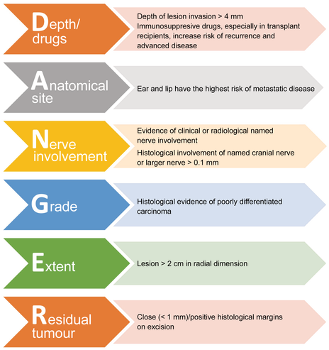
The DANGER acronym is an abbreviation of i) depth of invasion/drugs, ii) anatomical site, iii) nerve involvement, iv) grade, v) extent, and vi) residual tumour (Box 1). This section of the article discusses relevant published literature to each high risk feature included in the acronym.
Depth of invasion/drugs
Depth of invasion is an established risk factor for developing regional metastasis. Risk is thought to be proportional to depth of invasion. For instance, a cSCC with more than 4mm in depth of invasion is associated with increased risk of metastasis, with those invading the subcutaneous fat having a metastatic potential up to 46% (Box 2).5,9,10,11 However, the most reliable prospective data come from a study published in 2021 which reported that the rate of nodal metastases for the patients with a depth of invasion of 5mm or greater was 19.7% compared with 0% for the patients with a depth of invasion of less than 5mm (P = 0.01), where the rate of nodal metastases in the patients with a depth of invasion of 10mm or greater was 25%.11 Multiple guidelines concur that depth of invasion of more than 4mm for cSCC is considered a high risk factor and, therefore, tumours with high depth of invasion need ongoing surveillance for nodal metastases.9,12
In addition, patients who are taking immunosuppressive drugs have a higher risk of developing cSCC, which is associated with an increasing duration of immunosuppression. NMSCs in this patient group are often more aggressive, manifesting as recurrent, multifocal or metastatic disease, and are associated with significant morbidity and mortality.7,8 This is particularly relevant in patients who underwent solid organ transplants, with an about 100‐fold increase in risk of developing cSCC.13 With the increase in numbers and life expectancy of transplant recipients, the burden of disease in this patient group is likely to increase in the future.
Anatomical site
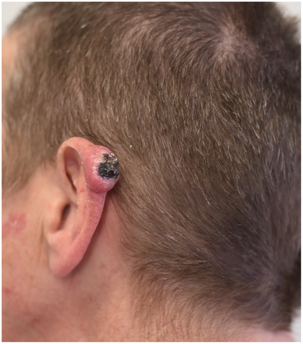
Most cSCCs develop on the sun‐exposed head and neck. The mid‐face accounts for 25% of all cutaneous malignancies, followed by the external ear and the surrounding skin.14 Systematic reviews of 71 studies15 and 36 studies16 identified that lesions of the ear and lip had the highest risk of metastases compared with lesions from other sites. These findings were echoed by a large prospective study, which saw 26% of patients with lesions on the lip and 13% on the ear developing nodal metastatic disease,5 and by a retrospective cohort study,17 which found that lesions localised to the ear had three times the risk for nodal metastasis (Box 3). This is presumably due to the particularly high ultraviolet exposure to the ear and lip and their proximity to draining nodal basins.16,18
Nerve involvement
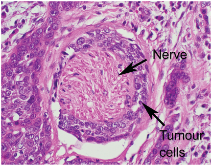
Perineural invasion is an infrequent but clinically important phenomenon that may occur in patients with head and neck cSCC.19 The incidence of PNI in this patient group is estimated to be between 2.5% and 14%.20 PNI represents tumour extension along the perineural space occurring over months to years (Box 4). It may be associated with larger primary lesions, higher risk of recurrence and metastatic spread, and reduced survival rates.21 Most PNI are termed “incidental PNI” and involve small peritumoural cutaneous nerves, which are only identified through histopathological examination without symptoms at presentation. However, a small proportion of PNI are termed “clinical PNI” and reflect clinically evident deficits in the distribution of the involved nerve (most commonly the facial or trigeminal nerve) at presentation. The latter type portends a much poorer prognosis.20 This disease process is often clinically and histopathologically under‐recognised.20,21 Patients with clinical PNI should be referred to an expert multidisciplinary team for further management, with strong consideration for referral in those with incidental PNI involving large (>0.1mm) or named nerves such as cervical branch of facial nerve or ophthalmic branch of trigeminal nerve (as opposed to small perforating branches at the end of named nerves).
Grade
Histological grading in cSCC can have a significant impact on assessing the risk of developing metastasis and death. cSCC may be graded as well differentiated, moderately differentiated, or poorly or undifferentiated according to the degree of cellular atypia histologically. A study with a median follow‐up period of 2.4years reported cure rates of 37% for poorly differentiated tumours, in comparison to cure rates of 59% and 88% in moderate and well differentiated tumours respectively.22 A review echoed these findings, reporting a 33% risk of metastasis in patients with poorly differentiated cSCC.15
Extent of lesion
The extent of lesion in the radial dimension is a predictive factor for advanced disease, as many studies report a threshold size of more than 2cm in which patients have an increased propensity to develop nodal metastasis (Box 3 and Box 5). For instance, a study examining 200 patients with cSCC found that 12.5% of patients developed metastatic disease. Of those developing metastatic disease, only 13% had lesion size smaller than 2cm.23 In addition, a large literature review reported that the incidence of metastatic disease in cSCC was 30% in primary lesions larger than 2cm, and 9% in primary lesions smaller than 2cm.15 Extent of lesion of more than 2cm is considered high risk by the AJCC cancer staging manual, 8th edition12 — although less predictive compared with depth of lesion invasion, as supported by a prospective study in 2019.11
Residual tumour
About 30–40% of patients with incompletely excised cSCC (ie, close/positive margins) will locally recur and have an increased risk of developing nodal metastasis. In contrast, cSCC with clear excision margins has excellent outcomes even compared with those with unreported margins.10 Obtaining oncological excisions margins in the head and neck can be challenging due to the proximity to structures important for maintaining form and function. Consequently, the management of inadequately excised lesions is debated in the literature with no clear consensus, but with the options including re‐excision, local adjuvant radiotherapy, or close observation. In most cases, patients with inadequately excised cSCC should be recommended further treatment. Recent studies, however, have shown improved survival and reduced local recurrence rates in patients with head and neck cSCC with a histological margin of 4mm or more.24,25
Referral to a multidisciplinary head and neck centre
Ongoing surveillance is recommended for patients with features outlined in the DANGER acronym, as they are more likely to develop recurrence and/or nodal metastases. At this stage, the acronym should be used as a clinical alert system; therefore, the threshold for referral should be based on individual patient risk. Strong consideration should be given to referring patients with multiple high risk factors, particularly in tumours with high depth of invasion or PNI on histopathological analysis. Future studies exploring the ranking of each feature and how the combination of different features increases risk would be worthwhile.
Patients who develop locally advanced disease, nodal metastatic disease and/or clinical PNI should be referred promptly. This can be especially challenging within the rural or regional setting, where access to expert care is often limited. It is therefore vital for primary care physicians to identify experts with whom they can closely collaborate. It has been the focus of our group at Chris O'Brien Lifehouse to develop a regional head and neck cancer access program, setting up satellite clinics staffed by experts to assist local practitioners when managing these challenging cases.
Conclusion
High risk head and neck cSCC, can be identified through the simple, evidence‐based DANGER acronym. Patients who present with DANGER signs are more likely to develop locally recurrent disease with PNI, local metastasis to parotid and cervical nodes, and distant metastasis. Recognition and understanding of these high risk features are critical in triggering prompt referral to a high volume unit and expert multidisciplinary management to achieve best outcomes in patients with aggressive head and neck cSCC.
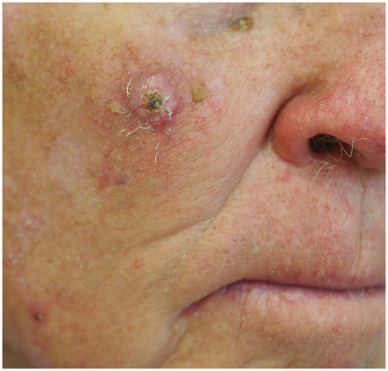
Box 2 – Macroscopic and microscopic histopathological images of cutaneous squamous cell carcinoma (cSCC)
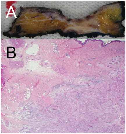
Image (A) represents the macroscopic appearance of a cut surface of skin excised from the cheek. There is a central area of ulceration with an underlying grey‐white firm tumour. Image (B) represents highly infiltrative, poorly differentiated cSCC infiltrating through the dermis and the subcutaneous tissue. Haematoxylin and eosin stains are used, visualised in 100×magnification.
Competing interests
No relevant disclosures.
References
- Kim RH, Armstrong AW. Nonmelanoma skin cancer. Dermatol Clin 2012; 30: 125–139.
- Australian Institute of Health and Welfare. Cancer in Australia: an overview. 2008 [Cancer Series no 42; Cat. No. CAN 32]. https://www.aihw.gov.au/getmedia/98bf9bd8‐4492‐4465‐9175‐7efb886756cb/ca08.pdf.aspx?inline=true (viewed June 2022).
- Madan V, Lear JT, Szeimies RM. Non‐melanoma skin cancer. Lancet 2010; 375: 673–685.
- Didona D, Paolino G, Bottoni U, Cantisani C. Non melanoma skin cancer pathogenesis overview. Biomedicines 2018; 6: 6.
- Brantsch KD, Meisner C, Schönfisch B, et al. Analysis of risk factors determining prognosis of cutaneous squamous‐cell carcinoma: a prospective study. Lancet Oncol 2008; 9: 713–720.
- Hasmat S, Ebrahimi A, Luk PP, et al. Positive survival trend in metastatic head and neck cutaneous squamous cell carcinoma over four decades: multicenter study. Head Neck 2019; 41: 3826–3832.
- Ramsay HM, Reece SM, Fryer AA, et al. Seven‐year prospective study of nonmelanoma skin cancer incidence in UK renal transplant recipients. Transplantation 2007; 84: 437–439.
- Long MD, Herfarth HH, Pipkin CA, et al. Increased risk for non‐melanoma skin cancer in patients with inflammatory bowel disease. Clin Gastroenterol Hepatol 2010; 8: 268–274.
- Newlands C, Currie R, Memon A, et al. Non‐melanoma skin cancer: United Kingdom national multidisciplinary guidelines. J Laryngol Otol 2016; 130: S125‐S132.
- Veness MJ. High‐risk cutaneous squamous cell carcinoma of the head and neck. J Biomed Biotechnol 2007; 2007: 80572.
- Mooney CP, Clark JR, Shannon K, et al. The significance of regional metastasis location in head and neck cutaneous squamous cell carcinoma. Head Neck 2021; 43: 2705–2711.
- Amin MB, Greene FL, Edge SB, et al. The eighth edition AJCC cancer staging manual: continuing to build a bridge from a population‐based to a more “personalized” approach to cancer staging. CA Cancer J Clin 2017; 67: 93–99.
- Stoff B, Salisbury C, Parker D, O'Reilly Zwald F. Dermatopathology of skin cancer in solid organ transplant recipients. Transplant Rev (Orlando) 2010; 24: 172–189.
- Rhee JS, Matthews BA, Neuburg M, et al. Creation of a quality of life instrument for nonmelanoma skin cancer patients. Laryngoscope 2005; 115: 1178–1185.
- Rowe DE, Carroll RJ, Day CL. Prognostic factors for local recurrence, metastasis, and survival rates in squamous cell carcinoma of the skin, ear, and lip. Implications for treatment modality selection. J Am Acad Dermatol 1992; 26: 976–990.
- Thompson AK, Kelley BF, Prokop LJ, et al. Risk factors for cutaneous squamous cell carcinoma recurrence, metastasis, and disease‐specific death: a systematic review and meta‐analysis. JAMA Dermatol 2016; 152: 419–428.
- Ross AS, Whalen FM, Elenitsas R, et al. Diameter of involved nerves predicts outcomes in cutaneous squamous cell carcinoma with perineural invasion: an investigator‐blinded retrospective cohort study. Dermatol Surg 2009; 35: 1859–1866.
- Jennings L, Schmults CD. Management of high‐risk cutaneous squamous cell carcinoma. J Clin Aesthet Dermatol 2010; 3: 39.
- Gupta A, Veness M, De'Ambrosis B, et al. Management of squamous cell and basal cell carcinomas of the head and neck with perineural invasion. Australas J Dermatol 2016; 57: 3–13.
- Warner GC, Gandhi M, Panizza B. Slowly progressive cranial nerve palsies. Med J Aust 2006; 184: 641. https://www.mja.com.au/journal/2006/184/12/slowly‐progressive‐cranial‐nerve‐palsies
- McCord MW, Mendenhall WM, Parsons JT, et al. Skin cancer of the head and neck with clinical perineural invasion. Int J Radiat Oncol Biol Phys 2000; 47: 89–93.
- Mullen JT, Feng L, Xing Y, et al. Invasive squamous cell carcinoma of the skin: defining a high‐risk group. Ann Surg Oncol 2006; 13: 902–909.
- Cherpelis B, Marcusen C, Lang P. Prognostic factors for metastasis in squamous cell carcinoma of the skin. Dermatol Surg 2008; 28: 268–273.
- Thiem DGE, Scharr K, Pabst AM, et al. Facial cutaneous squamous cell carcinoma — microscopic safety margins and their impact on developing local recurrences. J Craniomaxillofac Surg 2020; 48: 49–55.
- Phillips T, Harris B, Moore M, et al. Pathological margins and advanced cutaneous squamous cell carcinoma of the head and neck. J Otolaryngol Head Neck Surg 2019; 48: 55.
Provenance: Not commissioned; externally peer reviewed.
Diagnosing Acute Kava Dermopathy: A Case Report of a Characteristic Cutaneous Eruption
Ali Abid, Nicholas Allen, Abeer Hagelamin, Christopher Henderson, Artiene Tatian
Localised Herpes Simplex Following Midline Laparotomy
Jessica S. Bulluss, Paul Chee, Matthew J. Verheyden
No silver lining with health misinformation: argyria caused by intentional silver consumption
Luke Collins, Logesh Palanikumar, Stephen Bacchi
Lingual Raynaud phenomenon
Michael Taggart, Mina John
Melasma in the male: a less well recognised entity
Tim Aung, Rowland Noakes
Age group‐specific changes in keratinocyte cancer treatment rates in Australia, 2012–2021: a retrospective cohort study based on MBS claims data
Catherine M Olsen, Nirmala Pandeya, Rachel E Neale, David C Whiteman