




Medical education
Lessons from practice
Volume 217 - Issue 7
Acute lymphoblastic leukaemia presenting without significant blood count abnormalities: role of medical imaging in two cases
Authors: Royston Ponraj, Jenna Langfield, Louise Wong, Ian Kerridge and William S Stevenson
Med J Aust 2022; 217 (7): 346-347. || doi: 10.5694/mja2.51712
Published online: 3 October 2022
Published online: 3 October 2022
A 21-year-old man presented to the emergency department with a 2-week history of thoracic back pain
Clinical record
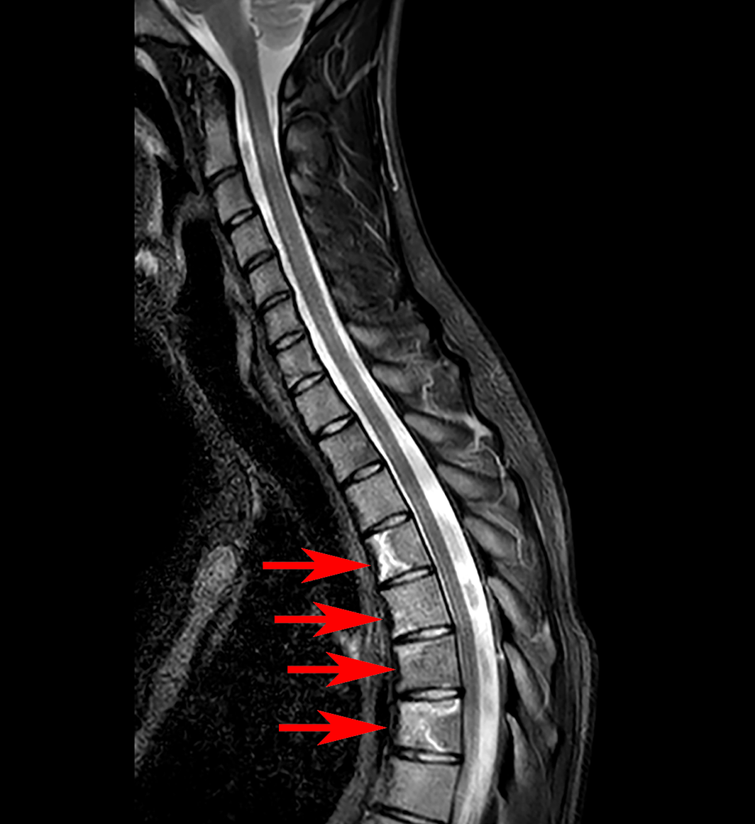
Patient A, a 21‐year‐old man, presented to the emergency department with a 2‐week history of thoracic back pain. This was initially attributed to mechanical injury. He had associated bilateral rib and chest wall pain, as well as low grade fever at 37.8°C. Examination was completely normal. A computed tomography (CT) pulmonary angiogram excluded pulmonary embolus and did not reveal any spinal pathology. His full blood count was unremarkable, with normal white cell count (8.4×10⁹/L; reference interval [RI], 4–10×109/L) and neutrophil count (6.1×10⁹/L; RI, 2–7×10⁹/L), slightly reduced haemoglobin 126g/L (RI for men, 130–170g/L), and normal platelet count (336×109/L; RI, 150–400×109/L). C reactive protein (CRP) was elevated at 157mg/L (RI, <4.9mg/L), with elevated lactate dehydrogenase (336U/L; RI, <250U/L). Given his symptoms, fever and elevated CRP, a magnetic resonance imaging (MRI) scan of the spine was performed. This MRI scan revealed diffuse T1 hypointensity and short tau inversion recovery (STIR) hyperintensity throughout the whole spine, consistent with diffuse marrow infiltration, along with multifocal infarcts (bright on STIR), indicating possible leukaemia (Box 1). The blood film showed occasional myeloid precursors, with no circulating blasts seen; however, bone marrow biopsy confirmed acute B‐cell lymphoblastic leukaemia with 86% blasts.
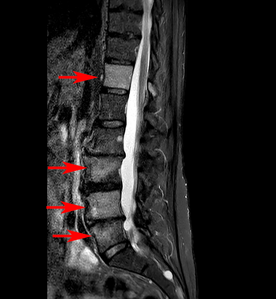
Patient B, a 66‐year‐old woman, presented to the clinic with multifocal musculoskeletal pain over 3 months, including lower back and bilateral lower limb pain with radicular features to the right groin and thigh. There were no red‐flag features such as fever or constitutional symptoms. Nevertheless, given the persistence of symptoms, an MRI scan was performed, showing diffuse T1 hypointense and STIR hyperintense abnormalities within multiple vertebrae, with a broad differential diagnosis of multiple myeloma, lymphoma, or metastatic malignancy (Box 2). A CT scan did not demonstrate corresponding bone lesions. However, bone scintigraphy also demonstrated multifocal abnormalities with widespread mildly increased irregular uptake, as well as focal increased uptake in both proximal humeri, pelvis, bilateral femurs, and vertebral spine. Again, in this patient, the full blood count was relatively normal, with haemoglobin 127g/L (RI for women, 120–150g/L), white cell count 10×109/L, and neutrophil count 7×10⁹/L. There was mild thrombocytosis, platelet count 524×109/L, which is non‐specific. Erythrocyte sedimentation rate and CRP were mildly elevated at 34mm/h (RI, 0–15mm/h) and 15mg/L respectively. Lactate dehydrogenase was normal at 213U/L. She had no lymphadenopathy or hepatosplenomegaly. Blood film demonstrated only mild thrombocytosis, without any blasts seen. However, given the imaging changes, a bone marrow biopsy was performed, confirming Philadelphia‐positive acute B‐cell lymphoblastic leukaemia with 77% blasts.
Discussion
Acute lymphoblastic leukaemia (ALL) is a life‐threatening haematological malignancy affecting both children and adults. Most cases (about 60%) occur in children and adolescents before the age of 20years, with a second smaller peak occurring in individuals aged over 60years, making up about 20% of cases.1,2 ALL often presents with symptoms related to pancytopenia (infections, fatigue, and easy bruising or bleeding) and with non‐specific constitutional symptoms, such as anorexia, weight loss, malaise, night sweats, and fever. Bone pain can be present due to bone marrow infiltration. Lymphadenopathy and hepatosplenomegaly can be present in up to half of patients.3 In addition, most patients with ALL present with evidence of blood count abnormalities, including neutropenia, anaemia and thrombocytopenia. Often, there are circulating lymphoblasts, detectable on blood film examination. These findings lead to further investigation and diagnosis.
Occasionally, however, as in the cases described above, ALL can present without significant cytopenias or detectable peripheral blasts, which may lead to delayed or missed diagnosis.4 For both patients, medical imaging for the investigation of bone pain played a key role in detecting bone marrow pathology, prompting further investigation with bone marrow biopsy and avoiding delay in diagnosis. CT was insensitive in detecting bone marrow infiltration without trabecular destruction. However, both MRI and bone scintigraphy detected the presence of marrow infiltration. MRI also demonstrated multifocal bone infarction. Findings on MRI include diffuse or multifocal loss of marrow T1 signal with corresponding high STIR signal, and with linear enhancement on post‐gadolinium T1 sequences in areas of infarction.5 It is important to note, however, that medical imaging alone cannot provide a specific diagnosis for ALL, and the sensitivity or negative predictive value is not known.
These cases are a reminder that leukaemia and other blood cancers can be a cause of bone pain. Musculoskeletal pain, along with red‐flag symptoms, or bone pain that is persistent and unexplained warrants medical imaging. In these cases, MRI and bone scan should be considered as they are useful in detecting bone marrow infiltration, whereas CT is insensitive. Upon detection of diffuse or multifocal bone marrow abnormality on imaging, further investigation and follow‐up are imperative, and bone marrow biopsy should be considered.5
Lessons from practice
- Acute lymphoblastic leukaemia can occasionally present without significant cytopenias or detectable circulating blasts in adults.
- Magnetic resonance imaging (MRI) and bone scintigraphy are useful tools in detecting bone marrow infiltration and infarction. Computed tomography is relatively insensitive.
- Diffuse or multifocal marrow infiltration on imaging warrants consideration of further investigation with bone marrow biopsy.
- Back pain with red‐flag symptoms always requires investigation. In the absence of red‐flag symptoms, imaging with MRI for persistent unexplained back pain may be appropriate.
Competing interests
No relevant disclosures.
References
- Malard F, Mohty M. Acute lymphoblastic leukaemia. Lancet 2020; 395: 1146‐1162.
- Samra B, Jabbour E, Ravandi F, et al. Evolving therapy of adult acute lymphoblastic leukemia: state‐of‐the‐art treatment and future directions. J Hematol Oncol 2020; 13: 70.
- Redaelli A, Laskin B, Stephens JM, et al. A systematic literature review of the clinical and epidemiological burden of acute lymphoblastic leukaemia (ALL). Eur J Cancer Care (Engl) 2005; 14: 53‐62.
- Kato M, Koh K, Kikuchi A, et al. Case series of pediatric acute leukemia without a peripheral blood abnormality, detected by magnetic resonance imaging. Int J Hematol 2011; 93: 787‐790.
- Navarro S, Matcuk G, Patel D, et al. Musculoskeletal imaging findings of hematologic malignancies. Radiographics 2017; 37: 881‐900.
Provenance: Not commissioned; externally peer reviewed.
Splenic injury in severe cases of the zoonoses Q fever and rickettsial infection: diagnostic challenges
Ashleigh Drury, Philippa Harrison, Aiveen Bannan
Health and economic benefits of improving pre‐hospital identification of stroke in Australian women: a modelling study
Thomas Gadsden, Lei Si, Emily R Atkins, Cheryl Carcel, Xia Wang, Stephen Jan, Mark Woodward, Laura E Downey
Updating the diagnosis and management of elevated serum ferritin levels in the era of routine ferritin testing of blood donors by Australian Red Cross Lifeblood
Gary D Zhang, James Chen, Daniel M Johnstone, Martin B Delatycki, Katie Allen, John K Olynyk
Early cardiovascular collapse after envenoming by snakes in Australia, 2005–2020: an observational study (ASP‐31)
Geoffrey K Isbister, Katherine Z Isoardi, Angela L Chiew, Shane Jenkins, Nicholas A Buckley
Updated recommendations for warfarin reversal in the setting of four‐factor prothrombin complex concentrate
Danielle Robinson, James McFadyen, Eileen Merriman, Chee Wee Tan, Ross Baker, Huyen Tran
Updating the diagnosis and management of iron deficiency in the era of routine ferritin testing of blood donors by Australian Red Cross Lifeblood
Gary D Zhang, Daniel Johnstone, Michael F Leahy, John K Olynyk