




Medical education
Lessons from practice
Volume 216 - Issue 3
Endogenous bacterial endophthalmitis: a case of infective endocarditis following a dental procedure
Authors: Peter ZX Cui, Elaine Chong and Thomas G Campbell
Med J Aust 2022; 216 (3): 124-125. || doi: 10.5694/mja2.51397
Published online: 21 February 2022
Published online: 21 February 2022
A 21-year-old white man presented with a 3-day history of central blurry vision in his
right eye in the context of 3 weeks of right
frontotemporal headaches and earaches, fevers, and drenching night sweats
Clinical record
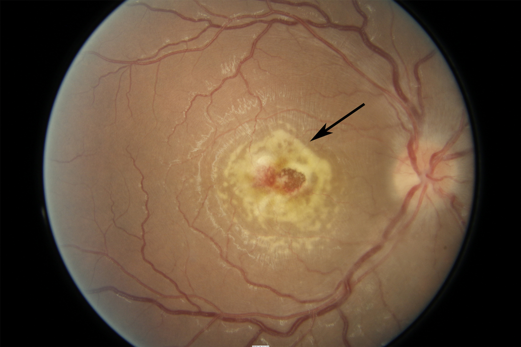
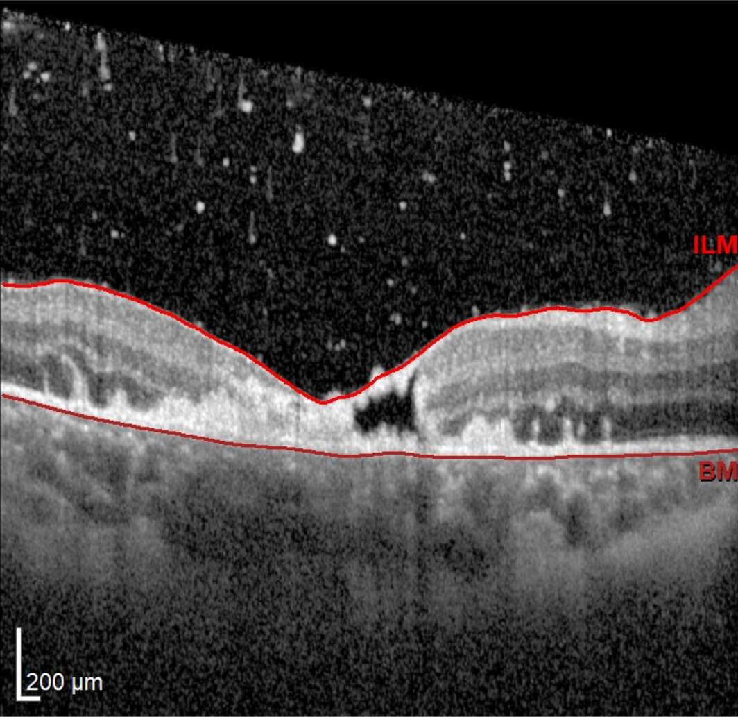
A 21‐year‐old white man presented with a 3‐day history of central blurry vision in his right eye in the context of 3 weeks of right frontotemporal headaches and earaches, fevers, and drenching night sweats. No murmurs or neurological deficits were found. He had no relevant medical or valvular disease history, apart from a dental extraction of his right upper molar 7 weeks prior. He had received two courses of oral antibiotics with minimal improvement (clarithromycin for 7 days, and phenoxymethylpenicillin plus metronidazole for the 3 days before presentation). Visual acuity was hand movements in the right eye and 6/5 in the left eye. There was a right relative afferent pupillary defect. The right eye demonstrated anterior chamber cells and a mild vitritis. Fundoscopy of the right eye revealed a striking area of yellow chorioretinal inflammation at the fovea with retinal haemorrhage, disc swelling, arteriovenous nipping along the superior arcade, tortuous dilated vessels, and cotton wool spots superior to the disc (Box 1). The left eye exhibited mild vitritis and a raised disc without any focal chorioretinal lesions. Optical coherence tomography of the right foveal lesion demonstrated full thickness necrosis of the retina (Box 2).
Inflammatory markers were raised, with a C‐reactive protein level of 144 mg/L (reference interval [RI], < 5 mg/dL) and erythrocyte sedimentation rate of 87 mm/h (RI, 1–12 mm/h). Tests for syphilis, bartonellosis, brucellosis, toxocariasis and toxoplasmosis were negative. Double‐stranded DNA, antineutrophil cytoplasmic antibodies, rheumatoid factor, and angiotensin‐converting enzyme levels were normal. A lumbar puncture was performed and sent for cytology, microscopy, culture and sensitivity, and an infective meningitis and encephalitis screen was unremarkable. Extended blood cultures remained negative in the setting of antibiotic pre‐treatment within 24 hours before collection. A magnetic resonance imaging (MRI) scan of the brain showed multiple areas of restricted diffusion, enhancement, and abnormal T2 signal, suggestive of multiple scattered emboli. An MRI scan of the orbits and globes and a magnetic resonance angiography of the brain were unremarkable.
A transthoracic echocardiogram (TTE) showed an echogenic mass on the mitral valve, with a subsequent transoesophageal echocardiogram demonstrating a 0.8 cm vegetation attached to the P2 scallop, suggestive of mitral valve endocarditis.
Intravenous piperacillin–tazobactam (4 g/500 mg four times a day) was administered for one week, followed by ceftriaxone (2 g daily) and vancomycin (1.5 g daily) for 5 weeks. At the one‐month review, inflammation had settled in both eyes, and his visual acuity was 6/36 in the right eye with retained peripheral vision and 6/4 in the left eye.
Discussion
This case highlights the importance of a thorough medical history of even relatively distant events and a comprehensive work‐up for acute vision loss. Differential diagnoses included infectious (endogenous endophthalmitis or neuroretinitis), inflammatory (neuro‐Behçet or neurosarcoidosis) and infiltrative causes (leukaemia or lymphoma). An urgent TTE was arranged and an anterior chamber tap and vitreous biopsy were strongly recommended, as we suspected an infective process given his constitutional symptoms and history of the dental procedure. The patient refused intraocular procedures.
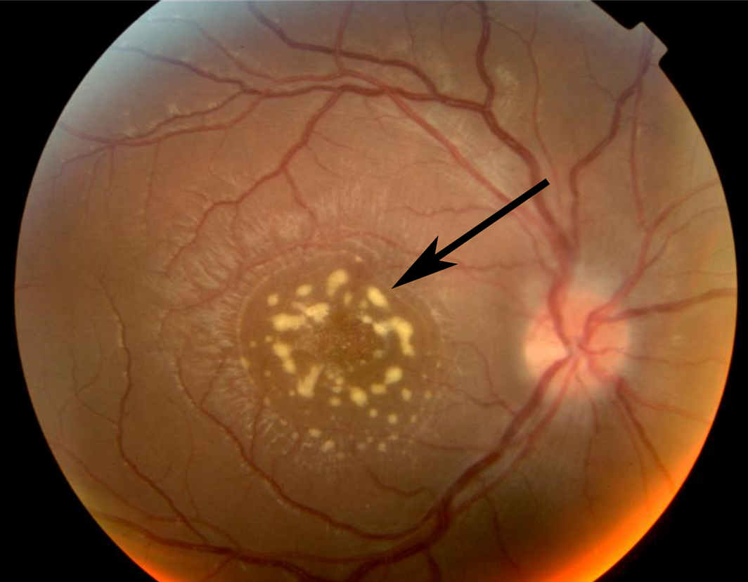
Given that transient bacteraemia has been reported in 15–55% of dental surgery cases,1 it is likely the molar extraction led to infective endocarditis with bacterial colonisation of his mitral valve. Septic emboli from his valvular vegetation then embolised to his brain and eye resulting in the formation of a retinal abscess (Box 3). Direct seeding of the retina with bacteria after dental surgery and routine teeth cleaning have also been reported.1,2,3 However, onset of symptoms through this mechanism is typically within 10 days after the procedure.1,2,3
Endogenous bacterial endophthalmitis (EBE) is an ophthalmic emergency and refers to a sight‐threatening intraocular infection resulting from haematogenous spread of bacteria to internal ocular structures.4 Prognosis of EBE largely depends on timely recognition and treatment,5 but it has an initial misdiagnosis rate of up to 63%.4 Retinal abscess represents a severe manifestation of EBE with poor visual prognosis.1,4 Prompt treatment with systemic antibiotics is paramount and the use of adjuvant intravitreal antibiotics should be considered.1,4,5 Given the patient’s refusal of intraocular procedures, he was treated with systemic antibiotics alone without intravitreal antibiotics or corticosteroids. Response to intravenous treatment alone has been reported indicating that a therapeutic level of antibiotics is achieved inside the eye.4,5
Close monitoring is essential as EBE may be complicated by poor response to systemic and intravitreal antibiotics, retinal tears or detachment, and may require a vitrectomy and/or retinal detachment repair.1,4 EBE from infective endocarditis is a life‐ and sight‐threatening disease and should be considered in all patients with vision loss and a history of invasive procedures, including intravenous drug use.
Lessons from practice
- • Endogenous bacterial endophthalmitis from infective endocarditis secondary to a dental procedure is a medical emergency; it is often misdiagnosed, and delayed recognition and treatment can lead to blindness and even death.
- • A thorough search for the source of endogenous bacterial endophthalmitis including brain imaging and echocardiography is mandatory.
- • A history of invasive procedures such as dental work and intravenous drug use could be the initial seeding event.
- • Management includes intravitreal and systemic antibiotics with close monitoring of response to treatment to guide the need for further surgical intervention.
Box 1 – Colour fundus photograph of the right eye showing a striking area of chorioretinal inflammation with retinal haemorrhage at the fovea (arrow)
Competing interests
No relevant disclosures.
Acknowledgements
Thomas Campbell is supported by the Hector Maclean Scholarship and the Centre for Eye Research Australia. The Hector Maclean Scholarship is awarded to a research student of the University of Melbourne in the department of Ophthalmology.
References
- Tsai TH, Yang CH, Yang CM, Chen MS. Endogenous endophthalmitis with subretinal abscess after dental procedures. J Formos Med Assoc 2005; 104: 47–49.
- Subramanian ML, Topping TM. Endogenous endophthalmitis after routine dental cleaning. Arch Ophthalmol 2003; 121: 576–577.
- Ziakas NG, Tzetzi D, Boboridis K, Georgiadis NS. Endogenous group G Streptococcus endophthalmitis following a dental procedure. Eur J Ophthalmol 2004; 14: 59–60.
- Fortun J, Modi YS, Bessette A, et al. Clinical features and management of subretinal abscesses secondary to methicillin‐resistant staphylococcus aureus endogenous endophthalmitis. Ophthalmic Surg Lasers Imaging Retina 2017; 48: 134–142.
- Prajapati R, Newton P, Ahmad S, Kelly S. Acute subretinal abscess in Staphylococcus aureus septicaemia with endophthalmitis showcased by multimodal retinal imaging and with 2‐year follow‐up. BMJ Case Rep 2018; 11: e227288.
Provenance: Not commissioned; externally peer reviewed.
A Diagnostic Headache
Thomas Glynn, Michelle T. Leech, Stacy K. Goergen, Emily Lin, Nadeem Toodayan, Ralph Junckerstorff
Neisseria meningitidis: an uncommon cause of conjunctivitis
Chloe Story, Tina Marinelli, Simeon Crawford, Amrita Ronnachit
Dismantling barriers to research and clinical care for individuals with a vision impairment
Eden G Robertson, Kate Hetherington, Meredith Prain, Julia Hall, Leighton Boyd AM, Rosemary Boyd OAM, Emily Shepard, Hollie Feller, Sally Karandrews, Fleur O'Hare, Kanae Yamamoto, Matthew P Simunovic, Robyn V Jamieson, Alan Ma, Lauren Ayton AM, Anai Gonzalez‐Cordero
The New South Wales Pharmacy Trial for herpes zoster: on the nose?
Christian P Pappas, Timothy R Holmes, Minas T Coroneo
Skin fragility disorder misdiagnosed as child abuse: a cautionary tale
Heather G Mack, Fred K Chen, John R Grigg
Health economic aspects of inherited retinal diseases: looking for cost‐effective treatments
Benjamin Kamien, Rachael Heath Jeffery, Fred K Chen