




Volume 215 - Issue 9
Eczema coxsackium
Authors: Hsing‐Jou Su and Chun‐Bing Chen
Med J Aust 2021; 215 (9): 403-403. || doi: 10.5694/mja2.51297
Published online: 1 November 2021
Published online: 1 November 2021
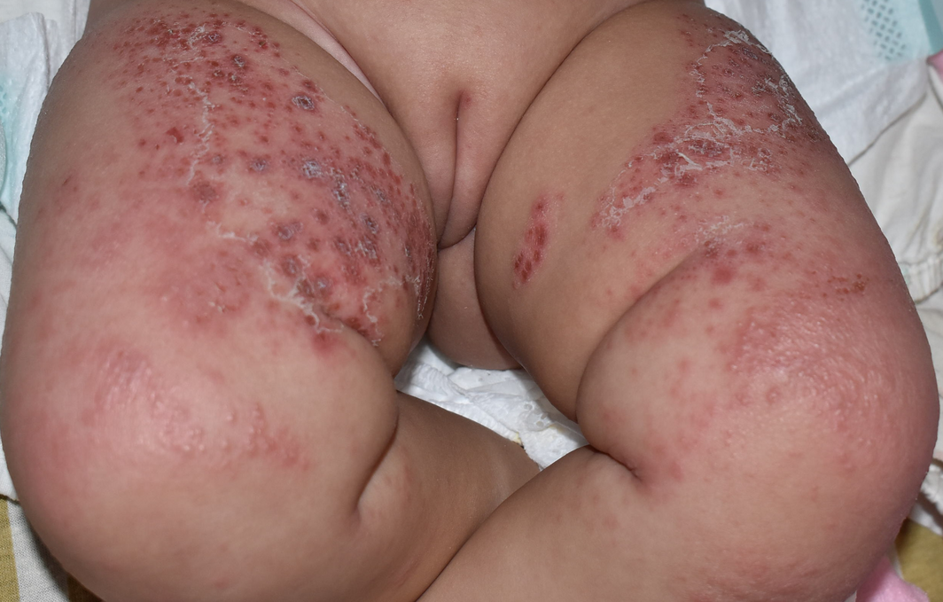
A 6-month-old female infant with atopic dermatitis presented with multiple erythematous papules and vesicles with crusts on the perioral area, trunk and extremities
A 6‐month‐old female infant with atopic dermatitis presented with multiple erythematous papules and vesicles with crusts on the perioral area, trunk and extremities (Figure). The distribution of the new skin eruptions was almost on the same areas of atopic dermatitis. She was afebrile with mildly decreased appetite and activity. The polymerase chain reaction (PCR) tests for throat and vesicle fluid were positive for coxsackievirus A6 but negative for herpes simplex virus, confirming diagnosis of eczema coxsackium. After topical corticosteroid, topical antibiotics, and adequate hydration, the skin lesions resolved within 2 weeks. Eczema coxsackium is commonly caused by coxsackievirus A6 in patients with atopic dermatitis.1 Differential diagnoses include varicella zoster infection, eczema herpeticum, and bullous impetigo. PCR testing for enterovirus detection of the vesicle fluid helps to differentiate eczema coxsackium from eczema herpeticum. Early diagnosis can prevent inappropriate use of antibiotics or acyclovir.2
Competing interests
No relevant disclosures.
References
- Horsten HH, Kemp M, Fischer TK, et al. Atypical hand, foot, and mouth disease caused by coxsackievirus A6 in Denmark: a diagnostic mimicker. Acta Derm Venereol 2018; 98: 350–354.
- Hoffmann AJ, Latrous M, Lam JM. Atypical hand‐foot‐and‐mouth disease. CMAJ 2020; 192: E69.
Reorienting Allied Health Into Community-Based Care for People Experiencing Trauma and Social Disadvantage
Simon Rosenbaum, Grace McKeon, Gulsah Kurt, Oscar Lederman, Kemi Wright, Sabuj Kanti Mistry, Jackie E. Curtis, Philip B. Ward, Zachary Steel, Hamish Fibbins, Rachel Morell, Melissa C. Eaton, Andrew Watkins, Ben Harris-Roxas, Brendan Goodger, Eleanor Beck, Megan Teychenne, Joseph Firth, Davy Vancampfort, David Burns, Russell Roberts, Tristan Favaloro, Danielle Weber, Rosanna Barbero, Vasili Maroulis, Melissa Holmes, Stefan Mackenzie, Chiara Mastrogiovanni, Afsana Anwar, Uzma Choudhry, Catherine Sherrington, Jane Currie, Thomas Gadsden, Scott Teasdale
Psychosocial Hazards for Healthcare Workers: Supporting the Second Victim Also Helps the Primary Victims
Sarah Michael
Shifting focus to adolescent wellbeing and inclusive participation in the digital age
Allyson R Todd, Elena Wang, Stephanie R Partridge
Shifting focus to adolescent wellbeing and inclusive participation in the digital age
Jodie Bailie, Helen Dickinson, Briony Lipton, Marissa Shields
The CURE Asthma roadmap
Gary P Anderson, Anthony Flynn, Phil G Bardin, John D Blakey, Shyamali C Dharmage, Paul Foster, Peter G Gibson, Adam Jaffe, Alan James, Christine R Jenkins, Sundram Sivamalai, Peter D Sly, Guy B Marks, Vanessa M McDonald, Judy Wetttenhall