




Volume 215 - Issue 8
Evaluation and management of rectal bleeding in pregnancy
Authors: Ralley Prentice, Aysha Al‐Ani, Tiffany Cherry, Julia Dixon‐Douglas, Jade Eccles‐Smith, Julia Matheson, Jeanne Tie, Iniyaval Thevathasan, Jacob J McCormick and Britt Christensen
Med J Aust 2021; 215 (8): 377-382. || doi: 10.5694/mja2.51267
Published online: 4 October 2021
Published online: 4 October 2021
Rectal bleeding is common in pregnancy, with a reported prevalence of 10-43%
Summary
- Rectal bleeding occurs in about 40% of pregnant women, and is predominantly attributed to benign perianal pathology (haemorrhoids or anal fissures).
- More sinister causes of rectal bleeding may be heralded by key red flag clinical and biochemical features. These features should be evaluated in all women with rectal bleeding. Imaging investigations or flexible sigmoidoscopy may be warranted. The latter can be performed safely by experienced operators in pregnant women.
- Women with evidence of haemodynamic compromise, elevated inflammatory markers, significant anaemia, signs of intestinal obstruction or compromise to the fetus should be evaluated urgently. Providers must be mindful of the changes in normal ranges for common haematological and biochemical parameters in pregnancy compared with the non‐pregnant state.
- Faecal calprotectin is an established tool for identification of intestinal inflammation and is valid in pregnancy. An elevated faecal calprotectin level (≥ 50 µg/g) signifies a need for further diagnostic evaluation.
- Inflammatory bowel disease may present initially, or with worsening disease activity, in pregnancy. Expedient diagnosis with the use of faecal calprotectin, sigmoidoscopy with or without intestinal ultrasound, exclusion of alternative or compounding infective aetiologies, and institution of appropriate therapy are critical. Medical therapies for management of inflammatory bowel disease can be safely instituted in pregnancy.
- Colorectal cancer incidence is increasing in younger age groups, but fortunately remains rare. When diagnosed in pregnancy, colorectal cancer can be successfully and safely managed with a collaborative multidisciplinary team approach. Early diagnosis is key to optimising outcomes.
Rectal bleeding is common in pregnancy, with a reported prevalence of 10–43%.1,2 In the vast majority of cases, it is due to benign perianal disease, specifically haemorrhoids and anal fissures.3,4 However, it is imperative to consider and exclude alternative causes for rectal bleeding, including inflammatory and neoplastic conditions.
This narrative review was collated by a multidisciplinary team of gastroenterologists, oncologists, colorectal and obstetric/maternal fetal medicine specialists from the Royal Melbourne Hospital, Peter MacCallum Cancer Centre and the Royal Women’s Hospital, Melbourne. The literature was reviewed and summarised (RP, AA, JM, TC and JES) utilising PubMed, EMBASE and MEDLINE. The respective sections were synthesised (RP, AA and BC) for final review and approval by all contributing authors. This review aims to guide clinicians in evaluating rectal bleeding in pregnancy. Management strategies for specific aetiologies are also discussed.
When to investigate
Perianal conditions become increasingly prevalent as pregnancy progresses, largely driven by constipation and progressively increasing abdominal pressure.2,3 Haemorrhoids arise from a dilated arteriovenous channel in the anal submucosa, and about 40% of cases will present with painless rectal bleeding associated with a bowel action.1 Perianal fissures result from a tear in the anal mucosa, typically causing pain exacerbated by defecation.3,4 Differentiating these conditions from a more sinister cause of rectal bleeding can be readily achieved using a combination of clinical and diagnostic tests, including assessing for red flag clinical features (Box 1).
How to investigate
Investigations must be tailored to the individual scenario and normal ranges in pregnancy, but may include:
- full blood examination;
- iron studies;
- albumin;
- C‐reactive protein;
- stool culture;
- faecal calprotectin;
- endoscopy; and
- intestinal ultrasound, computed tomography (CT) and magnetic resonance imaging (MRI).
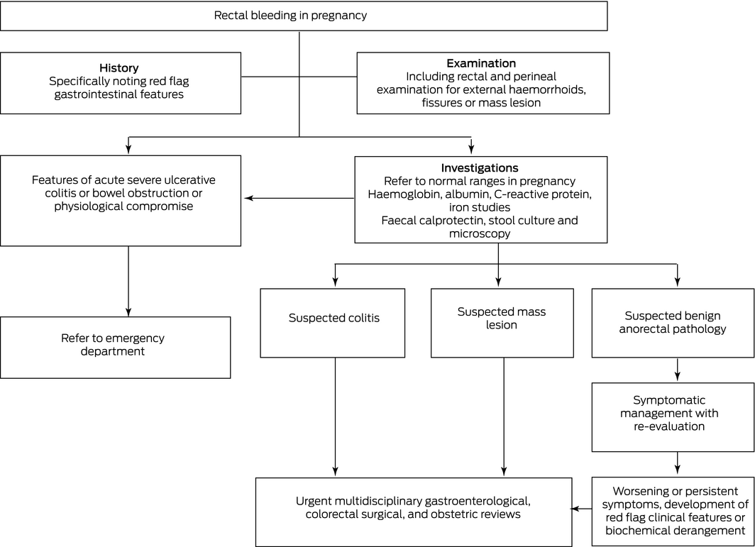
A flow chart summarising investigation and management of rectal bleeding in pregnancy is provided in Box 2.
Routine blood tests
Pregnancy results in many physiological changes that influence the interpretation of common biochemical and haematological investigations (Supporting Information). Pregnancy is associated with increased iron demands and relative dilutional anaemia.13 Increased renal plasma flow and glomerular filtration rate in the first trimester results in a 25% fall in serum creatinine, urea and urate from pre‐pregnancy values.14 Serum creatinine then rises in the second to third trimester.14 Additionally, pregnancy is associated with lower levels of albumin, alanine aminotransferase, asparate aminotransferase, γ ‐glutamyl transferase and bilirubin, while C‐reactive protein levels are unchanged but may increase following instrumental vaginal or caesarean delivery.14,15 Erythrocyte sedimentation rate varies substantially according to the presence of anaemia and gestational age.16
Faecal calprotectin
Faecal calprotectin, a granulocyte neutrophil‐predominant cytosolic protein, is an established marker of intestinal inflammation, used broadly in the diagnosis and monitoring of inflammatory bowel disease and as a discriminator between functional and inflammation‐driven gastrointestinal symptoms.17 Faecal calprotectin levels do not change with pregnancy, so can be used reliably in this setting as a diagnostic and monitoring tool.18,19 In women aged between 16–50 years presenting with gastrointestinal symptoms, a faecal calprotectin level of ≥ 50 µg/g can differentiate between inflammatory bowel disease and functional gastrointestinal disorders with a negative predictive value of 0.99.20
Importantly, the use of faecal calprotectin to avoid endoscopy can only be considered in those without alarm symptoms, which include rectal bleeding, bloody diarrhoea, nocturnal diarrhoea or faecal urgency, weight loss and anaemia.20 Thus, a normal faecal calprotectin level in a pregnant patient with rectal bleeding is not adequately reassuring to negate endoscopic evaluation but is useful when considering inflammatory bowel disease as a differential.
Additionally, average faecal calprotectin levels are higher in patients with colorectal cancer than comparable controls. The negative and positive predictive values of faecal calprotectin levels < 50 µg/g in a cohort of patients referred for endoscopic evaluation with suspected colorectal cancer were 98.6 (95% confidence interval [CI], 95.7–99.6) and 8.7 (95% CI, 6.3–11.9), respectively.21 Notably, this cohort included relatively high risk patients, being those with alarm symptoms qualifying the need for a colonoscopy within 2 weeks.21 Endoscopic evaluation should be considered in any pregnant patient with rectal bleeding and an elevated faecal calprotectin level, given the likelihood of an underlying organic cause warranting therapy. There is no role for faecal occult blood testing; by definition, this is a screening rather than diagnostic test and is of no additive value in the setting of reported overt rectal bleeding.
Endoscopy
In the setting of rectal bleeding, an unsedated flexible sigmoidoscopy performed by an experienced operator is prudent. Medical interventions during pregnancy are subject to intense scrutiny owing to the risk of maternal and fetal harm. Endoscopy in pregnancy has traditionally been avoided because of concerns about the impact of medications or bowel preparation; endoscopic intubation causing fetal trauma; and potential resultant maternal hypoxia, hypotension or cardiac events compromising the fetus.22 Although randomised controlled trial evidence is lacking in this area given the ethical and practical hurdles, there is mounting evidence to support the safety and utility of endoscopy in pregnancy, particularly when performed without sedation. A systematic review of pregnant women undergoing lower gastrointestinal endoscopy for any indication found a low risk to both mother and child in all three trimesters of pregnancy.23 A prospective study of 42 pregnant women with inflammatory bowel disease undergoing lower gastrointestinal endoscopy found no increase in adverse outcomes for the mother or newborn in any trimester compared with controls, although patients were only followed up until delivery.24 Ultimately, where there is an indication for endoscopy in a pregnant woman, it should be performed. Ideally, endoscopy should be deferred to the second trimester, but that will be dictated by clinical urgency.25 Moreover, when clearly indicated, lower gastrointestinal endoscopy may improve fetal outcomes through prompting changes in management.22 Experienced anaesthetic and obstetric support is essential in the context of patient instability and in the first and third trimesters.25
Imaging evaluation
Imaging evaluation of rectal bleeding is predominantly useful in suspected inflammatory bowel disease or staging of colorectal carcinoma, although with modest value for the former. Intestinal ultrasound can be accurately used by experienced operators in the first and second trimesters of pregnancy.18 Intestinal ultrasound can assess the colonic wall segments and small bowel for inflammatory changes; however, its utility in adequately assessing isolated rectal inflammation in pregnancy is limited.18
Abdominal CT scans are preferably avoided in pregnancy, given the radiation exposure for the fetus. This imparts an extremely low, but not negligible, risk of congenital malformations or childhood cancers.26 MRI can be performed for assessment of colonic inflammation or mass lesions.26 The use of non‐enhanced MRI is not associated with an attributable risk of stillbirth, neonatal death, congenital anomaly, neoplasm or hearing loss.27 Ongoing theoretical concerns regarding teratogenicity of gadolinium contrast in pregnancy preclude its use unless the benefit of contrast enhanced imaging is seen to clearly outweigh the risks.27
When to investigate urgently
Rectal bleeding should be investigated and managed urgently when it occurs in conjunction with physiological compromise or threat to ongoing viability of the pregnancy. Women with anaemia or clinically evident hypovolemia should be urgently assessed and managed in a multidisciplinary hospital setting. Women with large volume bleeding or with constitutional symptoms such as weight loss or failure to meet gestational weight gain recommendations should also be referred for inpatient management. Additionally, there are rare cases of rectal bleeding in pregnancy, such as abdominal ectopic pregnancy with invasion of the placenta into the bowel wall.12 The following causes of rectal bleeding require rapid assessment.
Acute severe ulcerative colitis
Acute severe ulcerative colitis is defined by one or more clinical and biochemical signs of toxicity (defined by tachycardia, fever, elevated C‐reactive protein level or erythrocyte sedimentation rate, or fever) with at least six bloody bowel actions daily.28 Patients may or may not have a preceding diagnosis of ulcerative colitis, with first presentation of acute severe ulcerative colitis in pregnancy being well described.29 Despite comparable responses to first line and salvage medical therapies, acute severe ulcerative colitis in pregnancy carries high rates of preterm delivery and low birth weights.29,30 Early evaluation and aggressive intervention is therefore warranted and is effective.29,30
Sexually transmitted proctitis
Sexually transmitted proctitis can present similarly to inflammatory bowel disease‐related proctocolitis, with fevers, tenesmus, urgency, and rectal bleeding or discharge.8 Taking a thorough sexual history, including of unprotected receptive anal intercourse, is prudent. Relevant investigations include rectal swabs for bacterial culture and herpes simplex virus types 1 and 2, Chlamydia trachomatis and Neisseria gonorrhoea polymerase chain reaction, and syphilis serology.8 Vaginal swabs for these organisms, as well as for oncogenic human papilloma virus types, should be carried out at the same time. Repeat serology testing for human immunodeficiency virus and hepatitis B and C in the second and third trimesters may potentially provide the opportunity to prevent mother–child transmission.31
Colorectal cancer
Diagnosis of colorectal cancer in pregnancy remains rare, with a calculated pool risk of 0.002%.32 Early symptoms of colorectal cancer such as bloating, nausea, vomiting and rectal bleeding can be difficult to differentiate from common pregnancy‐related complications. Rates of colorectal cancer are increasing in younger age groups, particularly rectal cancer in those aged 30–39 years.33 A review including 119 patients diagnosed with colorectal cancer during pregnancy found high rates of rectal primary cancer (44%), with bleeding present in 47%.36 Patients are commonly diagnosed with advanced disease, likely due in part to delayed evaluation in the setting of viable pregnancy.32 Expediently considering colorectal cancer as a differential in a pregnant woman with rectal bleeding may enable diagnosis at an earlier stage, with resultant decreased morbidity and mortality.32,34 Once diagnosed, colorectal cancer staging can occur with a combination of MRI and targeted liver ultrasound, rather than the traditional CT of chest, abdomen and pelvis. Carcinogenic embryonic antigen levels may be slightly raised in pregnancy, but levels before resection can still provide a baseline for ongoing cancer surveillance.34
Management of common and serious causes
Haemorrhoids and anal fissures
Haemorrhoid management in pregnancy is typically non‐operative, with a focus on avoiding constipation through dietary fibre supplementation, stool softeners, adequate fluid intake, and correct toileting position to minimise straining.2,35 Further symptomatic relief may be obtained using warm sitz baths and topical anaesthetic agents commonly used in non‐pregnant patients; however, specific data regarding safety and efficacy in pregnancy are not available.2 Similarly, anal fissure management should focus on prevention and symptom relief.36 Glyceryl trinitrate and calcium channel blocker ointments, and intersphincteric botulinum toxin injections should be avoided due to inadequate safety data in pregnancy. Surgical intervention is usually contraindicated.37
Inflammatory bowel disease
It is well established that uncontrolled inflammatory bowel disease at conception and during pregnancy is a risk factor for adverse pregnancy outcomes, including preterm birth, low birth weight, and newborns who are small for gestational age.30,38 Current inflammatory bowel disease guidelines highlight the importance of optimising disease control to avoid pregnancy and delivery complications.30,38 Acute inflammatory bowel disease flares should be treated expediently during pregnancy.25 5‐Aminosalicylic acid medications (except those formulated with dibutylphthalate coating), corticosteroids, and anti‐tumour necrosis factor drugs can all be safely instituted in pregnancy, while data are emerging to support the use of more recently introduced biologics.30,38,39 Surgery may be warranted during pregnancy in the setting of symptomatic partial or complete bowel obstruction, medically refractory acute severe ulcerative colitis, overt intestinal perforation, or toxic megacolon.30 This should ideally be performed in specialist centres.
Colorectal cancer
Surgical management
The diagnosis of colorectal cancer during pregnancy is challenging for both the patient and the treating clinicians. Timely management decisions involving an experienced multidisciplinary team are critical. Surgical treatment is influenced by the gestational age, the patient’s preference for termination or completion of pregnancy, plans for future pregnancies, tumour stage and location, and elective versus emergency presentation. Oncological and survival outcomes are equivalent to those for non‐pregnant patients, provided that diagnosis and subsequent management are not significantly delayed.32
Early‐stage colonic cancers diagnosed before 20 weeks’ gestation can be treated with surgical resection and primary anastomosis. Although not specific to colorectal surgery, a recent systematic review suggests that laparoscopy may be safer than an open approach in pregnancy; however, there are conflicting data on rates of fetal loss, particularly in the first trimester.40 Patients need to be made aware of the small risk of preterm labour or fetal death in utero, with anaesthetic risk considered highest in the first trimester. To mitigate anaesthetic risks, surgery may be deferred to the second trimester in those diagnosed with a colonic cancer in the first trimester.
For colon cancers diagnosed after 20 weeks’ gestation, it may be reasonable to delay surgical resection until after delivery. Induction of labour or planned caesarean delivery once fetal viability has been reached may reduce further delays to treatment.32
There are separate considerations when evaluating a patient with rectal cancer. Staging with MRI is important in guiding management. In early‐stage disease, primary resection is still recommended, despite the gravid uterus likely requiring retraction to enable visualisation of the pelvis. There is very limited research into the safety of restoring intestinal continuity versus forming a diverting stoma, but the risk of an anastomotic leak to the both the patient and their pregnancy must be considered.
For patients who present with an emergency complication of colorectal cancer such as obstruction or perforation, both the presenting issue and the required intervention will impart risk to the pregnancy. A diverting stoma, particularly with obstructing rectal cancers, will provide more gestational time while definitive intervention is planned.32
In advanced metastatic colorectal cancer with a poor prognosis, palliative measures such as stoma formation or colonic stenting may prolong maternal survival and the period of fetal gestation.41
Radiotherapy
Pre‐operative (neoadjuvant) radiotherapy aims to downstage locally advanced rectal cancer.42 In addition to allowing for a less extensive surgical resection, neoadjuvant radiotherapy decreases the risk of local recurrence for cancers less than 12 cm from the anal verge with high risk prognostic features, including extramural vascular invasion and MRI‐predicted circumferential resection margin ≤ 1 mm.42 Termination of pregnancy is required before pelvic radiotherapy for rectal cancer, owing to the significant risk of radiation‐induced fetal mortality or morbidity.34,40,43 However, there are viable options for delaying radiotherapy until post partum if the patient chooses to continue their pregnancy. A diverting stoma to prevent obstruction can be formed, with neoadjuvant radiotherapy and primary surgical resection undertaken following delivery.40,43 Alternatively, primary surgical resection can be performed during pregnancy, with radiotherapy deferred until post partum. Post‐operative radiotherapy does not compromise overall survival compared with preoperative radiotherapy, but is associated with an increased risk of anorectal dysfunction caused by iatrogenic sphincter damage.45 Because of the considerable risk of infertility following pelvic radiotherapy, ovarian preservation must also be considered and can be facilitated at the time of surgery.46
Chemotherapy
Due to the paucity of data regarding systemic therapies in pregnancy, indications and recommendations for treatment generally follow standard practice for non‐pregnant patients. However, teratogenicity of different anti‐cancer treatments varies and should be considered in addition to stage of cancer, burden of disease and expected benefit to the patient.
The majority of traditional chemotherapy agents have a molecular weight that is able to cross the placenta.47 Teratogenicity of all agents is highest in the first trimester, with rates of major congenital malformation of up to 20–30% compared with 8% and 6% in second and third trimesters, respectively.47 This compares with an incidence of congenital abnormalities of 3% of births in Australia in 2002–2003, with a reported incidence rate varying between 2% and 6% in international registry data.48,49 In a systematic review of patients with colorectal cancer diagnosed in pregnancy, 9.8% received chemotherapy during pregnancy. Of these pregnancies, 72% resulted in live births. Although there was one case of hypothyroidism, there were no cases of permanent disability.47
Fluoropyrimidines with or without oxaliplatin (and/or irinotecan in the metastatic setting) form the backbone of chemotherapy in colorectal cancer. By far the most experience in pregnancy exists for fluorouracil. Data from over 50 cases of fluorouracil use in pregnancy report a low rate of major congenital malformations of 1.2%.47,50 There are minimal data for the safety of capecitabine (one reported case); while oxaliplatin has been used in combination with fluorouracil in five human births, resulting in only one case of hypothyroidism.47,50 This exceeds experience with irinotecan, which is limited to two reported cases, with one of these cases complicated by intrauterine growth restriction.47 Other non‐chemotherapy systemic treatments, including anti‐angiogenic bevacizumab and epidermal growth factor receptor monoclonal antibodies, have no human data, although animal data show embryolethal, teratogenic and abortifacient potential.47
Pharmacokinetic changes associated with pregnancy may affect drug exposure, although the extent to which this affects efficacy or safety is unknown and no recommendations can be made regarding altered dosing.47 Pharmacodynamic effects of chemotherapy on the mother should also be considered, with peri partum myelosuppression raising concern for infection and bleeding risks. It is recommended that chemotherapy be administered no later than 33–35 weeks of gestation, or no later than 3 weeks before estimated delivery date.47,50
Chemotherapy should be offered to pregnant women where it would otherwise be clinically indicated, but should be avoided in the first trimester. Transparent discussion regarding the unknowns outlined is necessary. Fluorouracil either alone or in combination with oxaliplatin is the preferred regimen for the treatment of colorectal cancer in pregnancy, with some human data to support its safety.47,50
Conclusion
Although common, rectal bleeding in pregnancy may herald an underlying sinister cause. All cases warrant investigation. Associated symptoms, relevant personal and family history, and basic investigation results must be evaluated, and advanced interventions should be promptly facilitated. Despite unique challenges, consequential causes of rectal bleeding can be effectively managed in pregnancy when identified and referred for specialist evaluation in a timely fashion.
Box 1 – Red flag clinical features in the evaluation of rectal bleeding5,6
|
Clinical feature |
Differential diagnosis |
||||||||||||||
|
|
|||||||||||||||
|
Blood mixed in with bowel motions, with or without mucus |
Inflammatory bowel disease7 |
||||||||||||||
|
Family history of colorectal cancer (particularly at a young age), known polyposis syndrome or personal history of advanced colonic polyps |
Colorectal cancer or advanced neoplasia9 |
||||||||||||||
|
Abdominal pain, vomiting, abdominal distension |
Bowel obstruction with colonic mass10 |
||||||||||||||
|
Vaginal bleeding |
Pregnancy‐related complication: placental percreta;11 chronic ectopic pregnancy12 |
||||||||||||||
|
|
|||||||||||||||
|
|
|||||||||||||||
Competing interests
Britt Christensen has received speaking fees from Abbvie, Jannsen, Pfizer, Takeda and Ferring Pharmaceuticals, and research grants from Janssen and Ferring Pharmaceuticals, and has served on the advisory boards of Gilead and Novartis.
References
- Riss S, Weiser FA, Schwameis K, et al. The prevalence of hemorrhoids in adults. Int J Colorectal Dis 2012; 27: 215–220.
- Staroselsky A, Nava‐Ocampo AA, Vohra S, et al. Hemorrhoids in pregnancy. Can Fam Physician 2008; 54: 189–190.
- Poskus T, Buzinskienė D, Drasutiene G, et al. Haemorrhoids and anal fissures during pregnancy and after childbirth: a prospective cohort study. BJOG 2014; 121: 1666–1671.
- Ferdinande K, Dorreman Y, Roelens K, et al. Anorectal symptoms during pregnancy and postpartum: a prospective cohort study. Colorectal Dis 2018; 20: 1109–1116.
- Poston GJ, Tait D, O’Connell S, et al. Diagnosis and management of colorectal cancer: summary of NICE guidance. BMJ 2011; 343: d6751.
- Story L, Rafique S, Samadi N, et al. Lower gastrointestinal bleeding in pregnancy: Differential diagnosis, assessment and management. Obstet Med 2020; https://doi.org/10.1177/1753495X20948300 [online ahead of print].
- Koslowsky B, Grisaru‐Granovsky S, Livovsky DM, et al. Pregnancy‐onset inflammatory bowel disease: a subtle diagnosis. Inflamm Bowel Dis 2018; 24: 1826–1832.
- Assi R, Hashim PW, Reddy VB, et al. Sexually transmitted infections of the anus and rectum. World J Gastroenterol 2014; 20: 15262–15268.
- Gausman V, Dornblaser D, Anand S, et al. Risk factors associated with early‐onset colorectal cancer. Clin Gastroenterol Hepatol 2020; 18: 2752–2759.e2.
- Zachariah SK, Fenn MG. Acute intestinal obstruction complicating pregnancy: diagnosis and surgical management. BMJ Case Rep 2014; 2014: bcr2013203235.
- Alshammary E, Elmuzaini MF. Successful conservative treatment of multiorgan infiltrating placenta percreta by uterine embolization and followup with serial magnetic resonance. A case report. Int J Surg Case Rep 2019; 58: 216–219.
- Saravanane C, Robinson Smile S, Chandra SS, et al. Rectal bleeding: a rare complication of abdominal pregnancy. Aust N Z J Obstet Gynaecol 1997; 37: 124–125.
- World Health Organization. Haemoglobin concentrations for the diagnosis of anaemia and assessment of severity. Geneva: WHO, 2011. https://apps.who.int/iris/bitstream/handle/10665/85839/WHO_NMH_NHD_MNM_11.1_eng.pdf?sequence=22&isAllowed=y (viewed June 2020).
- Larsson A, Palm M, Hansson LO, et al. Reference values for clinical chemistry tests during normal pregnancy. BJOG 2008; 115: 874–881.
- Romem Y, Artal R. C‐reactive protein in pregnancy and in the postpartum period. Am J Obstet Gynecol 1985; 151: 380–383.
- van den Broe NR, Letsky EA. Pregnancy and the erythrocyte sedimentation rate. BJOG 2001; 108: 1164–1167.
- Walsham NE, Sherwood RA. Fecal calprotectin in inflammatory bowel disease. Clin Exp Gastroenterol 2016; 9: 21–29.
- Flanagan E, Wright EK, Begun J, et al. Monitoring inflammatory bowel disease in pregnancy using gastrointestinal ultrasonography. J Crohns Colitis 2020; 14: 1405–1412.
- Julsgaard M, Hvas CL, Gearry RB, et al. Fecal calprotectin is not affected by pregnancy: clinical implications for the management of pregnant patients with inflammatory bowel disease. Inflamm Bowel Dis 2017; 23: 1240–1246.
- Kennedy NA, Clark A, Walkden A, et al. Clinical utility and diagnostic accuracy of faecal calprotectin for IBD at first presentation to gastroenterology services in adults ged 16–50 Years. J Crohns Colitis 2015; 9: 41–49.
- Turvill J, Aghahoseini A, Sivarajasingham N, et al. Faecal calprotectin in patients with suspected colorectal cancer: a diagnostic accuracy study. Br J Gen Pract 2016; 66: e499.
- Cappell MS, Fox SR, Gorrepati N. Safety and efficacy of colonoscopy during pregnancy: an analysis of pregnancy outcome in 20 patients. J Reprod Med 2010; 55: 115–123.
- De Lima A, Galjart B, Wisse PHA, et al. Does lower gastrointestinal endoscopy during pregnancy pose a risk for mother and child? – a systematic review. BMC Gastroenterol 2015; 15: 15.
- De Lima A, Zelinkova Z, van der Woude CJ. A prospective study of the safety of lower gastrointestinal endoscopy during pregnancy in patients with inflammatory bowel disease. J Crohns Colitis 2015; 9: 519–524.
- Shergill AK, Ben‐Menachem T, Chandrasekhara V, et al. Guidelines for endoscopy in pregnant and lactating women. Gastrointest Endosc 2012; 76: 18–24.
- American College of Radiology. ACR–SPR practice parameter for imaging pregnant or potentially pregnant adolescents and women with ionizing radiation. 2018. https://www.acr.org/‐/media/acr/files/practice‐parameters/pregnant‐pts.pdf (viewed June 2020).
- Ray JG, Vermeulen MJ, Bharatha A, et al. Association between MRI exposure during pregnancy and fetal and childhood outcomes. JAMA 2016; 316: 952–961.
- Truelove SC, Witts LJ. Cortisone in ulcerative colitis; final report on a therapeutic trial. Br Med J 1955; 2: 1041–1048.
- Ollech JE, Avni‐Biron I, Glick L, et al. Effective treatment of acute severe ulcerative colitis in pregnancy is associated with good maternal and fetal outcomes. Clin Gastroenterol Hepatol 2020; S1542‐3565(20)31493‐2 [online ahead of print].
- Mahadevan U, Robinson C, Bernasko N, et al. Inflammatory bowel disease in pregnancy clinical care pathway: a report from the American Gastroenterological Association IBD parenthood project working group. Gastroenterology 2019; 156: 1508–1524.
- Budeus B, Kibler A, Brauser M, et al. Human neonatal B cell immunity differs from the adult version by conserved Ig repertoires and rapid, but transient response dynamics [preprint]. bioRxiv 2020; https://doi.org/10.1101/2020.08.11.245985.
- Pellino G, Simillis C, Kontovounisios C, et al. Colorectal cancer diagnosed during pregnancy: systematic review and treatment pathways. Eur J Gastroenterol Hepatol 2017; 29: 743–753.
- Araghi M, Soerjomataram I, Bardot A, et al. Changes in colorectal cancer incidence in seven high‐income countries: a population‐based study. Lancet Gastroenterol Hepatol 2019; 4: 511–518.
- Saif MW. Management of colorectal cancer in pregnancy: a multimodality approach. Clin Colorectal Cancer 2005; 5: 247–256.
- Rungsiprakarn P, Laopaiboon M, Sangkomkamhang US, et al. Interventions for treating constipation in pregnancy. Cochrane Database Syst Rev 2015; (9): CD011448.
- Stewart DB, Sr., Gaertner W, Glasgow S, et al. Clinical practice guideline for the management of anal fissures. Dis Colon Rectum 2017; 60: 7–14.
- Saleeby RG, Rosen L, Stasik JJ, et al. Hemorrhoidectomy during pregnancy: risk or relief? Dis Colon Rectum 1991; 34: 260–261.
- Bell SJ, Flanagan EK. Updates in the management of inflammatory bowel disease during pregnancy. Med J Aust 2019; 210: 276–280. https://www.mja.com.au/journal/2019/210/6/updates‐management‐inflammatory‐bowel‐disease‐during‐pregnancy
- Flanagan E, Gibson PR, Wright EK, et al. Infliximab, adalimumab and vedolizumab concentrations across pregnancy and vedolizumab concentrations in infants following intrauterine exposure. Aliment Pharmacol Ther 2020; 52: 1551–1562.
- Sachs A, Guglielminotti J, Miller R, et al. Risk factors and risk stratification for adverse obstetrical outcomes after appendectomy or cholecystectomy during pregnancy. JAMA Surg 2017; 152: 436–441.
- Alonso‐Lazaro N, Bustamante‐Balen M, Pous‐Serrano S, et al. Insertion of self‐expanding metal stent for treatment of malignant obstruction in a pregnant woman. Rev Esp Enferm Dig 2014; 106: 216–219.
- Glynne‐Jones R, Wyrwicz L, Tiret E, et al. Rectal cancer: ESMO clinical practice guidelines for diagnosis, treatment and follow‐up. Ann Onc 2017; 28: 22–40.
- Mazzola R, Corradini S, Eidemüeller M, et al. Modern radiotherapy in cancer treatment during pregnancy. Crit Rev Oncol Haematol 2019; 136: 13–19.
- Caforio L, Draisci G, Ciampelli M, et al. Rectal cancer in pregnancy: a new management based on blended anesthesia and monitoring of fetal well being. Eur J Obstet Gynecol Reprod Biol 2000; 88: 71–74.
- Sauer R, Liersch T, Merkel S, et al. Preoperative versus postoperative chemoradiotherapy for locally advanced rectal cancer: results of the German CAO/ARO/AIO‐94 randomized phase III trial after a median follow‐up of 11 years. J Clin Oncol 2012; 30: 1926–1933.
- Mariani S, Chiloiro G, Villa P, et al. Fertility preservation in chemo‐radiotherapy for rectal cancer: a combined approach. Clin Transl Radiat Oncol 2019; 19: 77–79.
- Rogers JE, Dasari A, Eng C. The treatment of colorectal cancer during pregnancy: cytotoxic chemotherapy and targeted therapy challenges. Oncologist 2016; 21: 563–570.
- Abeywardana S, Sullivan EA. Congenital anomalies in Australia 2002–2003 (AIHW Cat. No. PER 41; Birth Anomalies Series No. 3). Canberra: AIHW, 2008. https://www.aihw.gov.au/getmedia/fe8e4da8‐3983‐4d1c‐8af5‐e0d9a3bef956/Congenital%20anomalies%20in%20Australia%202002‐2003.pdf.aspx?inline=true (viewed June 2020).
- Boyle B, Addor M‐C, Arriola L, et al. Estimating global burden of disease due to congenital anomaly: an analysis of European data. Arch Dis Child Fetal Neonatal Ed 2018; 103: 22.
- Koren G, Carey N, Gagnon R, et al. Cancer chemotherapy and pregnancy. J Obstet Gynaecol Can 2013; 35: 263–278.
Provenance: Not commissioned; externally peer reviewed.
Sovereignty and Survival: Understanding Lung Cancer Outcomes for Aboriginal and Torres Strait Islander People in Australia, a Case–Control Study
Alice R. T. Bergin, Luc te Marvelde, Roger L. Milne, Benjamin J. Solomon, Kris Ivanova, Peter Savas, Stephen J. Luen, Jay Hamann, Andrea Casey, Nicole Watt, Sue M. Evans, Gail Garvey, Sherene Loi
The Changing Indications for Adult Liver Transplantation in Australia: A Narrative Review
Fadak Mohammadi, Alan J. Wigg, Ken Liu, Simone I. Strasser
Uneven Ground: Survival Differences Among Victorian Lung Cancer Patients by Location of Residence (2011–2023): A Retrospective Cohort Study
Evangeline Samuel, Eldho Paul, Mike Lloyd, Sanuki Tissera, Craig Underhill, Sagun Parakh, Phillip Parente, Inger Olesen, Javier Torres, Katharine See, Gavin M. Wright, David Langton, Thomas John, Matthew Conron, James Bartlett, Nicola Atkin, Nikolajs Zeps, Susan V. Harden, Wasek Faisal, John R. Zalcberg, Rob G. Stirling
Early-Onset Colorectal Cancer With Liver-Only Metastases: A Retrospective Cohort Study Integrating Prospectively Collected Real-World Clinical and Molecular Data From an Australian National Database (2009–2024) to Guide Treatment Planning
Savio G. Barreto, Christos S. Karapetis, Shahid Ullah, Matthew Burge, Susan Caird, Angus Campbell, Azim Jalali, Ross Jennens, Muhammad A. Khattak, Belinda Lee, Stephanie H. Lim, Shehara Mendis, Louise Nott, Timothy J. Price, Jeremy D. Shapiro, Jeanne Tie, Javier Torres, Colin Williams, Rachel Wong, Vanessa Wong, Peter Gibbs
Australian Pathways for Specialist Pain Management and Early Palliative Care for People With Pancreatic Cancer: Developed Using a Community Consensus Approach
Jennifer Philip, Melanie R. Lovell, Kylee Bellingham, Gail Garvey, Gregory B. Crawford, Nicole M. Rankin, Kara Burns, Isabel Young, Vivienne Milch, Dorothy Keefe, Katrina Anderson, James Lawson, Meinir Krishnasamy
Program Guidelines for the National Lung Cancer Screening Program: Targeted Lung Cancer Screening in High-Risk Individuals in Australia
Nicole M. Rankin, Rebecca Zosel, Lisa J. Whop, Raglan Maddox, Annette McWilliams, Miranda Siemienowicz, Jon Emery, Maria A. R. Lantin, Georgia Bartlett, Mikayla Wolfe, Abbey Diaz, Katrina Anderson, Lillian Liu, Cindy Toms, Sarah McDermott, Peter Bligh, Jeremy Chalke, Stephen Melsom, Claire E. Nightingale, Alison Brown, Sam Pope, Julia Brotherton, Anne Fidler, Michel Itel, Mark Brooke, Diane M. Pascoe, Fraser Brims, Tracy L. Leong, Emily Stone, Dorothy Keefe, Vivienne Milch