




Volume 215 - Issue 2
A case of central Horner syndrome after haemorrhagic stroke
Authors: Julia Lim and Yi Chao Foong
Med J Aust 2021; 215 (2): 70-70.e1. || doi: 10.5694/mja2.51130
Published online: 19 July 2021
Published online: 19 July 2021
A 75-year-old man presented with right hemiparesis
secondary to a left thalamic haemorrhage
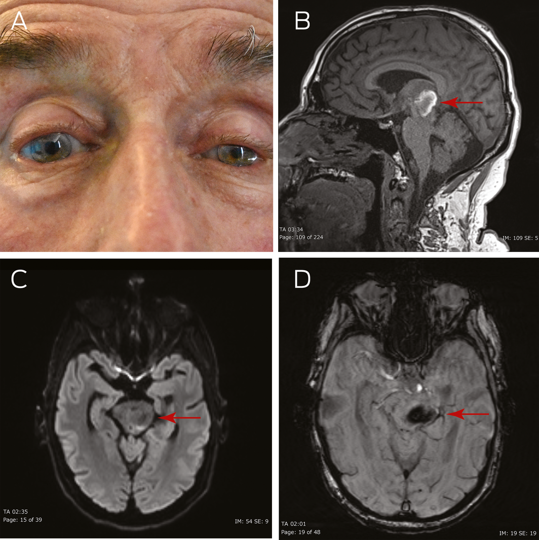
A 75‐year‐old man presented with right hemiparesis secondary to a left thalamic haemorrhage. Five days after presentation, he developed left Horner syndrome. He had left ptosis (Figure, A) with palpebral aperture measured at 3 mm on the left (7 mm on the right). In a well lit room, the left pupil measured 2 mm, while in a dark room, it was 3 mm, confirming miosis. Relative afferent pupillary defect was absent. Left‐sided anhidrosis was also present. A magnetic resonance imaging scan revealed extension of the haemorrhage into the thalamo‐mesencephalic junction (Figure, B–D).
Horner syndrome is caused by disruption to the three‐neuron sympathetic pathway. The first order neuron begins in the hypothalamus and synapses at the cervical cord. The second order neuron travels through the brachial plexus and over the lung apex to synapse in the superior cervical ganglion. The third order neuron ascends with the carotid artery to the iris dilator and Muller muscle.
Clinically, third order Horner can be differentiated by lack of anhidrosis and response to hydroxyamphetamine.1 This is due to the sparing of sudomotor fibres that branch off at the superior cervical ganglion and the lack of noradrenaline release from the postganglionic neuron respectively.
Competing interests
No relevant disclosures.
References
- Johnson D, Sharma S. Answer: can you identify this condition? Can Fam Physician 2010; 56: 443.
A Diagnostic Headache
Thomas Glynn, Michelle T. Leech, Stacy K. Goergen, Emily Lin, Nadeem Toodayan, Ralph Junckerstorff
Neisseria meningitidis: an uncommon cause of conjunctivitis
Chloe Story, Tina Marinelli, Simeon Crawford, Amrita Ronnachit
Dismantling barriers to research and clinical care for individuals with a vision impairment
Eden G Robertson, Kate Hetherington, Meredith Prain, Julia Hall, Leighton Boyd AM, Rosemary Boyd OAM, Emily Shepard, Hollie Feller, Sally Karandrews, Fleur O'Hare, Kanae Yamamoto, Matthew P Simunovic, Robyn V Jamieson, Alan Ma, Lauren Ayton AM, Anai Gonzalez‐Cordero
The New South Wales Pharmacy Trial for herpes zoster: on the nose?
Christian P Pappas, Timothy R Holmes, Minas T Coroneo
Skin fragility disorder misdiagnosed as child abuse: a cautionary tale
Heather G Mack, Fred K Chen, John R Grigg
Health economic aspects of inherited retinal diseases: looking for cost‐effective treatments
Benjamin Kamien, Rachael Heath Jeffery, Fred K Chen