




Volume 213 - Issue 2
The risks of medical complacency towards poliomyelitis
Authors: Meryta May, David Durrheim, Jason A Roberts and Rhonda Owen
Med J Aust 2020; 213 (2): 61-63.e1. || doi: 10.5694/mja2.50681
Published online: 6 July 2020
Published online: 6 July 2020
Australia needs to improve vigilance in the global endeavour to eradicate poliomyelitis
In 1988, there were over 350 000 cases of paralytic poliomyelitis globally.1 In 2018, there were 29 cases and in 2019 there were 112 cases2 — all in the only two remaining countries in the world where wild poliovirus (WPV) is endemic (Afghanistan and Pakistan). We are tantalisingly close to global eradication.
What is poliomyelitis? Poliovirus is an enterovirus and exists as three serotypes: WPV types 1, 2 and 3. Spread via the faecal–oral route, poliomyelitis results in subclinical or self‐limited infection in most patients, but causes acute flaccid paralysis (AFP) due to anterior horn cell damage in about one in 200 cases.3 Ubiquitous distribution of polioviruses and epidemics of paralysis caused widespread panic throughout the world in the early 20th century.
With an ambitious and unprecedented level of international public and private collaboration and funding, the Global Polio Eradication Initiative (GPEI) was launched in 1988.2 In recent years, significant achievements have been recorded (Box 1), but the target of global eradication is yet to be reached. The GPEI currently faces two main global issues. Firstly, addressing the eradication of WPV1 in Afghanistan and Pakistan, and secondly, dealing with the growing issue of vaccine‐derived poliovirus (VDPV).1 Vaccine strain virus can slowly accumulate mutations over time, which eventually result in reversion to neurovirulence — these strains are known as VDPV. Although extremely uncommon, this phenomenon becomes increasingly prominent in areas where there are long term low vaccination rates, allowing continued circulation of the attenuated poliovirus contained in the Sabin vaccine. Ironically, the modern prominence of VDPVs is a consequence of the GPEI's successful endeavours to reduce WPV.
Poliomyelitis close to home
VDPVs are appearing in areas with low immunisation rates in Africa, and recent emergence in closer neighbours puts poliomyelitis back on our doorstep. In 2018, there was an outbreak in Papua New Guinea involving 26 VDPV type 1 AFP cases, including a death.4 In late 2019, the Philippines reported 15 VDPV cases, and Malaysia reported three cases in 2019 and one in 2020.5 These countries had previously been declared poliomyelitis‐free.6 Between 2012–13 and 2017–18, the median number of annual arrivals for Philippine citizens to Australia was 141 813, with 8% of these arrivals being children younger than 15 years.7 Screening individuals at our borders is not an economically viable option to prevent poliomyelitis, thus highlighting the importance of optimal immunisation and high quality surveillance.
Australia's commitment to World Health Organization targets
Australia and all other Western Pacific region countries were certified as poliomyelitis‐free on 29 October 2000.6 As a signatory to the World Health Organization's International Health Regulations (2005),8 Australia reports annually on its compliance, with obligations to prevent and respond to acute public health risks of international consequence. This includes observing temporary recommendations issued when the WHO declared the risk of international spread of poliovirus a public health emergency of international concern in 2014, poliovirus containment activities, and reporting to the WHO Regional Certification Commission providing evidence that Australia's poliomyelitis‐free status has been maintained. This evidence requires Australia to meet WHO‐specified surveillance standards. The Australian National Enterovirus Reference Laboratory plays an important role in providing enterovirus testing and environmental surveillance for Australia and the Western Pacific region to meet these requirements.
Environmental surveillance for polioviruses is costly and labour‐intensive and involves sampling sewage for detection and then characterisation of enteroviruses. There is currently inadequate capacity to routinely conduct environmental surveillance throughout Australia. Therefore, this capacity is currently directed at monitoring during high risk episodes; for example, when there is a cluster of AFP cases or after the importation of a confirmed case. The detection of any poliovirus in Australia is considered a likely importation event, as Australia stopped the use of the oral polio vaccine in 2005.
Adequate clinical surveillance is based on two key WHO indicators. Firstly, achieving an AFP detection rate of at least one case per 100 000 children younger than 15 years. Secondly, the WHO requires enterovirus culture on two stool samples collected at least 24 hours apart, both within 14 days of onset of paralysis, for at least 80% of reported AFP cases.9 Submission of two samples ensures adequate sensitivity, required due to intermittent viral shedding.10 Meeting these targets provides national and international reassurance that there is timely investigation that excludes poliomyelitis as the cause of AFP. However, for Australian clinicians, awareness of this surveillance and its purpose is often not well understood. An overview of the AFP surveillance structure is provided in Box 2. Importantly, AFP cases need to be notified and investigated even if another diagnosis (eg, Guillain–Barré syndrome) is likely.
Australia's performance in meeting World Health Organization targets
While Australia has met the surveillance target for AFP notification for the past 11 years, we consistently fail to reach the WHO benchmark for stool submissions (Box 3).11 This is in marked contrast to many of our closest neighbours. Only New Zealand, the small Pacific Island countries and Papua New Guinea have a similarly low performance over recent years. In 2018, adequate stool collection was achieved in only 44% of Australian AFP cases and 2019 results are currently at 65%.12 The most populous states of New South Wales and Victoria consistently underperform, with rates of 33% and 42% respectively for 2018 (Bruce Thorley, Head of Victorian Infectious Diseases Reference Laboratory, Australia, personal communication, September 2019). In 2018, three cases of AFP and anterior horn cell abnormality on magnetic resonance imaging in young children were reported to WHO by Australia as “poliomyelitis compatible” because of a lack of adequate clinical information and appropriate stool sample collection (David Isaacs, Chair of Polio Expert Panel, Australia, personal communication, September 2019).
In addition to providing robust public health surveillance, ensuring adequate investigation of AFP can produce relevant diagnostic information for an individual. A 3‐year‐old child with permanent significant disability following AFP in 2018 had the neuropathic enterovirus D68 (EV‐D68) in faeces sent for AFP surveillance purposes.13,14 The converse may also apply. Detection of a non‐polio enterovirus by polymerase chain reaction (PCR) in a clinical sample does not preclude the possibility of dual infection with poliovirus. Co‐infection and subsequent recombination of species C non‐polio enteroviruses with Sabin‐like poliovirus is an important precursor event in the development of VDPVs.15
Barriers to improvement
A number of logistical issues affect successful stool sample collection; for example, late presentation of patients, discharge before sample collection, and constipation may all have an impact on stool collection rates.16
In some instances, pre‐examination by microbiology laboratories using enterovirus reverse transcriptase PCR (RT‐PCR) may occur. This does not exclude poliovirus infection and testing at the WHO reference laboratory is still required.
Due to the extended viral shedding in the gastrointestinal tract, stool samples are the specimen type most likely to facilitate enterovirus identification. The collection of rectal or throat swabs is discouraged by WHO due to reduced sensitivity compared with faeces samples. Pragmatism may dictate that the former may be preferable to no testing at all in a particular child if barriers to faeces collection exist.
Recognising poliomyelitis in a low prevalence community
Cases of poliomyelitis present as acute and often painful weakness in affected limbs. The weakness is often asymmetrical, affecting lower limbs more frequently than upper limbs, with rapid onset and usually no further progression after 48 hours. Sometimes patients may present atypically, reinforcing the need for any AFP to be reported and investigated. Alternative presentations may include dyspnoea or dysphagia due to weakness of bulbar or respiratory muscles. Cerebrospinal fluid findings are suggestive of viral meningitis. There are usually no systemic symptoms, although a recent history of a mild upper respiratory tract infection with or without headache may be elicited.3 A history of exposure to a high risk area (eg, Central Africa, Pakistan, Papua New Guinea or Afghanistan) and/or lack of previous immunisation is important. A history of distant past immunisation will not exclude the diagnosis, particularly if this was received overseas.10
Call to action
There appears to be a level of complacency among physicians due to the rarity of clinical poliomyelitis in Australia. In addition, there is a lack of awareness in the diagnostic chain regarding the importance of laboratory surveillance. In 2020, a comprehensive action plan was implemented by the Paediatric Active Enhanced Disease Surveillance (PAEDS) network to improve faeces collections across the country.
Clinicians should not fear that they are being alarmist in notifying AFP cases that they believe have negligible risk of poliomyelitis. The emphasis on detection and investigation of AFP cases despite an alternative diagnosis may seem pointless for an individual case, but at a national level, it allows confidence in the integrity of surveillance and, ultimately, achievement of poliomyelitis eradication.
Conclusion
The recent VDPV outbreaks in Papua New Guinea and the Philippines and the ongoing WPV1 circulation in Pakistan and Afghanistan emphasise the possibility of poliomyelitis re‐introduction into Australia. Clinical acumen is unlikely to provide a timely diagnosis.
Clinicians are reminded that poliomyelitis as a diagnosis should be excluded in all cases of AFP; faeces collection from all AFP cases independent of age should be viewed as a priority to ensure the country remains poliomyelitis‐free and as an opportunity to maintain surveillance, even when another diagnosis is confirmed or highly likely.
Box 1 – Selected achievements relevant for Australia in the history of the Global Polio Eradication Initiative2
|
Year |
Milestone |
||||||||||||||
|
|
|||||||||||||||
|
2000 |
Australia declared poliomyelitis‐free |
||||||||||||||
|
2005 |
Inactivated polio vaccine replaces oral polio vaccine in Australia |
||||||||||||||
|
2014 |
South‐East Asia declared poliomyelitis‐free |
||||||||||||||
|
2015 |
Wild poliovirus type 2 declared eradicated |
||||||||||||||
|
2017 |
99% of poliomyelitis eradicated globally |
||||||||||||||
|
2019 |
Wild poliovirus type 3 declared eradicated |
||||||||||||||
|
|
|||||||||||||||
|
|
|||||||||||||||
Box 2 – Schematic overview of acute flaccid paralysis surveillance structure in Australia
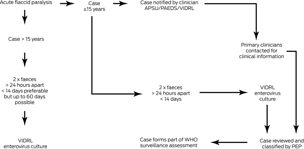
APSU = Australian Paediatric Surveillance Unit (www.apsu.org.au); PEP = Polio Expert Panel; PAEDS = Paediatric Active Enhanced Disease Surveillance Network (www.paeds.org.au); VIDRL = Victorian Infectious Disease Reference Laboratory (https://www/vidrl.org.au/surveillance/afp-surveillance1); WHO = World Health Organization.
Box 3 – Percentage of acute flaccid paralysis notification with adequate stool sample collection, Australia, 1995–2018*
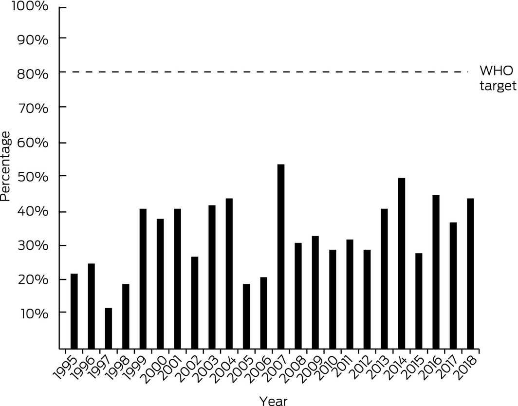
WHO = World Health Organization. * Data reproduced, with permission, from Roberts et al.11
Competing interests
No relevant disclosures.
Acknowledgements
We thank David Isaacs and Bruce Thorley for their assistance in providing relevant information for this manuscript.
References
- World Health Organization. Polio endgame strategy 2019–2023: eradication, integration, certification and containment [WHO/Polio/19.04]. WHO, 2018. https://www.who.int/publications/i/item/polio-endgame-strategy-2019-2023-eradication-integration-certification-and-containment (viewed June 2020).
- Global Polio Eradication Initiative. Polio this week as of 2 June 2020. http://polioeradication.org/polio-today/polio-now/this-week (viewed June 2020).
- Poliovirus infections. In: Pickering LK, Baker CJ, Long SS, McMillan JA, editors. Red book: 2006 report of the Committee on Infectious Diseases; 27th ed. Elk Grove Village, IL: American Academy of Pediatrics, 2006; p. 542.
- Hall JJ, Ambang T, Asante A, et al. Poliomyelitis outbreak in Papua New Guinea: health system and health security implications for PNG and Australia. Med J Aust 2019; 211: 161–163. https://www.mja.com.au/journal/2019/211/4/poliomyelitis-outbreak-papua-new-guinea-health-system-and-health-security
- Global Polio Eradication Initiatve. Where we work: Malaysia. http://polioeradication.org/where-we-work/malaysia (viewed June 2020).
- D'Souza RM, Kennett M, Watson C. Australia declared polio free. Commun Dis Intell Q Rep 2002; 26: 253–260.
- Australian Government Department of Home Affairs. Visa statistics https://www.homeaffairs.gov.au/research-and-statistics/statistics/visa-statistics/visit (viewed Oct 2019).
- World Health Organization. International Health Regulations (2005); 3rd ed. WHO, 2016. https://www.who.int/ihr/publications/9789241580496/en/ (viewed Dec 2019).
- World Health Organization. WHO Vaccine‐Preventable Diseases Surveillance Standards: poliomyelitis; updated 5 Sept 2018. WHO, 2018. https://www.who.int/immunization/monitoring_surveillance/burden/vpd/WHO_SurveillanceVaccinePreventable_18_Polio_R2.pdf?ua (viewed Aug 2019).
- Carnie JA, Lester R, Moran R, et al. Public health response to imported case of poliomyelitis Australia, 2007. Emerg Infect Dis 2009; 15: 1733–1737.
- Roberts JA, Hobday LK, Ibrahim A, Thorley BR. Australian National Enterovirus Reference Laboratory annual report 2018. Commun Dis Intell 2018; 2020: 44. https://doi.org/10.33321/cdi.2020.44.26.
- World Health Organization. Polio Bulletin 2020, issue 6, week 18 (5 May 2020). WHO, 2020. https://apps.who.int/iris/bitstream/handle/10665/330696/Polio-Bulletin-2020-No-06-Week-18.pdf (viewed June 2020).
- Levy A, Roberts J, Lang S, et al. Enterovirus D68 disease and molecular epidemiology in Australia. J Clin Virol 2015; 69: 117–121.
- Uprety P, Curtis D, Elkan M, et al. Association of enterovirus D68 with acute flaccid myelitis, Philadelphia, Pennsylvania, USA, 2009–2018. Emerg Infect Dis 2019; 25: 1676–1682.
- Muslin C, Mac Kain A, Bessaud M, et al. Recombination in enteroviruses, a multi‐step modular evolutionary process. Viruses 2019; 11: 859.
- Dinsmore N, McRae J, Saravanos G, et al. Acute flaccid paralysis surveillance for polio: challenges of stool collection in Australia. 16th Immunisation Conference of the Public Health Association of Australia; Adelaide (Australia), 5–7 June 2018.
Provenance: Not commissioned; externally peer reviewed.
Dousing the burning inequity of global warming for people experiencing homelessness
Timothy English, Alejandro Vásquez Hernández, Glenn Miller, Nuala Fogarty, Charles Cosgrove, Paul Rosenthal, Matthew Larkin, Jon Swain, Danielle Austin, Jo River
The contribution of evidence‐based practice and the practice‐based evidence approaches to contemporary Australian psychology: implications for culturally safe practice
Paul Gray (Wiradjuri), Dawn Darlaston‐Jones, Pat Dudgeon AM (Bardi), Kate Derry, Joanna Alexi, William Smith (Wiradjuri and Wemba Wemba), Tanja Hirvonen (Jaru and Bunuba), David Badcock, Shraddha Kashyap, Belle Selkirk (Noongar)
Explaining risk in chronic conditions: the Yolŋu science of signs
Emma Haynes, Alison Mitchell, Minitja Marawili (Yolŋu), Dawn C Bessarab (Bardi)
Is it time to retire the label “CALD” in public health research and practice?
Sabine Allida, Della Maneze
What do Aboriginal people in the Northern Territory value during the operation journey? A qualitative study
Edith B Waugh, Marita Hefler, Sophie Pascoe, Mark Mayo, Matthew JL Hare, David A Story, Neil Wilkshire, Peter Henwood, David Croker, Jampijinpa Ross
Fulfilling First Nations health, cultural safety and equity accreditation standards in primary medical education: reflections from a First Nations desktop review team
Paul Saunders, Nicole Mercer, Maria Mackay, Ian Lee, Madelyne Hudson‐Buhagiar, Miriam Cavanagh, Emma Milliss, Melody Muscat, Kathleen Martin, Adam Shipp, Melissa Johnson, Belinda Gibb