




Medical education
Clinical skills
Volume 212 - Issue 4
How to perform a skin biopsy
Authors: Kirsty JL Wark, Saxon D Smith and Deshan F Sebaratnam
Med J Aust 2020; 212 (4): 156-158.e1. || doi: 10.5694/mja2.50473
Published online: 2 March 2020
Published online: 2 March 2020
The skin has more disease
processes than any other
organ system in medicine,
with over 3000 dermatological
conditions described
The skin has more disease processes than any other organ system in medicine, with over 3000 dermatological conditions described.1 Teaching of dermatology is often neglected in medical training internationally, and most doctors feel ill‐equipped to diagnose cutaneous pathology.2 Compounding this, limitations in access to specialist dermatologists in Australia are well recognised.3 Fortunately, biopsy of the skin is a simple skill to learn which can greatly help with the diagnosis of dermatological diseases. For cutaneous malignancies, the diagnosis is principally based on histopathological findings. However, for rashes, the correlation between clinical and pathological findings is paramount. For instance, observation of a lichenoid reaction pattern on skin biopsy may reflect lichen planus, lupus, dermatomyositis, lichen sclerosus, cutaneous T cell lymphoma, or graft‐versus‐host disease. To maximise the diagnostic yield of a skin biopsy, an understanding of the different types of biopsy, their indications and limitations is vital (Box 1 and Box 2).
Common types of skin biopsy
Punch biopsy
A punch biopsy is a hollow cylindrical blade that varies in diameter from 2 mm to 8 mm (Supporting Information, video 1). It provides a skin sample from the epidermis to superficial fat and is the most commonly employed biopsy type. In most settings, a 3 mm or 4 mm punch biopsy can be used.
Shave biopsy
A shave biopsy is a flexible rectangular blade used to sample skin to dermis (Supporting Information, video 2). It is useful for epidermal and superficial dermal pathology offering a larger surface area and obviates the need for suturing. Haemostasis may be achieved with pressure, aluminium chloride or electrosurgery.
Incisional biopsy
An incisional biopsy is performed with a scalpel. A representative portion of the lesion is excised with the defect and then sutured. It is useful for sampling pathology down to the level of the fat and is favoured for deeper processes such as vasculitis and panniculitis.
Excisional biopsy
An excisional biopsy is usually performed with a scalpel and involves the extirpation of a skin lesion in its entirety. Removal of the entire lesion helps reduce the risk of sampling error and is favoured for the investigation of pigmented lesions.4,5 In certain settings, such as in large pigmented macules or when there is a low likelihood of malignancy, a shave biopsy or incisional biopsy may be considered. These biopsy techniques may mitigate unnecessary morbidity and improve diagnostic yield. In this case, dermoscopy should be employed to direct biopsy site.
Curettage
Curettage uses a surgical tool similar to a sharp spoon to scrape away samples of skin. In specific settings, it may be used to treat low grade cutaneous malignancies. Given these are generally more friable than normal skin, the treating doctors can scrape away the tumour, with the difference in texture being a guide to tumour margins clinically. The procedure is operator‐dependent and the histological interpretation of the curettings may be challenging.
Investigation
Histopathology
Histopathology is interpretation of the skin under the microscope, generally employing haematoxylin and eosin staining. It is the main study of skin biopsy and helpful in inflammatory, neoplastic and structural pathology.
Direct immunofluorescence
Direct immunofluorescence involves the application of fluorophore‐antibody complexes, which bind to epitopes present within specific dermatoses. It is useful for selected autoimmune blistering diseases, vasculitis and genodermatoses.
Microbial studies
Tissue is examined using microscopy or incubated under specific laboratory conditions to let pathogens multiply, allowing for identification and antimicrobial sensitivity analysis. Polymerase chain reaction can also be performed to identify culprit pathogens.
Cell culture
Cell culture involves incubation of skin samples in special mediums to facilitate growth of specific cells, such as fibroblasts, which may then be employed for further specialised testing.
Transport mediums
Formalin
Formalin is the saturated form of formaldehyde in water. It is both a fixative and a transport medium, used for transport of tissue to be analysed for histopathology.
Normal saline
Saline‐soaked gauze is a commonly used transport medium for tissue samples for direct immunofluorescence, frozen sections, and tissue culture.
Michel medium
This transport medium preserves immunoreactants within the skin, enabling direct immunofluorescence to be performed.
Procedure for a punch biopsy
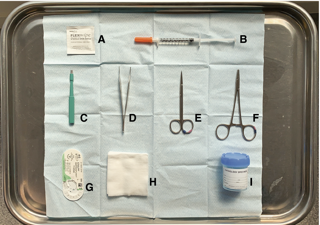
The patient should first consent for the procedure, and photography is recommended to document the site of the biopsy and lesion of interest. In almost all cases, anticoagulation can be continued, as skin biopsy is minimally invasive and haemostasis is achieved easily. The site to be biopsied should be marked with a sterile surgical marker, and then only infiltrated with local anaesthetic (lest vasoconstriction obscure the lesion). Once the area is anaesthetised, the punch biopsy should be performed by placing the biopsy blade perpendicular to the skin, then rotating it clockwise and counterclockwise until the superficial subcutis is reached (Supporting Information, video 1). The specimen can be removed gently with forceps and cut at the base with scissors or by tilting the biopsy blade 45°, before being placed into the appropriate transport medium. In general, biopsies greater than 2 mm require a suture to achieve optimal haemostasis and cosmesis (Box 3). Follow‐up should be organised for removal of sutures and discussion of pathology.
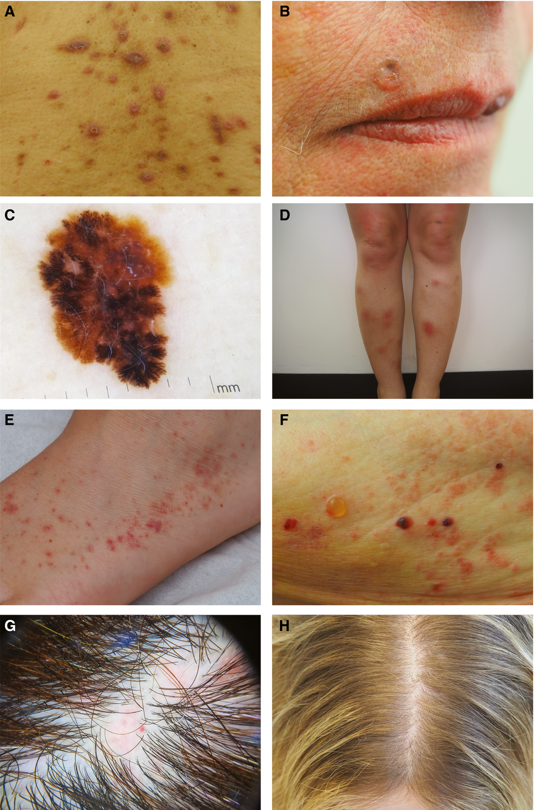
Box 1 – Range of skin pathology requiring different biopsy techniques: (A) rash (acquired perforating collagenosis), (B) non‐melanoma skin cancer (pigmented basal cell carcinoma), (C) melanoma, (D) panniculitis (erythema nodosum), (E) vasculitis (leucocytoclastic vasculitis), (F) bullous dermatosis (bullous pemphigoid), (G) scarring alopecia (cutaneous lupus erythematosus), and (H) non‐scarring alopecia (androgenetic alopecia)
Box 2 – Skin biopsy for specific clinical scenarios
Type of lesion |
Biopsy | ||||||||||||||
Rash |
3–4 mm punch biopsy for HE detailing as much clinical information as possible |
||||||||||||||
Non‐melanoma skin cancer (eg, SCC, BCC) |
2–3 mm punch biopsy or shave biopsy for HE |
||||||||||||||
Pigmented lesions (eg, when melanoma suspected) |
Excisional biopsy with margins for HE |
||||||||||||||
Panniculitis |
Deep incisional biopsy to subcutaneous fat for HE |
||||||||||||||
Vasculitis |
4 mm punch biopsy of established lesion (> 72 h) for HE |
||||||||||||||
And |
|||||||||||||||
3 mm punch biopsy of a new lesion (< 24 h) for DIF |
|||||||||||||||
Blistering eruption (eg, bullous pemphigoid, pemphigus) |
3mm punch biopsy at the edge of an intact blister for HE |
||||||||||||||
And |
|||||||||||||||
3 mm punch biopsy of perilesional skin (within 1 cm of a blister) for DIF |
|||||||||||||||
Scarring alopecia |
4 mm punch biopsy of an established area of involvement for HE and consider another biopsy for DIF if connective tissue disease suspected5 |
||||||||||||||
Non‐scarring alopecia |
4 mm punch biopsy of an established area of involvement for HE for horizontal sectioning |
||||||||||||||
And |
|||||||||||||||
4 mm punch biopsy of an established area of involvement for HE for vertical sectioning |
|||||||||||||||
When androgenetic alopecia is suspected, a 4 mm biopsy from an affected site and from an unaffected site (ie, the occiput) may be advisable |
|||||||||||||||
BCC = basal cell carcinoma; DIF = direct immunofluorescence; HE = haematoxylin and eosin staining; SCC = squamous cell carcinoma. | |||||||||||||||
Competing interests
No relevant disclosures.
Acknowledgements
We thank Alicia O'Connor, Anes Yang, Victoria Venning, Imogen Faulds and our patients for their contributions to the clinical images.
References
- Bickers DR, Lim HW, Margolis D, et al. The burden of skin diseases: 2004: a joint project of the American Academy of Dermatology Association and the Society for Investigative Dermatology. J Am Acad Dermatol 2006; 55: 490–500.
- Hussain W, Hafiji J, Stanley AG, Khan KM. Dermatology and junior doctors: an evaluation of education, perceptions and self‐assessed competencies. Br J Dermatol 2008; 159: 505–506.
- Sebaratnam DF, Murrell DF. Dermatology training and practice in Australia. Int J Dermatol 2014; 53: 1259–1264.
- de Menezes SL, Kelly JW, Wolfe R, et al. The increasing use of shave biopsy for diagnosing invasive melanoma in Australia. Med J Aust 2019; 211: 213–218. https://www.mja.com.au/journal/2019/211/5/increasing-use-shave-biopsy-diagnosing-invasive-melanoma-australia
- Elston DM, Stratman EJ, Miller SJ. Skin biopsy: biopsy issues in specific diseases. J Am Acad Dermatol 2016; 74: 1–16.
Provenance: Commissioned; externally peer reviewed.
Diagnosing Acute Kava Dermopathy: A Case Report of a Characteristic Cutaneous Eruption
Ali Abid, Nicholas Allen, Abeer Hagelamin, Christopher Henderson, Artiene Tatian
Localised Herpes Simplex Following Midline Laparotomy
Jessica S. Bulluss, Paul Chee, Matthew J. Verheyden
No silver lining with health misinformation: argyria caused by intentional silver consumption
Luke Collins, Logesh Palanikumar, Stephen Bacchi
Lingual Raynaud phenomenon
Michael Taggart, Mina John
Melasma in the male: a less well recognised entity
Tim Aung, Rowland Noakes
Age group‐specific changes in keratinocyte cancer treatment rates in Australia, 2012–2021: a retrospective cohort study based on MBS claims data
Catherine M Olsen, Nirmala Pandeya, Rachel E Neale, David C Whiteman