




Medical education
Lessons from practice
Volume 211 - Issue 9
Orbital rhabdomyosarcoma: a rare ophthalmic condition
Authors: Jiyeon Kim and Jonathan G Ussher
Med J Aust 2019; 211 (9): 398-399.e1. || doi: 10.5694/mja2.50365
Published online: 4 November 2019
Published online: 4 November 2019
Correction(s) for this article:
Erratum | Published online: 13 January 2020
A previously well 6-year-old girl presented to her doctor with a mildly injected right eye and upper eyelid swelling
Clinical record
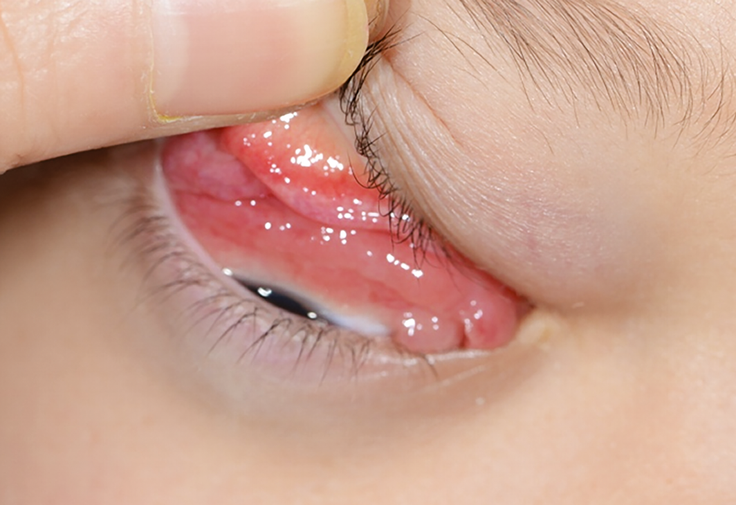
A previously well 6‐year‐old girl with an unremarkable past medical and family history presented to her doctor with a mildly injected right eye and upper eyelid swelling. Her mother initially thought it was due to a minor contusion from a recent fall. Signs developed overnight, having been completely normal the day before. She had normal vision, denied discomfort and was systemically well. She was diagnosed with conjunctivitis and chloramphenicol ointment was prescribed. However, her condition progressed and she was referred to the eye clinic. Her visual acuity was 6/6 in each eye, with normal intraocular pressures, pupil reactions and colour vision. Her right upper eyelid was visibly swollen. When the upper eyelid was everted, there was granulomatous‐like tissue extending from the superior aspect of the globe. The rest of the eye examination, including dilated fundal examination, was unremarkable. With the history of recent trauma, she was initially diagnosed and treated as pyogenic granuloma.
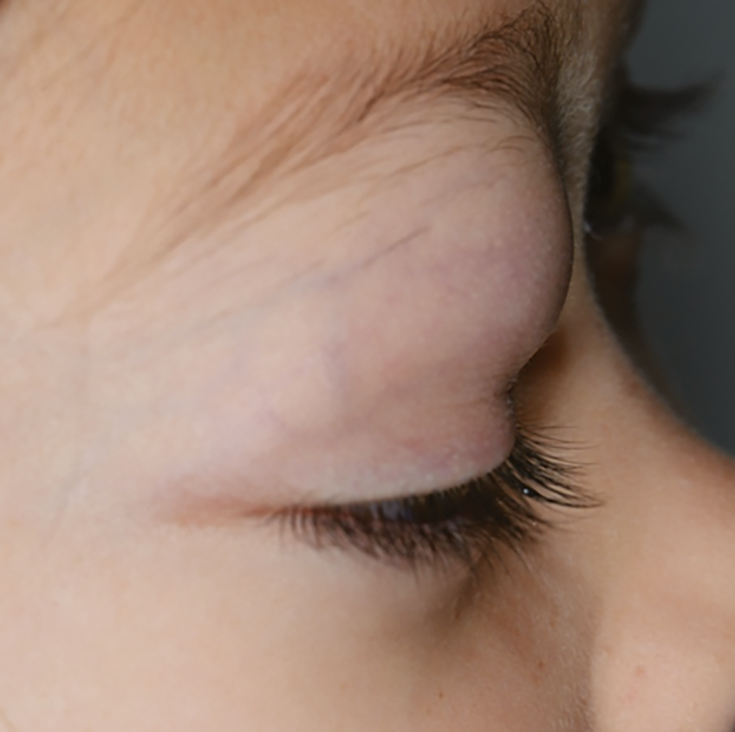
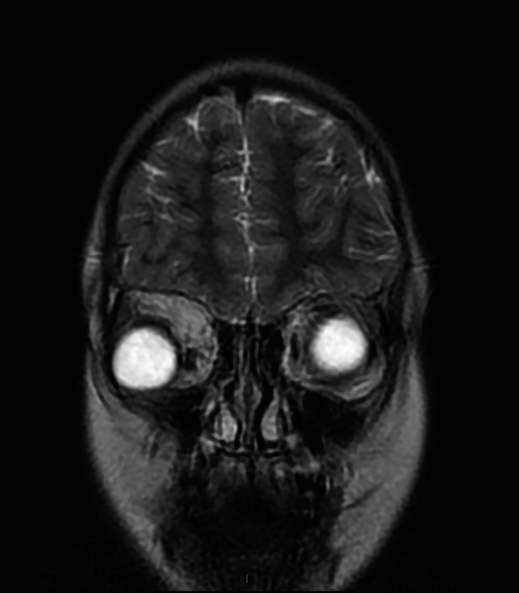
Her condition continued to progress rapidly. At 4‐day follow‐up, the right upper eyelid swelling was much worse (Box 1). The patient now had proptosis and exophthalmos. An increase in the granulomatous‐like tissue was observed (Box 2). The visual acuity was still 6/6 in each eye, with normal pupillary reactions. Other than restricted range of eye movement on the right, the cranial nerve examination was normal. A suspicion for orbital rhabdomyosarcoma was raised and urgent computed tomography imaging was performed. A tumour extending from the right superior rectus muscle displacing the adjacent tissues was seen (Box 3). She then underwent a tissue biopsy, which confirmed the diagnosis of rhabdomyosarcoma. Following histological diagnosis and a multidisciplinary team review, she progressed to treatment with combined radiotherapy and chemotherapy under the direction of the paediatric oncology team.
Discussion
Rhabdomyosarcoma is a rare connective tissue cancer with an incidence rate of 4.3 per million population.1 Nevertheless, it is the most common primary orbital malignancy that affects children, with about 50% of cases occurring in the first decade of life. The mean age at diagnosis is 8 years and higher rates are seen in males.
Patients typically present with proptosis and ophthalmoplegia.1 Globe displacement to the inferotemporal quadrant is also common due to two‐thirds of the tumours arising from the superonasal quadrant area.2 The rate of progression varies, but significant progression can occur within just days to weeks. Overnight onset has also been described in several cases,2 which is perhaps the main misleading factor that confuses many health practitioners from not considering malignancy as a differential diagnosis at initial consultation. Potential causes for proptosis can be divided by the presence or absence of pain. Most inflammatory conditions such as orbital cellulitis, preseptal cellulitis and orbital myositis typically present with pain. Orbital rhabdomyosarcoma, on the other hand, presents with rapid painless swelling in an otherwise well child. Other potential causes for painless proptosis include orbital neurofibroma, orbital haemangioma and orbital dermoid cyst.3
Although uncommon, both local and systemic metastases can occur if the condition is left untreated. Common metastatic sites include bone, bone marrow and lungs.2 The overall survival rate for rhabdomyosarcoma has improved significantly over time thanks to medical advancements. Until the late 1960s, surgery was the primary treatment for this condition and the overall survival rate was about 25–30%. Later, adjuvant treatment of chemotherapy and radiotherapy was introduced and the overall survival rate improved to about 90%.2
Today, chemotherapy and radiotherapy are the recommended first line treatments. Surgery is mainly reserved for patients with recurrent disease.1,2 A study showed that primary orbital rhabdomyosarcoma had significantly better prognosis than paranasal rhabdomyosarcoma, with a 5‐year survival rate of 100% and 29% respectively.5 The overall globe conservation rate for primary orbital rhabdomyosarcoma group varied between 71% and 90%.4,5
Lessons from practice
- Orbital rhabdomyosarcoma is a rare connective tissue malignancy that mainly affects children in their first decade, and a higher incidence rate is noted in males.
- The onset of orbital rhabdomyosarcoma can vary significantly but often ranges from days to weeks.
- At early stage, patients often present with an acute episode of red eye, with or without swollen eyelid and ptosis.
- If suspicion is raised, urgent imaging studies such as computed tomography or magnetic resonance imaging scan should be organised and the patient should be referred to an appropriate service for an urgent tissue biopsy.
- In contrast to inflammatory conditions, orbital rhabdomyosarcoma presents with painless proptosis with rapid swelling in an otherwise well child.
Competing interests
No relevant disclosures.
References
- Karcioglu ZA, Hadjistilianou D, Rozans M, DeFrancesco S. Orbital rhabdomyoscarcoma. Cancer Control 2004; 11: 328–333.
- Jurdy L, Merks JH, Pieters BR, et al. Orbital rhabdomyosarcomas: a review. Saudi J Ophthalmol 2013; 27: 167–175.
- Sindhu K, Downie J, Ghabrial R, Martin F. Aetiology of childhood proptosis. J Paediatr Child Health 2003; 34: 374–376.
- Boutroux H, Levy C, Mosseri V, et al. Long‐term evaluation of orbital rhabdomyosarcoma in children. Clin Exp Opthalmol 2015; 43: 9–12.
- Eade E, Tumuluri K, Do H, et al. Visual outcomes and late complications in paediatric orbital rhabdomyosarcoma. Clin Exp Ophthalmol 2016; 45: 168–173.
Provenance: Not commissioned; externally peer reviewed.
A Diagnostic Headache
Thomas Glynn, Michelle T. Leech, Stacy K. Goergen, Emily Lin, Nadeem Toodayan, Ralph Junckerstorff
Neisseria meningitidis: an uncommon cause of conjunctivitis
Chloe Story, Tina Marinelli, Simeon Crawford, Amrita Ronnachit
Dismantling barriers to research and clinical care for individuals with a vision impairment
Eden G Robertson, Kate Hetherington, Meredith Prain, Julia Hall, Leighton Boyd AM, Rosemary Boyd OAM, Emily Shepard, Hollie Feller, Sally Karandrews, Fleur O'Hare, Kanae Yamamoto, Matthew P Simunovic, Robyn V Jamieson, Alan Ma, Lauren Ayton AM, Anai Gonzalez‐Cordero
The New South Wales Pharmacy Trial for herpes zoster: on the nose?
Christian P Pappas, Timothy R Holmes, Minas T Coroneo
Skin fragility disorder misdiagnosed as child abuse: a cautionary tale
Heather G Mack, Fred K Chen, John R Grigg
Health economic aspects of inherited retinal diseases: looking for cost‐effective treatments
Benjamin Kamien, Rachael Heath Jeffery, Fred K Chen