




Volume 211 - Issue 7
Perioperative considerations in the management of obstructive sleep apnoea
Authors: Nicolette R Holt, Glenn Downey and Matthew T Naughton
Med J Aust 2019; 211 (7): 326-332. || doi: 10.5694/mja2.50326
Published online: 16 September 2019
Published online: 16 September 2019
The increasing age and obesity rates of the Australian population are contributing to the higher prevalence of patients with obstructive sleep apnoea presenting for surgery
Summary
- Obstructive sleep apnoea (OSA) is characterised by repetitive compromise of the upper airway, causing impaired ventilation, sleep fragmentation, and daytime functional impairment. It is a heterogeneous condition encompassing different phenotypes.
- The prevalence of OSA among patients presenting for elective surgery is growing, largely attributable to an increase in age and obesity rates, and most patients remain undiagnosed and untreated at the time of surgery. This condition is an established risk factor for increased perioperative cardiopulmonary morbidity, heightened in the presence of concurrent medical comorbidities. Therefore, it is important to perform preoperative OSA screening and risk stratification — using the STOP‐Bang screening questionnaire, nocturnal oximetry, and ambulatory and in‐laboratory polysomnography, for example.
- Postoperative risk assessment is an evolving process that encompasses evaluation of upper airway compromise, ventilatory control instability, and pain–sedation mismatch.
- Optimal postoperative OSA management comprises continuation of regular positive airway pressure, a multimodal opioid‐sparing analgesia strategy to limit respiratory depression, avoidance of supine position, and cautious intravenous fluid administration.
- Supplemental oxygen does not replace a patient's regular positive airway pressure therapy and should be administered cautiously to avoid risk of hypoventilation and worsening of hypercapnia. Continuous pulse oximetry monitoring with specified targets of peripheral oxygen saturation measured by pulse oximetry is encouraged.
The prevalence of obstructive sleep apnoea (OSA) among patients presenting for elective surgery is growing, in part due to risk factors attributable to increased age and obesity rates.1 OSA is an established risk factor for increased perioperative cardiovascular and pulmonary morbidity. Such risks are heightened in the setting of concurrent medical comorbidities.2
The domain of the perioperative clinician is expanding, as more surgery on complex patients with OSA is performed in smaller and regional facilities. This narrative review is targeted at clinicians involved in the perioperative care of patients with suspected or confirmed OSA who undergo procedures under conscious sedation or under general anaesthesia. It highlights perioperative considerations of OSA, including changes in sleep architecture and physiology, and the salient features that can predictably exacerbate previously stable OSA in the postoperative setting. The scope of this review does not encompass the intraoperative management of these patients.
Methods
We have identified relevant literature via PubMed from 1995 to 2019, including original articles, topic reviews, societal guidelines and recent conference abstract publications, to formulate an evidence‐based synopsis of the perioperative management of OSA.
Epidemiology
The global prevalence of OSA in the general adult population is estimated to range from 9% to 38%, with variations depending on the population sampled and diagnostic criteria applied.3 The overall prevalence of OSA among adult surgical patients ranges from 24% to 41%, depending on surgery type;4 for example, the prevalence of OSA can exceed 70% in patients undergoing bariatric surgery.5
Given the increasing number of patients with OSA in the Australian population, it is concerning that most cases remain undiagnosed, and therefore untreated, among adult patients at the time of presentation for elective surgery.6
Risk factors for obstructive sleep apnoea
Non‐modifiable risk factors for OSA include male gender, post‐menopausal status, family history of OSA, and advancing age. The primary modifiable risk factor for the development of OSA is increased body weight, which demonstrates a direct association with the risk and severity of OSA.7 OSA is almost twice as likely in patients with obesity (body mass index [BMI] ≥ 30 kg/m2) compared with non‐obese adults. A large longitudinal study determined that a 10% increase in body weight translated to a sixfold increase in the odds of developing at least moderate to severe OSA and that weight loss can attenuate OSA severity.8
It is important to note that up to 25% of patients with OSA are not obese (BMI < 25 kg/m2).9 Anatomical features of craniofacial structure (eg, retrognathia) and certain ethnic appearances (eg, Asian) are important to recognise.10,11 Non‐anatomical features, such as low respiratory arousal threshold, are important in the pathogenesis of OSA in affected non‐obese patients.9 Dismissing the possibility of OSA in adults with healthy weight must be avoided if symptoms or other risk factors are indicated on preoperative screening questionnaires or clinical assessment (Box 1).12
Pathophysiology and diagnosis of obstructive sleep apnoea
OSA involves the cyclical collapse of the pharyngeal airway and resultant airflow limitation during sleep, either partially, producing hypopnoea, or completely, resulting in apnoea.13 The pathophysiological consequences include hypoxaemia and hypercapnia, activation of the sympathetic nervous system, cortical arousal, and fragmented sleep architecture, with subsequent daytime functional impairment. Presence of OSA is an independent risk factor for systemic hypertension, atrial fibrillation, pulmonary hypertension and insulin resistance.14,15
The diagnosis of OSA is via polysomnography, either at a laboratory or ambulatory.13 Its presence is established via the Apnoea–Hypopnoea Index (AHI) — the sum of apnoeas and hypopnoeas recorded per hour of sleep — with a score of five or more events per hour being confirmatory in adult patients.13 An AHI of 30 or more events per hour is indicative of severe OSA. Associated nocturnal hypoxaemic burden is established via several parameters, including arterial oxygen nadir (lowest peripheral oxygen saturation measured by pulse oximetry [Spo2] level), time spent with an oxygen saturation below Spo2 90%, and the Oxygen Desaturation Index (ODI) — quantity of subthreshold desaturations per hour. In addition, hypoventilation and resultant hypercapnia can be assessed via transcutaneous carbon dioxide monitoring and arterial blood gas sampling at the beginning and end of the polysomnography.
Management of OSA depends on severity, symptomatology and patient comorbidities, but it commonly comprises an oral appliance (mandibular advancement splint), upper airway surgery, or continuous positive airway pressure (CPAP), in addition to conservative measures such as weight loss and positional modification when appropriate.16
Obstructive sleep apnoea phenotypes and implications for the perioperative period
A recent and evolving aspect of OSA pathophysiology is the concept of an OSA phenotype. In addition to upper airway anatomical predisposition, individual variability in the following factors is a key influencing mechanism in OSA pathogenesis:
- upper airway neuromuscular impairment — reduced upper airway dilator muscle tone, primarily genioglossus, reducing patency;
- respiratory arousal threshold — high thresholds require greater respiratory stimuli to restore patency and flow to the upper airway; and
- ventilatory control instability — fluctuation in ventilatory output signals.17
This variability has led to an increased focus on specific OSA phenotypes potentially predisposing to increased perioperative vulnerability to OSA exacerbation. For example, patients with high arousal thresholds (low propensity to arouse to stimuli) may be at heightened postoperative risk of respiratory depression with sedative and opioid administration. Conversely, in patients with an exaggerated ventilatory response following an apnoeic episode, targeted use of postoperative oxygen therapy may assist in stabilising breathing patterns.18
Physiology of postoperative obstructive sleep apnoea
Anaesthesia, analgesics and surgery can significantly disrupt ventilation and sleep architecture in patients with and without OSA.19 The AHI increases significantly from baseline across the third postoperative night in patients with OSA.19 This exacerbation corresponds to the recovery of rapid eye movement (REM) sleep, which is significantly reduced across the first postoperative night; apnoea and hypopnoea events and associated oxygen desaturation are typically more pronounced during the REM phase of sleep. Arterial oxygen desaturation is correspondingly greatest on the third postoperative night, during which time supplemental oxygen therapy may have an adjuvant role to regular OSA treatment.19
This is a notable trend that remains underappreciated and is relevant, as monitoring is often discontinued before Day 3. Clinician awareness of such disruptions in postoperative physiology, combined with the milieu of anaesthesia and analgesia, is critical to understanding some of the contributing factors that can destabilise previously stable OSA, and have implications when considering the duration and intensity of postoperative monitoring.
Important conditions in the perioperative period
In addition to OSA, there are several other related conditions that warrant consideration during the perioperative period due to the heightened risk of complications.
Upper airway resistance syndrome
Upper airway resistance syndrome can be conceptualised as being at the milder end of the OSA spectrum and typically occurs in younger, non‐obese individuals who snore and have fragmented sleep with an AHI under five events per hour.13 This syndrome can transition to overt hypopnoeas or apnoeas after surgery, and may account for the 26% of patients without preoperative sleep apnoea who develop at least moderate grade OSA between the first and third postoperative nights.20
Obesity hypoventilation syndrome
Obesity hypoventilation syndrome (OHS) is the demonstration of daytime hypercapnia (arterial partial pressure of carbon dioxide ≥ 45 mmHg) in a person with obesity (BMI ≥ 30 kg/m2).13 While OHS is estimated to be present in up to 0.3% of the general population,21 prevalence is as high as 8% among patients presenting for bariatric surgery.22 In one review, OHS occurred in approximately 10%, 15% and 25% of patients grouped by BMI 30–35 kg/m2, 35–40 kg/m2 and > 40 kg/m2, respectively.23 An estimated 90% of patients with OHS have concurrent OSA.22
In comparison with patients with isolated OSA, patients with hypercapnia from OHS are more likely to experience postoperative complications, including respiratory failure, cardiac failure and unplanned transfer to an intensive care unit (ICU).24 OHS should be suspected in a patient with obesity presenting for surgery with resting hypoxaemia, restrictive ventilatory defect on spirometry, or elevated serum bicarbonate (≥ 27 mEq/L).22 Therapeutic management for OHS can comprise continuous or bilevel positive airway pressure, with both showing similar efficacy in managing ventilatory failure, in addition to weight loss strategies.25 Preoperative identification of OHS affords the opportunity for greater vigilance to avoid common management pitfalls that can exacerbate hypercapnia, including indiscriminate supplemental oxygen administration, sedation or respiratory depression from pharmacotherapy, and excessive diuresis increasing serum bicarbonate.26
Obstructive sleep apnoea and perioperative morbidity
A meta‐analysis of postoperative outcomes after non‐cardiac surgery highlighted the significantly increased odds of any cardiac event after surgery (odds ratio [OR], 2.07; 95% confidence interval [CI], 1.23–3.50) among patients with OSA, including an increased incidence of postoperative cardiac arrhythmia, myocardial ischaemia and cardiac arrest.2 Patients with severe unrecognised OSA undergoing major non‐cardiac surgery experienced a significantly increased 30‐day risk of cardiovascular complications.27
Similarly, OSA was associated with significantly higher odds of pulmonary complications, including respiratory failure (OR, 2.43; 95% CI, 1.34–4.39) and oxygen desaturation (OR, 2.27; 95% CI, 1.20–4.26), and increased transfer to an ICU (OR, 2.81; 95% CI, 1.46–5.43).2
These complications are obviously in addition to the increased perianaesthetic risk of difficulty in intubation, mask ventilation, and maintaining airway patency after extubation. OSA is also associated with a significantly increased risk of unplanned transfer to an ICU.28
At present, patients at greatest risk of postoperative cardiopulmonary complications from OSA remain poorly defined due to limited high grade evidence.29 It remains unclear what precise proportion of perioperative complications can be attributable to OSA per se, rather than to obesity or associated cardiac, respiratory and metabolic comorbid conditions.30,31
Preoperative screening and risk stratification
Given that most people with OSA remain undiagnosed, many patients presenting for elective surgery are similarly undiagnosed.6 The heightened perioperative morbidity highlights the importance of preoperative screening for OSA. The major international societal guidelines on perioperative OSA assessment and management, developed by the American Society of Anesthesiologists (ASA)32 and the Society of Anesthesia and Sleep Medicine (SASM),33 support OSA screening as part of standard pre‐anaesthetic evaluation. The optimal preoperative OSA screening and confirmatory test remains unclear. There are currently no Australian‐based consensus guidelines for the perioperative evaluation or management of OSA; rather, individual hospital networks often develop internal guidelines for use in patients admitted to hospital.
The OSA screening questionnaires commonly used include the STOP‐Bang Questionnaire, the Berlin Questionnaire and the Epworth Sleepiness Scale (ESS) — the STOP‐Bang and ESS questionnaires have recently become essential requirements for the Medicare funding of sleep studies requested in primary care. In Australia, the STOP‐Bang Questionnaire (Box 2) is routinely employed for preoperative screening of OSA, with a score of 3 or greater raising concern for OSA.34,35
While in‐laboratory polysomnography is the most comprehensive confirmatory test for OSA, ambulatory sleep studies and nocturnal oximetry are alternatives due to practical considerations in the preoperative period. Surrogate markers such as polycythaemia, hypercapnia or elevated serum bicarbonate, and associated comorbid features such as hypertension and atrial fibrillation may be present, although they are non‐specific for OSA. The current evidence is not sufficient to support delaying or cancelling surgery for confirmatory OSA testing.33
Perhaps one of the greatest advantages in preoperative OSA screening is the identification of at‐risk patients to guide risk stratification and modification in perioperative care. Box 3 outlines predictive factors for postoperative exacerbations of OSA related to the patient, surgery, medication administration, and monitoring conditions than can be used to assist with perioperative risk stratification of the surgical patient with suspected or confirmed OSA.
Role of perioperative positive airway pressure
Among patients with established OSA adherent with CPAP, the SASM guideline supports the postoperative continuation of CPAP therapy, unless specifically contraindicated (GRADE: Strong, according to the Grading of Recommendations Assessment, Development and Evaluation [GRADE] system).33 If a patient has an oral appliance, this should similarly be used postoperatively. A recent systematic review did not identify increased incidence of anastomotic dehiscence when CPAP was implemented immediately after bariatric surgery.36 Continuation of CPAP therapy after surgery has been shown in two recent large observational studies to reduce incidence of postoperative cardiopulmonary complications compared with patients with untreated OSA.37,38
Postoperative positive airway pressure should be continued at the patient's preoperative settings. However, pressure adjustments may be required to account for postoperative changes in physiology, pharmacotherapy and upper airway oedema after extubation. An auto‐titrating positive airway pressure device can be used when pressure requirements are unknown, and has proposed advantages over a fixed pressure device in being able to adjust to the dynamic postoperative changes caused by sedation from anaesthesia and analgesia, fluid shifts, and supine positioning.39
The perioperative management of untreated or suspected OSA is far less certain. Once OSA is detected, the SASM guidelines recommend extending preoperative assessment to evaluate for comorbid ventilatory or gas exchange conditions, such as pulmonary hypertension, hypoventilation syndromes, and resting hypoxaemia in the absence of other cardiopulmonary pathology (GRADE: Weak).33
Proceeding with surgery in untreated patients must encompass evaluation of the severity of OSA and any comorbid impairment of ventilation or gas exchange and the invasiveness of the surgical procedure. Surgical patients with suspected or confirmed OSA who remain untreated with CPAP therapy can proceed to scheduled surgery if mitigating strategies to limit postoperative complications are employed (GRADE: Weak).33
While both the ASA and SASM guidelines recommend consideration of implementation of CPAP for suspected or confirmed severe OSA, high quality evidence informing such recommendations is currently lacking.32,33 It similarly remains uncertain if the preoperative initiation and optimisation of positive airway pressure in newly diagnosed patients translate into reduced postoperative cardiopulmonary complications.29 However, it is pertinent that facilities performing surgery on such high risk patients should be suitably equipped with positive airway pressure machines and skilled staff to be able to implement therapy across the perioperative period should ventilatory support be required.
Postoperative adjuvant strategies
Role of supplemental oxygen therapy
Among patients with OSA, continuous supplemental oxygen is recommended as an adjuvant to effective CPAP to manage early postoperative hypoxaemia (Spo2 < 92%), resulting in improvements in oxygenation and a reduction in AHI.40 Supplemental oxygen therapy should be continued until the patient's baseline preoperative oxygen saturations are achieved while breathing room air.32 It is important to note that supplemental oxygen does not replace the role of a patient's regular positive airway pressure therapy.
Supplemental oxygenation can confer risk by masking hypoventilation and apnoea, leading to hypercapnia. This risk is heightened in the setting of concurrent OSA and OHS. Supplemental oxygen should be administered cautiously, with reduced Spo2 targets (eg, 85–90%) and attention to avoid hyperoxia, and thereby avoid hypoventilation and worsening of hypercapnia.41
An understanding of the distinct roles of ventilation and oxygenation and of the pathophysiology of type 1 and type 2 respiratory failure is essential to limit risk. Ultimately, titrated oxygen therapy with clearly specified Spo2 targets should always remain the therapeutic goal, with caution regarding prolongation of apnoea and insight regarding potential for impaired detection of hypoventilation via pulse oximetry.
Postoperative analgesia
Acute postoperative systemic opiate therapy is associated with increased incidence of ventilatory depression, especially in patients with OSA. This risk is particularly prominent within the first 12–24 hours after surgery.42 Most deaths and hypoxic ischaemic encephalopathy secondary to perioperative opioid‐induced respiratory depression have been determined to be preventable, and occurred in a setting of pain–sedation mismatch and inadequate monitoring conditions.43,44 It is thought that a reduced chemoreflex ventilatory responsiveness and an elevated arousal threshold in a subset of patients with OSA may predispose to opiate‐induced ventilatory depression.45 Both the ASA and SASM guidelines recommend reducing or eliminating systemic opioids in patients with confirmed or suspected OSA.32,33
A multimodal analgesia approach utilising different classes and administration routes should be employed, with a focus on opioid‐sparing strategies. These include the use of regional analgesia techniques and systemic non‐opioid analgesia, incorporating non‐steroidal anti‐inflammatory drugs, COX‐2 inhibitors, paracetamol, gabapentin and pregabalin. If stronger analgesia is required, consideration should be given to buprenorphine, a less potent μ‐receptor agonist, or the atypical opioids tramadol and tapentadol, which are weaker μ‐agonists than standard opioids and have less respiratory depressive effects.46,47 Sedating agents, such as benzodiazepines and barbiturates, may also cause respiratory depression and should be used with caution. When opioid analgesia is administered, continuous pulse oximetry monitoring is encouraged as well as close monitoring of pain–sedation mismatch.
Patient position
OSA related to the supine position occurs in 20–60% of the general population, where AHI is at least twice as great in supine compared with lateral position.48 During the postoperative period, AHI is significantly higher during supine compared with non‐supine sleep.49 Perioperative awareness of such positional exacerbation of OSA may offer therapeutic utility, with avoidance of prolonged postoperative supine recovery recommended.
Fluid management
Perioperative administration of excessive intravenous fluid, particularly with high salt content, combined with supine positioning, immobilisation, compression stockings and rostral fluid shifts has the potential to worsen severity of apnoea and hypopnoea events. Intravenous infusion of normal saline (0.9% sodium chloride) has been shown to substantially increase neck circumference and result in a significant (up to threefold) increase in AHI.50
Postoperative monitoring: getting the balance right
The optimal method for detecting patients at heightened risk of OSA‐related perioperative complications remains poorly defined, and an individualised and evolving risk evaluation is required. Balance must ensure, as best possible, patient safety while utilising the finite hospital resources judiciously. The human and financial costs of potentially preventable OSA‐related complications justify increased investment in perioperative and other preventive strategies for such patients.
Postoperative risk stratification of patients with confirmed or suspected OSA are summarised via the algorithm in Box 4, comprising OSA severity but also invasiveness of surgery and analgesia requirements.32,33,51,52 Performing a respiratory assessment in the post‐anaesthesia care unit has been demonstrated as an effective strategy for early identification of patients at risk of upper airway compromise (loud snoring, apnoea) and ventilatory vulnerability (bradypnoea, hypoxia, hypercapnia), indicating requirement for a monitored bed and continuous pulse oximetry, and unsuitability for routine ward‐based care.53,54 If ward‐based care is considered appropriate, continued vigilance via “line‐of‐sight” nursing is imperative during the initial 72‐hour postoperative period, when most respiratory events occur.
Conclusion
The increasing age and obesity rates of the Australian population are contributing to the higher prevalence of patients with OSA presenting for surgery. Preoperative screening and risk stratification, particularly in patients with comorbidities, increase awareness of aspects of care that can exacerbate OSA.
OSA is a heterogeneous condition, and greater knowledge and understanding of OSA phenotypes may provide a pathway to targeted personalised perioperative management. The utility of AHI in predicting increased risk of postoperative compromise from sleep disordered breathing is likely an imperfect measure. An important preoperative metric translating into significantly higher rates of postoperative compromise is the hypoxaemic burden.55
Most cases of morbidity and mortality from respiratory depression and ventilatory compromise after surgery are preventable. Balancing requirements for increased postoperative monitoring against the associated increase in health care costs requires ongoing research to formulate optimal evidenced‐based recommendations.
Box 1 – Risk factors, pathogenic mechanisms, and treatment for obstructive sleep apnoea12
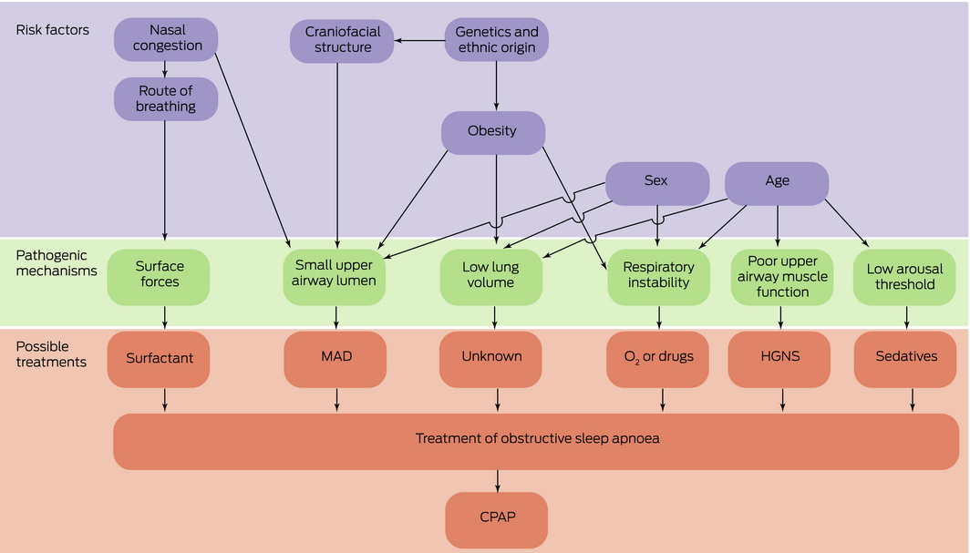
CPAP = continuous positive airway pressure; HGNS = hypoglossal nerve stimulation; MAD = mandibular advancement device.
Source: figure reproduced, with permission, from Jordan et al.12
Box 2 – STOP‐Bang screening questionnaire for obstructive sleep apnoea (OSA)35
|
|
Questions |
||||||||||||||
|
|
|||||||||||||||
|
Snoring |
Do you snore loudly (loud enough to be heard through closed doors)? |
||||||||||||||
|
Tired |
Do you often feel tired, fatigued or sleepy during the day? |
||||||||||||||
|
Observed apnoea |
Has anyone observed you stop breathing, chocking or gasping during your sleep? |
||||||||||||||
|
Blood pressure |
Do you have or are being treated for high blood pressure? |
||||||||||||||
|
BMI > 35 |
Is the BMI > 35 kg/m2? |
||||||||||||||
|
Age |
Is the patient aged > 50 years? |
||||||||||||||
|
Neck circumference |
Is the patient's neck circumference > 41 cm (women) or > 43 cm (men)? |
||||||||||||||
|
Gender |
Is the patient male? |
||||||||||||||
|
Scoring criteria for risk of OSA (1 point for each question):
|
|||||||||||||||
|
|
|||||||||||||||
|
BMI = body mass index |
|||||||||||||||
Box 3 – Factors predictive of postoperative exacerbation of obstructive sleep apnoea (OSA)
|
|
Factors |
||||||||||||||
|
|
|||||||||||||||
|
Patient features preoperatively |
|
||||||||||||||
|
Patient features postoperatively |
|
||||||||||||||
|
Medication affecting ventilatory drive and/or upper airway patency |
|
||||||||||||||
|
Surgical features |
|
||||||||||||||
|
Postoperative monitoring |
|
||||||||||||||
|
|
|||||||||||||||
|
AHI = Apnoea–Hypopnoea Index; BMI = body mass index; COPD = chronic obstructive pulmonary disease; CPAP = continuous positive airway pressure; Paco2 = arterial partial pressure of carbon dioxide; Spo2 = peripheral oxygen saturation measured by pulse oximetry. |
|||||||||||||||
Box 4 – Postoperative risk stratification and management of patients in post‐anaesthesia care unit (PACU) with confirmed or suspected obstructive sleep apnoea (OSA)*
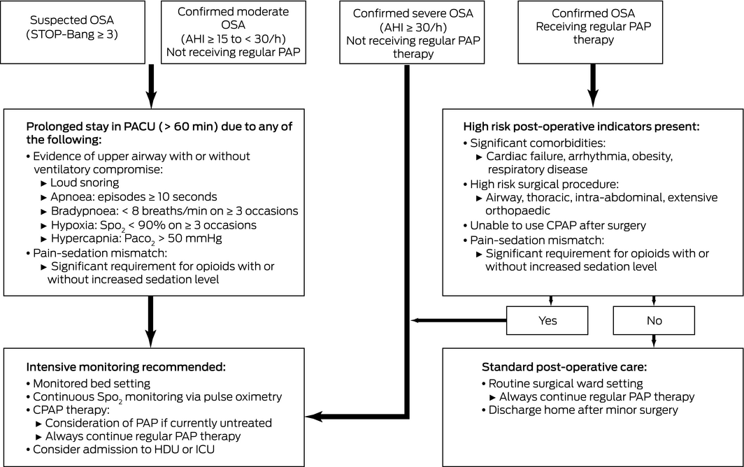
AHI = Apnoea–Hypopnoea Index; BMI = body mass index; CPAP = continuous positive airway pressure; HDU = high dependency unit; ICU = intensive care unit; Paco2 = arterial partial pressure of carbon dioxide; Pao2 = arterial partial pressure of oxygen; PAP = positive airway pressure; SpO2 = peripheral oxygen saturation measured by pulse oximetry. *Adapted from Seet and Chung51 and Hillman and Chung52 and from the ASA32 and SASM33 guidelines.
Competing interests
No relevant disclosures.
References
- Peppard PE, Young T, Barnet JH, et al. Increased prevalence of sleep‐disordered breathing in adults. Am J Epidemiol 2013; 177: 1006–1014.
- Kaw R, Chung F, Pasupuleti V, et al. Meta‐analysis of the association between obstructive sleep apnoea and postoperative outcome. Br J Anaesth 2012; 109: 897–906.
- Senaratna CV, Perret JL, Lodge CJ, et al. Prevalence of obstructive sleep apnea in the general population: a systematic review. Sleep Med Rev 2017; 34: 70–81.
- Vasu TS, Grewal R, Doghramji K. Obstructive sleep apnea syndrome and perioperative complications: a systematic review of the literature. J Clin Sleep Med 2012; 8: 199–207.
- Reed K, Pengo MF, Steier J. Screening for sleep‐disordered breathing in a bariatric population. J Thorac Dis 2016; 8: 268–275.
- Singh M, Liao P, Kobah S, et al. Proportion of surgical patients with undiagnosed obstructive sleep apnoea. Br J Anaesth 2013; 110: 629–636.
- Romero‐Corral A, Caples SM, Lopez‐Jimenez F, Somers VK. Interactions between obesity and obstructive sleep apnea: implications for treatment. Chest 2010; 137: 711–719.
- Peppard PE, Young T, Palta M, et al. Longitudinal study of moderate weight change and sleep‐disordered breathing. JAMA 2000; 284: 3015–3021.
- Gray EL, McKenzie DK, Eckert DJ. Obstructive sleep apnea without obesity is common and difficult to treat: evidence for a distinct pathophysiological phenotype. J Clin Sleep Med 2017; 13: 81–88.
- Watanabe T, Isono S, Tanaka A, et al. Contribution of body habitus and craniofacial characteristics to segmental closing pressures of the passive pharynx in patients with sleep‐disordered breathing. Am J Respir Crit Care Med 2002; 165: 260–265.
- Chen X, Wang R, Zee P, et al. Racial/ethnic differences in sleep disturbances: the Multi‐Ethnic Study of Atherosclerosis (MESA). Sleep 2015; 38: 877–888.
- Jordan AS, McSharry DG, Malhotra A. Adult obstructive sleep apnoea. Lancet 2014; 383: 736–747.
- American Academy of Sleep Medicine. International Classification of Sleep Disorders. 3rd ed. Darien, IL: American Academy of Sleep Medicine; 2014.
- Hamilton GS, Naughton MT. Impact of obstructive sleep apnoea on diabetes and cardiovascular disease. Med J Aust 2013; 199: S27–S30. https://www.mja.com.au/journal/2013/199/8/impact-obstructive-sleep-apnoea-diabetes-and-cardiovascular-disease
- Drager LF, McEvoy RD, Barbe F, et al; INCOSACT Initiative. Sleep apnea and cardiovascular disease: lessons from recent trials and need for team science. Circulation 2017; 136: 1840–1850.
- Epstein LJ, Kristo D, Strollo PJ, et al. Clinical guideline for the evaluation, management and long‐term care of obstructive sleep apnea in adults. J Clin Sleep Med 2009; 5: 263–276.
- Eckert DJ. Phenotypic approaches to obstructive sleep apnoea ‐ New pathways for targeted therapy. Sleep Med Rev 2018; 37: 45–59.
- Subramani Y, Singh M, Wong J, et al. Understanding phenotypes of obstructive sleep apnea: applications in anesthesia, surgery, and perioperative medicine. Anesth Analg 2017; 124: 179–191.
- Chung F, Liao P, Yegneswaran B, et al. Postoperative changes in sleep‐disordered breathing and sleep architecture in patients with obstructive sleep apnea. Anesthesiology 2014; 120: 287–298.
- Chung F, Liao P, Yang Y, et al. Postoperative sleep‐disordered breathing in patients without preoperative sleep apnea. Anesth Analg 2015; 120: 1214–1224.
- Mokhlesi B. Obesity hypoventilation syndrome: a state‐of‐the‐art review. Respir Care 2010; 55: 1347–1362; discussion 1363‐1365.
- Chau EH, Lam D, Wong J, et al. Obesity hypoventilation syndrome: a review of epidemiology, pathophysiology, and perioperative considerations. Anesthesiology 2012; 117: 188–205.
- Mokhlesi B, Tulaimat A. Recent advances in obesity hypoventilation syndrome. Chest 2007; 132: 1322–1336.
- Kaw R, Bhateja P, Paz Y, Mar H, et al. Postoperative complications in patients with unrecognized obesity hypoventilation syndrome undergoing elective noncardiac surgery. Chest 2016; 149: 84–91.
- Howard ME, Piper AJ, Stevens B, et al. A randomised controlled trial of CPAP versus non‐invasive ventilation for initial treatment of obesity hypoventilation syndrome. Thorax 2017; 72: 437–444.
- Manthous CA, Mokhlesi B. Avoiding management errors in patients with obesity hypoventilation syndrome. Ann Am Thorac Soc 2016; 13: 109–114.
- Chan MT, Wang CY, Seet E, et al. Association of unrecognized obstructive sleep apnea with postoperative cardiovascular events in patients undergoing major noncardiac surgery. JAMA 2019; 321: 1788–1798.
- Liao P, Yegneswaran B, Vairavanathan S, et al. Postoperative complications in patients with obstructive sleep apnea: a retrospective matched cohort study. Can J Anaesth 2009; 56: 819–828.
- Ayas NT, Laratta CR, Coleman JM, et al. Knowledge gaps in the perioperative management of adults with obstructive sleep apnea and obesity hypoventilation syndrome. An official American Thoracic Society workshop report. Ann Am Thorac Soc 2018; 15: 117–126.
- Ortiz VE, Kwo J. Obesity: physiologic changes and implications for preoperative management. BMC Anesthesiol 2015; 15: 97.
- Sood A, Abdollah F, Sammon JD, et al. The effect of body mass index on perioperative outcomes after major surgery: results from the National Surgical Quality Improvement Program (ACS‐NSQIP) 2005–2011. World J Surg 2015; 39: 2376–2385.
- American Society of Anesthesiologists Task Force on Perioperative Management of Patients With Obstructive Sleep Apnea. Practice guidelines for the perioperative management of patients with obstructive sleep apnea: an updated report by the American Society of Anesthesiologists Task Force on Perioperative Management of Patients with Obstructive Sleep Apnea. Anesthesiology 2014; 120: 268–286.
- Chung F, Memtsoudis SG, Ramachandran SK, et al. Society of Anesthesia and Sleep Medicine Guidelines on Preoperative Screening and Assessment of Adult Patients with Obstructive Sleep Apnea. Anesth Analg 2016; 123: 452–473.
- Nagappa M, Liao P, Wong J, et al. Validation of the STOP‐Bang Questionnaire as a screening tool for obstructive sleep apnea among different populations: a systematic review and meta‐analysis. PLoS One 2015; 10: e0143697.
- Chung F, Abdullah HR, Liao P. STOP‐Bang questionnaire: a practical approach to screen for obstructive sleep apnea. Chest 2016; 149: 631–638.
- Tong S, Gower J, Morgan A, et al. Noninvasive positive pressure ventilation in the immediate post‐bariatric surgery care of patients with obstructive sleep apnea: a systematic review. Surg Obes Relat Dis 2017; 13: 1227–1233.
- Mutter TC, Chateau D, Moffatt M, et al. A matched cohort study of postoperative outcomes in obstructive sleep apnea: could preoperative diagnosis and treatment prevent complications? Anesthesiology 2014; 121: 707–718.
- Abdelsattar ZM, Hendren S, Wong SL, et al. The impact of untreated obstructive sleep apnea on cardiopulmonary complications in general and vascular surgery: a cohort study. Sleep 2015; 38: 1205–1210.
- Chung F, Nagappa M, Singh M, Mokhlesi B. CPAP in the perioperative setting: evidence of support. Chest 2016; 149: 586–597.
- Liao P, Wong J, Singh M, et al. Postoperative oxygen therapy in patients with OSA: a randomized controlled trial. Chest 2017; 151: 597–611.
- Mokhlesi B, Tulaimat A, Parthasarathy S. Oxygen for obesity hypoventilation syndrome: a double‐edged sword? Chest 2011; 139: 975–977.
- Ramachandran SK, Haider N, Saran KA, et al. Life‐threatening critical respiratory events: a retrospective study of postoperative patients found unresponsive during analgesic therapy. J Clin Anesth 2011; 23: 207–213.
- Gupta K, Prasad A, Nagappa M, et al. Risk factors for opioid‐induced respiratory depression and failure to rescue: a review. Curr Opin Anaesthesiol 2018; 31: 110–119.
- Lee LA, Caplan RA, Stephens LS, et al. Postoperative opioid‐induced respiratory depression: a closed claims analysis. Anesthesiology 2015; 122: 659–665.
- Lam KK, Kunder S, Wong J, et al. Obstructive sleep apnea, pain, and opioids: is the riddle solved? Curr Opin Anaesthesiol 2016; 29: 134–140.
- Tarkkila P, Tuominen M, Lindgren L. Comparison of respiratory effects of tramadol and oxycodone. J Clin Anesth 1997; 9: 582–585.
- van der Schrier R, Jonkman K, van Velzen M, et al. An experimental study comparing the respiratory effects of tapentadol and oxycodone in healthy volunteers. Br J Anaesth 2017; 119: 1169–1177.
- Dieltjens M, Braem MJ, Van de Heyning PH, et al. Prevalence and clinical significance of supine‐dependent obstructive sleep apnea in patients using oral appliance therapy. J Clin Sleep Med 2014; 10: 959–964.
- Chung F, Liao P, Elsaid H, et al. Factors associated with postoperative exacerbation of sleep‐disordered breathing. Anesthesiology 2014; 120: 299–311.
- Yadollahi A, Gabriel JM, White LH, et al. A randomized, double crossover study to investigate the influence of saline infusion on sleep apnea severity in men. Sleep 2014; 37: 1699–1705.
- Seet E, Chung F. Management of sleep apnea in adults — functional algorithms for the perioperative period: continuing professional development. Can J Anaesth 2010; 57: 849–864.
- Hillman DR, Chung F. Anaesthetic management of sleep‐disordered breathing in adults. Respirology 2017; 22: 230–239.
- Gali B, Whalen FX, Schroeder DR, et al. Identification of patients at risk for postoperative respiratory complications using a preoperative obstructive sleep apnea screening tool and postanesthesia care assessment. Anesthesiology 2009; 110: 869–877.
- Weingarten TN, Kor DJ, Gali B, Sprung J. Predicting postoperative pulmonary complications in high‐risk populations. Curr Opin Anaesthesiol 2013; 26: 116–125.
- Chung F, Zhou L, Liao P. Parameters from preoperative overnight oximetry predict postoperative adverse events. Minerva Anestesiol 2014; 80: 1084–1095.
Provenance: Not commissioned; externally peer reviewed.
Non-Technical Errors Associated With Deaths in Surgical Care, Australia, 2012–2019, by Surgical Specialty (Australian and New Zealand Audit of Surgical Mortality)
Jesse Ey, Guy J. Maddern
Localised Herpes Simplex Following Midline Laparotomy
Jessica S. Bulluss, Paul Chee, Matthew J. Verheyden
Non‐technical errors associated with deaths in surgical care, Australia, 2012–2019, by surgical specialty (Australian and New Zealand Audit of Surgical Mortality): a retrospective cohort study
Jesse Ey, Victoria Kollias, Octavia Lee, Kelly Hou, Matheesha Herath, John B North, Ellie Treloar, Suzanne Edwards, Martin Bruening, Adam J Wells, Guy J Maddern
Changes in patient management after preoperative MRI for newly diagnosed breast cancer: a multicentre prospective observational study
Michael L Marinovich, Nehmat Houssami, Andrew Spillane, Gregory B Mann, Donna Taylor, Michelle Reintals, Nadine Phillips, Max K Bulsara, Patsy Siok Hwa Soon, Tracey Dickens, Christobel M Saunders
The first Australian evidence‐based guidelines on male infertility
Darren J Katz, Liza O’Donnell, Robert I McLachlan, Tim J Moss, Clare V Boothroyd, Veena Jayadev, Sarah R Catford
The first live term birth following uterus transplantation in Australia
Rebecca Deans, Brigitte Gerstl, Antonia W Shand, Sarah Lyons, Aaron Budden, Helen L Barrett, Grant Luxton, Mangalee Fernando, Kenneth Yong, Karen Keung, Kaushalya Arulpragasam, Henry Pleass, King Man Wan, Eva Kehag, Jana‐Emily Pittman, Mianna Lotz, Maria Fenn, Erin Nesbitt‐Hawes, Lily Byun, Katrina Tang, Mats Brannstrom, Jason Abbott