




Medical education
Lessons from practice
Volume 211 - Issue 6
5‐Fluorouracil‐induced acute coronary syndrome
Authors: Souvik K Das, Avik K Das and Maged William
Med J Aust 2019; 211 (6): 255-257.e1. || doi: 10.5694/mja2.50317
Published online: 16 September 2019
Published online: 16 September 2019
A 70- year- old man was admitted to Gosford District Hospital with exertional chest pain
Clinical record
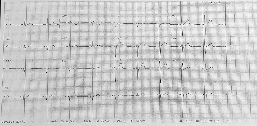
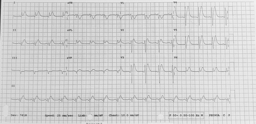
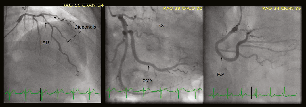
A 70‐year‐old man was admitted to Gosford District Hospital with exertional chest pain on his way back home after starting receiving 5‐fluorouracil (5‐FU) infusion for treatment of recently diagnosed stage III nasopharyngeal cancer. He did not have a significant cardiovascular history. His risk factors included a 17 pack‐year smoking history, hypertension and hyperlipidaemia. His medications on presentation were amlodipine, rosuvastatin and fenofibrate. His cardiovascular examination was unremarkable. His initial electrocardiograms (ECGs) in the emergency department (ED) revealed a sinus rhythm without any ST segment or T wave abnormalities (Box 1). Serial high sensitivity troponins were 39 ng/L and 69 ng/L, respectively. His chest x‐ray showed normal cardiac silhouette and lung vasculature. Based on his risk factors and mild troponin rise, he was diagnosed with non‐ST‐elevation myocardial infarction. He received loading doses of aspirin and ticagrelor, started taking therapeutic low molecular weight heparin, and was admitted to a monitored cardiology bed. Shortly after admission to the wards, he developed severe chest pain radiating to the left arm, with associated diaphoresis. His ECG revealed anterolateral ST segment elevation consistent with transmural ischaemia (Box 2). He was given sublingual glyceryl trinitrate (GTN), with immediate relief. Two hours later, he developed further chest pain after which he was commenced on GTN infusion, with optimal pain relief. He then underwent a coronary angiogram that revealed only minor coronary artery disease (Box 3). A transthoracic echocardiogram on the same day did not reveal any major wall motion abnormality, with an overall mildly impaired systolic function with an ejection fraction of 45–50%. Subsequently, a presumptive diagnosis of coronary vasospasm secondary to 5‐FU was made. The patient was reviewed by his regular oncologist and 5‐FU was ceased from his chemotherapy regimen.
Discussion
Fluoropyrimidines, which include 5‐FU and capecitabine, form the backbone of chemotherapy regimens for a variety of cancers. In fact, 5‐FU is the third most commonly used chemotherapeutic agent for the treatment of solid malignancies.1 Unfortunately, 5‐FU is also the second most common chemotherapeutic agent causing cardiotoxicity, with a reported incidence between 2% and 18%. Some of the reported difference in incidence can be accounted for by the differences in treatment duration, dose, additional agents, and patient characteristics.2 Capecitabine, which is an oral pro‐drug of 5‐FU, has also been associated with cardiotoxicity, with a reported incidence between 3% and 9%.2 While the most common manifestation of 5‐FU‐induced cardiotoxicity is chest pain, presentations can vary from arrhythmias, myocarditis and pericarditis, heart failure and even death.2 Of note is that 5‐FU cardiotoxicity tends to occur most commonly during the first cycle of administration. The median time to symptoms is 12 hours following an infusion, but symptoms could occur any time after commencing 5‐FU, even up to 1–2 days after infusion, thus complicating causal analysis. Clinical studies have shown that infusional regimens are associated with greater risk compared with bolus therapies.3
The potential mechanisms via which 5‐FU causes cardiotoxicity remain a matter of debate. While multiple pathways have been implicated, the two most likely are coronary vasospasm leading to myocardial ischaemia and direct drug‐related myocardial toxicity. Indeed, both experimental and physiological studies have hinted at these being the primary pathways. However, further vigorous prospective investigations in appropriate clinical settings as well as experimental models are required to provide evidence.2
Further complicating the use of a potentially life‐saving drug is the lack of predictive, clinically relevant risk factors that would enable clinicians to stratify risk and prescribe appropriately. It is interesting that previous history of cardiac diseases does not necessarily predict 5‐FU‐related toxicity, as a review demonstrated that, out of the 377 cases, only 14% had a previous history of cardiac disease.4
Despite 5‐FU‐associated cardiotoxicity being well documented, there is no widely established treatment strategy. While it is universally agreed that the first step should be to cease the infusion immediately and then administer medical anti‐anginal therapy aimed at symptomatic relief with calcium channel blockers (CCBs) and/or nitrates, the challenge of establishing a causal relationship between exposure and symptoms remains daunting. Fortunately, CCBs and nitrates have been shown to abort symptoms in up to 69% of affected patients.2 However, there remains a distinct lack of a definitive test to establish 5‐FU as the cause. Temporal relationship, while a necessary condition, does not exclude other possible contributory factors.
If the patient is at high risk of coronary artery disease, as in our example, it is advised to perform a prompt coronary angiogram to identify an alternative pathological process driving the patient's symptoms. Patients without conventional cardiac risk factors may be screened using a non‐invasive test; for example, a computed tomography coronary angiography. 5‐FU‐induced cardiotoxicity can be presumed in patients with clinically insignificant coronary disease or normal arteries.
However, presence of significant coronary stenosis does not necessarily exclude the possibility of superimposed 5‐FU‐related toxicity. To further investigate causality, invasive pharmacological provocation using acetylcholine or nitroglycerin during coronary angiography may be useful by demonstrating excessive vasospasm, thereby suggesting either coronary endothelial or primary smooth muscle dysfunction. However, this is not widely available and its role in this setting has not been validated. Moreover, there is no evidence to establish that positive provocation tests predict cardiotoxicity.2
Furthermore, rechallenging these patients with 5‐FU drugs remains controversial, as randomised clinical trials comparing different strategies are lacking.2 Rates of recurrence of cardiotoxicity as high as 90% have been reported, with death up to 13%.2 Considering the potential implications of rechallenge, it is advised to have a detailed discussion with the patient, the cardiologist and the oncologist regarding the risk–benefit analysis, taking into account the original probability of 5‐FU‐related cardiotoxicity, availability of alternative non‐5‐FU drugs, intent of therapy (ie, curative versus palliative) and the patient's own expectations. Since rechallenge may be fatal, it should only be pursued in a subset of patients for whom the benefits are thought to significantly outweigh the risk, and even then, only in a carefully monitored setting with a low threshold for discontinuation. One example would be in patients with chest pain after exposure to 5‐FU who are found to have significant coronary artery disease and undergo revascularisation and for whom no other reasonable treatment options are available.
Lessons from practice
- 5‐Fluorouracil (5‐FU) is the second most common drug associated with cardiotoxicity and can commonly present as acute coronary syndrome including ST‐elevation myocardial infarction.
- 5‐FU‐related cardiotoxicity occurs most often during the first administration, and while the median time of onset of symptoms is 12 hours, it can be delayed up to 1–2 days after exposure.
- If the history on presentation reveals current or recent exposure to 5‐FU, the first step should be to cease the infusion and subsequently administer anti‐anginal therapy with calcium channel blockers and/or nitrates. 5‐FU exposure should be flagged to the medical oncologist and the cardiologist.
- Depending on the pre‐test probability of coronary artery disease, as evaluated by the presence or absence of conventional cardiovascular risk factors, the patient should undergo either invasive testing with coronary angiography or non‐invasive imaging with computed tomography coronary angiography. 5‐FU‐related cardiotoxicity is presumed to be the cause of the cardiac injury in the absence of clinically significant coronary artery disease.
- Once a patient has been diagnosed with 5‐FU‐related cardiotoxicity, rechallenge is not advised in most patients, as it may cause death. However, a careful weighing of the risks against the potential benefits of re‐treatment is advised for each individual patient.
Competing interests
No relevant disclosures.
References
- Grem JL. 5‐Fluorouracil: forty‐plus and still ticking. A review of its preclinical and clinical development. Invest New Drugs 2000; 18: 299–313.
- Sara JD, Kaur J, Khodadadi R, et al. 5‐fluorouracil and cardiotoxicity: a review. Ther Adv Med Oncol 2018; 10: 175883591878014.
- Basselin C, Fontanges T, Descotes J, et al. 5‐Fluorouracil‐induced Takotsubo‐like syndrome. Pharmacotherapy 2011; 31: 226.
- Saif MW, Shah MM, Shah AR. Fluoropyrimidine‐associated cardiotoxicity: revisited. Expert Opin Drug Saf 2009; 8: 191–202.
Provenance: Not commissioned; externally peer reviewed.
Fatty Liver Disease in Australia: A Narrative Review on the Epidemiology, Natural History, Prognostication and Management in People With Metabolic Dysfunction
Karl Vaz, Daniel Clayton-Chubb, William W. Kemp, Stuart K. Roberts, Ammar Majeed
Sex Differences in Management and Outcomes of People With ST-Elevation Myocardial Infarction, New South Wales, 2011–2020: A Retrospective Cohort Study
Adam C. Livori, Jedidiah I. Morton, Dianna J. Magliano, Jonathan E. Shaw
Rethinking Rheumatic Fever Diagnosis: Progress, Pitfalls and the Path Towards a Definitive Test
Anna P. Ralph, Rachel Webb, Tom Parks, Nicole J. Moreland, Asha C. Bowen, Benjamin Jones, Emma Ndagire, Andrea Z. Beaton
Sex Differences in Management and Outcomes of People With ST-Elevation Myocardial Infarction, New South Wales, 2011–2020: A Retrospective Cohort Study
Samia Kazi, Simone Marschner, Clara K. Chow
Distribution of Cardiovascular Disease Risk Based on the Updated 2023 Guideline-Recommended Australian Cardiovascular Disease Risk Algorithm and Comparison With the 2012 Algorithm: An Observational Study
Nina Lazarevic, Meghana Bhat, Grace Joshy, Danielle C. Butler, Mark Woodward, Anushka Patel, Rod T. Jackson, Garry Jennings, Rosemary Wyber, Ellie Paige, Emily Banks
They Brought the Hospital to Me
Hugh N. Burrill