




Volume 211 - Issue 2
Pulmonary embolism: update on diagnosis and management
Authors: Paul C Kruger, John W Eikelboom, James D Douketis and Graeme J Hankey
Med J Aust 2019; 211 (2): 82-87. || doi: 10.5694/mja2.50233
Published online: 15 July 2019
Published online: 15 July 2019
Pulmonary embolism is a potentially life-threatening condition, mandating urgent diagnosis and treatment
Summary
- Pulmonary embolism (PE) is a potentially life‐threatening condition, mandating urgent diagnosis and treatment.
- The symptoms of PE may be non‐specific; diagnosis therefore relies on a clinical assessment and objective diagnostic testing.
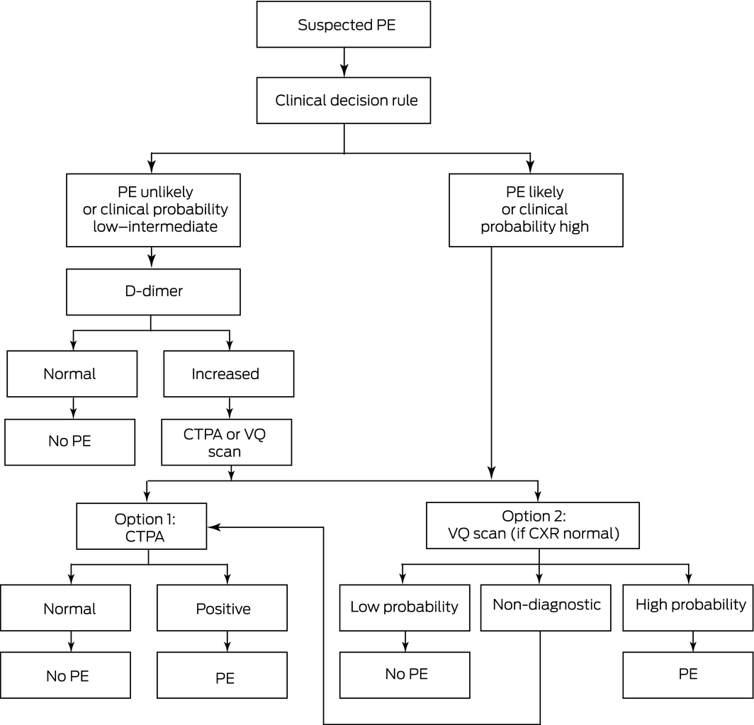
- A clinical decision rule can determine the pre‐test probability of PE. If PE is “unlikely”, refer for a D‐dimer test. If the D‐dimer result is normal, PE can be excluded. If D‐dimer levels are increased, refer for chest imaging. If PE is “likely”, refer for chest imaging.
- Imaging with computed tomography pulmonary angiogram is accurate and preferred for diagnosing PE, but may detect asymptomatic PE of uncertain clinical significance.
- Imaging with ventilation–perfusion (VQ) scan is associated with lower radiation exposure than computed tomography pulmonary angiogram, and may be preferred in younger patients and pregnancy. A low probability or high probability VQ scan is helpful for ruling out or confirming PE, respectively; however, an intermediate probability VQ scan requires further investigation.
- The direct oral anticoagulants have expanded the anticoagulation options for PE. These are the preferred anticoagulant for most patients with PE because they are associated with a lower risk of bleeding, and have the practical advantages of fixed dosage, no need for routine monitoring, and fewer drug interactions compared with vitamin K antagonists. Initial parenteral treatment is required before dabigatran and edoxaban.
Pulmonary embolism (PE) is characterised by embolic occlusion of one or more pulmonary arteries. The incidence of symptomatic PE is estimated to be about 0.5–1 per 1000 people per year, and is increasing as the population ages. PE may also be asymptomatic and undetected.1 It has different clinical manifestations from deep vein thrombosis (DVT), and may result in chronic thromboembolic pulmonary hypertension (CTEPH) and mortality.2 This article focuses on the diagnosis and management of PE, and refers to an accompanying MJA article on DVT regarding evidence that is common to both DVT and PE.3
We have used clinical guidelines from the Thrombosis and Haemostasis Society of Australia and New Zealand (THANZ) to formulate evidenced‐based recommendations.4 Evidence supporting the treatment recommendations has been graded according to National Health and Medical Research Council criteria (Box 1).5
Diagnosis
Symptoms and signs of PE
The clinical manifestations of PE range from no symptoms to sudden death, depending on the degree of obstruction of the pulmonary vasculature, and the cardiovascular reserve of the patient. Symptoms include dyspnoea (82% of patients), central or pleuritic chest pain (49%), cough (20%), syncope (14%) and haemoptysis (7%).1 Clinical examination may reveal tachycardia, cyanosis, tachypnoea, low grade fever, and signs of right ventricular dysfunction that include distended jugular veins, tricuspid regurgitation and accentuated pulmonary component of the second heart sound. Up to 50% of PEs are asymptomatic.2
Pre‐test clinical decision rule: the simplified Wells rule to exclude PE
In patients with suspected PE, clinical decision rules are helpful to determine the pre‐test probability of PE and guide further investigation.6 For example, the simplified Wells rule assigns one point to various clinical factors to determine an “unlikely” (≤ 1 point) or “likely” (≥ 2 points) clinical pre‐test probability, as shown in Box 5 of the THANZ guidelines.4
Laboratory testing
The utility of the D‐dimer test for diagnosis of PE is similar to DVT,3 with a median sensitivity of 95–97% and median specificity of 39–43%.7
Medical imaging
In patients with suspected PE, a chest x‐ray may help identify alternative diagnoses such as pneumonia, heart failure or pneumothorax. Two objective non‐invasive imaging modalities — multidetector row computed tomography pulmonary angiogram (CTPA) and ventilation–perfusion (VQ) scan — are used to assess patients with suspected PE.8,9 Deciding which modality is most suitable depends on availability of the imaging equipment, chest x‐ray findings (if normal, VQ scan is reasonable, but if abnormal, CTPA is superior to VQ for finding alternative diagnoses), contraindications to iodinated contrast media used in CTPA, and concerns about radiation exposure (which are higher for CTPA than VQ scan), especially in women of childbearing age.
CTPA involves peripheral intravenous injection of iodinated contrast followed by a multidetector row computed tomography scan of the chest. A filling defect within a pulmonary artery indicates a PE. CTPA can visualise the pulmonary arteries down to the subsegmental level. The sensitivity of CTPA is 83% (95% CI, 76–92%) and the specificity is 96% (95% CI, 93–97%) in the largest trial to date.10 In patients with a “likely” PE, the positive predictive value of CTPA is 96%.10 In patients with an “unlikely” PE, the negative predictive value is 96% (92–98%); a negative CTPA can therefore accurately rule out PE (evidence level A).9,10
CTPA is the preferred imaging modality for most patients with suspected PE as it is highly accurate and can detect alternative diagnoses. Potential disadvantages of CTPA include radiation exposure and allergic reactions to iodinated contrast (anaphylaxis in about 1 in 100 000 patients), and acute kidney injury. Severe renal impairment is not an absolute contraindication to establishing the diagnosis by CTPA because the potential mortality associated with undiagnosed, untreated PE exceeds the treatable risk of acute kidney injury.11
A VQ scan involves administering aerosolised radiolabelled xenon to visualise bronchial ventilation and radiolabelled intravenous albumin to visualise the pulmonary circulation. The scan result is classified as low, intermediate or high probability for a diagnosis of PE.8 A low probability VQ scan reliably excludes PE because it has a high negative predictive value (evidence level A).12 An intermediate probability VQ scan neither confirms nor excludes PE, and indicates the need for bilateral lower limb compression ultrasound to identify an embolic source, or consideration of a CTPA. A high probability VQ scan, which is characterised by absent perfusion in well ventilated lung segments, has a positive predictive value for PE of > 90%.13 An advantage of a VQ scan is that the exposure to ionising radiation is lower than in CTPA, which makes it safer for younger patients and women of childbearing age or who are pregnant (see below). VQ scan limitations include a non‐diagnostic or “intermediate probability” result, which is as high as 50% in some studies, and inability to identify alternative diagnoses.12
Diagnosing first PE using integrated information from clinical presentation, clinical decision rule and investigations
Symptoms and signs of PE are neither sensitive nor specific. An integrated diagnostic approach using a clinical decision rule and confirmatory investigations assists in diagnosis (Box 2).
If PE is “unlikely” (simplified Wells rule ≤ 1) a D‐dimer test is indicated. In patients with “unlikely” PE, a negative D‐dimer result has a negative predictive value of 99.7% and effectively rules out PE without chest imaging (evidence level A).14 A positive D‐dimer result should prompt a CTPA or VQ scan.
If PE is “likely” (simplified Wells rule ≥ 2), rapid CTPA or VQ imaging is indicated. In this risk group, an abnormality in a segmental or larger vessel on CTPA has a high positive predictive value for PE.10 VQ scan has similar diagnostic accuracy to CTPA, provided that the chest x‐ray is normal; however, VQ scans have a higher risk of being non‐diagnostic in patients with an abnormal chest x‐ray. In a high clinical risk patient with a negative CTPA or VQ scan showing a low probability of PE, anticoagulation is not indicated because the negative predictive value of objective testing is high.
Investigations to exclude other diagnoses
Electrocardiogram and chest x‐ray can help identify some differential diagnoses of PE, including myocardial infarction, pericarditis, congestive heart failure, pneumothorax and pneumonia. The most common but non‐specific electrocardiogram manifestation of PE is sinus tachycardia, whereas other electrocardiogram features such as right ventricular strain (deep S wave in lead I, Q wave in lead III, T wave inversion in leads III and aVF) are less common.15 Similarly, chest x‐ray features of PE, which include pleural effusion, atelectasis and consolidation related to pulmonary infarction, are non‐specific for PE.3
Right ventricular systolic dysfunction, as suggested by increased blood concentrations of troponin T or I, or brain natriuretic peptide or echocardiographic features of right ventricular hypokinesis, typically correlates with the extent of pulmonary vascular obstruction and is a predictor of all‐cause mortality.16,17,18 However, acute right ventricular dysfunction is common after PE and typically resolves within a few weeks in most patients. If right ventricular dysfunction is identified when PE is diagnosed, a repeat echocardiogram after 3 months of anticoagulation is warranted whether the patient has cardiorespiratory symptoms or not, because CTEPH can be asymptomatic. Persistent right ventricular dysfunction may indicate lung imaging and right heart catheterisation to further investigate for CTEPH.19
Prognosis
Survival
PE is associated with a mortality risk of about 15% at 3 months.2 The simplified Pulmonary Embolism Severity Index (PESI) tool estimates survival and can help guide the decision for hospitalisation (Box 3). A simplified PESI score of 0 denotes low risk (30‐day mortality about 1%), whereas a score of 1 or more denotes high risk (30‐day mortality about 10.9%).20 Survival risk is also influenced by the pulmonary artery pressure. At the time of diagnosis of PE, mean pulmonary artery pressure ≥ 30 mmHg is associated with an increased risk of progressive pulmonary hypertension, and mean pulmonary artery pressure ≥ 50 mmHg is associated with < 20% survival at 2 years.21
Chronic thromboembolic pulmonary hypertension
CTEPH should be considered in patients who have been diagnosed with PE and have ongoing dyspnoea, fatigue and/or chest pain 3–6 months after diagnosis. Patients with suspected CTEPH should undergo echocardiography to identify right ventricular dysfunction and if this is present, a VQ scan is indicated. The presence of persistent VQ mismatches warrants further assessment in a specialised pulmonary hypertension clinic to consider right heart catheterisation and pulmonary thromboendarterectomy.22
Management
Treatment of PE aims to prevent thrombus progression and recurrent embolisation, and decrease the risk of mortality and CTEPH. Patients with a simplified PESI score of 0 (and selected patients with a simplified PESI score ≥ 1; for example, a patient with cancer but no other risk factors) may be considered for outpatient management or early hospital discharge with therapeutic anticoagulation, provided they have adequate post‐discharge follow‐up.23 Hospitalisation is considered in patients with a simplified PESI score ≥ 1 or with either haemodynamic instability, need for oxygen or parenteral analgesics, or comorbidities.24
Anticoagulation
The anticoagulant management for acute PE is generally the same as for DVT (see Box 4 of the accompanying MJA article),3 although thrombolytic therapy is considered more frequently. In patients being considered for thrombolytic therapy, treatment may be initiated with intravenous unfractionated heparin or low molecular weight heparin instead of a direct oral anticoagulant, because heparins have a shorter half‐life.25
When PE is suspected and there is a delay in objective testing, empirical anticoagulation is recommended if there is a moderate to high clinical suspicion of PE. When clinical suspicion of PE is low, it is reasonable to wait up to 24 hours for the results.26
Duration of anticoagulation
As with DVT, the duration of anticoagulation for PE is determined by the underlying cause (eg, risk factors) and the estimated risk of recurrence after stopping anticoagulation. Three months of anticoagulation is recommended for treatment of provoked PE associated with surgery (evidence level A), PE associated with non‐surgical transient risk factors (eg, oestrogen therapy, pregnancy and puerperium, being confined to bed with an acute illness, leg injury with associated reduced mobility for at least 3 days), and unprovoked PE in patients at high risk of bleeding. At least 6 months of anticoagulation is recommended for PE that is associated with active cancer (evidence level A). Extended anticoagulation therapy is recommended for unprovoked PE if the long term risk of bleeding is acceptable (low or moderate), and for patients with PE and active cancer.27 All patients receiving extended therapy should have anticoagulation reassessed at least annually to re‐evaluate their individual risk of thrombosis and bleeding, monitor for anticoagulant‐related adverse effects, and assess renal and hepatic function, which can affect anticoagulant safety.
Thrombolysis
Systemic thrombolysis involves intravenous administration of a thrombolytic agent which accelerates the resolution of PE compared with anticoagulation but increases the bleeding risk. There are two situations where thrombolytic therapy should be considered. The first is patients with clinically massive PE, manifest by systemic hypotension (systolic blood pressure < 90 mmHg or cardiogenic shock) or impending respiratory failure and acceptable bleeding risk (evidence level C). The second is clinically apparent haemodynamic deterioration after starting anticoagulation in conjunction with a bleeding risk that is not unacceptably high.27 However, there are relative contraindications to thrombolysis in selected patients; for example, those with recent (within 2 weeks) bleeding, an intracranial mass or vascular malformation, or recent (within 2 weeks) surgery.28 Hence, the potential benefits of thrombolysis should be weighed against the bleeding risks on a case‐by‐case basis (eg, type of recent surgery and post‐surgical haemostasis, clinical severity of PE).29,30 Moreover, thrombolytic therapy can be adjusted according to bleeding risk; for example, using a lower dose parenterally or direct administration by pulmonary catheter.
Catheter‐directed thrombolysis involves inserting a catheter into the pulmonary artery and infusing a thrombolytic directly into the PE. The treatment appears to be as effective as systemic thrombolysis for thrombus removal but is more difficult to undertake.27 In an observational study of 2060 patients with PE, the risk of in‐hospital mortality and major bleeding was lower with catheter‐directed thrombolysis compared with systemic thrombolysis (7.9% v 15.5%; P < 0.001), and the risk of intracranial haemorrhage was not significantly different (1.0% v 0.9%; P = 0.849).31 Limited evidence suggests that, if available, catheter‐directed thrombolysis is preferred to systemic thrombolysis in patients with risk factors for bleeding.27,32
Thrombus removal strategies
Evidence for catheter‐based thrombus removal for acute PE is limited because it has not been evaluated without thrombolytic therapy. However, it may be an option when a patient is hypotensive and has a high bleeding risk, failed systemic thrombolysis, or shock that is likely to cause death before systemic thrombolysis can take effect.28 Surgical pulmonary embolectomy may be considered in patients with a massive PE that is surgically accessible and who have absolute contraindications to thrombolysis.
Special circumstances that may influence diagnosis and management
Pregnancy may normally cause dyspnoea and increase D‐dimer levels, which can obscure the diagnosis of PE.33 Clinical judgement and a high index of suspicion for diagnosing PE is therefore required.34 Ionising radiation from CTPA and VQ scan carries risks for mother and fetus. For the mother, the average whole body dose of radiation associated with CTPA ranges from 2 to 10 mSv, and with VQ scan from 0.6 to 1.5 mSv.35 The lifetime risk of breast cancer may be increased after exposure to radiation from CTPA.35,36 For the developing fetus, exposure to ionising radiation has teratogenic and carcinogenic risks which are dependent on the stage of fetal development (highest risk during first trimester) and fetal absorbed dose.37,38 The risk of miscarriage or major fetal malformations is negligible when the fetus is exposed to < 50 mGy of ionising radiation.38 The estimated fetal doses of radiation from CTPA and VQ scan are within acceptable limits at about 0.66 mGy and 0.6 mGy, respectively.39,40 The carcinogenic risk for the fetus is controversial. The International Commission on Radiological Protection estimates that exposure to 30 mGy is associated with one cancer per 500 fetuses.41 Despite these risks, it is important for pregnant or post partum women with suspected PE to undergo prompt diagnostic work‐up, including imaging, because the risk of ionising radiation from imaging is lower than the risks of undetected PE.42 VQ scan is considered the chest imaging investigation of first choice for a young woman, and radiation dose reduction technologies should be used if available.42 CTPA should be used in pregnant women with an abnormal chest x‐ray, or when compression ultrasound or VQ are non‐diagnostic.43 As for DVT, treatment for PE during pregnancy typically involves low molecular weight heparin, which does not cross the placenta.44
Unresolved issues
Incidental, asymptomatic PE has been disclosed by multidetector CT, performed increasingly for other indications, with prevalence of about 2.6% overall and 3.1% in patients with malignancy.45 Up to 50% of incidental PEs may be found in the main and lobar pulmonary arteries.46 Management is uncertain due to a lack of high quality evidence. Anticoagulation is recommended if the patient has symptoms that could be attributable to the PE; the PE is located in the main, lobar and/or segmental pulmonary arteries; concurrent DVT is present; or the patient has active cancer.26,47,48,49 Anticoagulation may not be necessary if the patient has a single subsegmental PE and no additional VTE risk factors.49,50
Isolated symptomatic subsegmental pulmonary embolism (SSPE) also poses a management challenge and has become more frequently detected following the introduction of CTPA.51 While it has been suggested that SSPE represents an overdiagnosis of PE in which the risks of anticoagulation outweigh the benefits,52,53 two studies report that patients with SSPE have a higher risk of recurrence and mortality than patients without PE,54 thus supporting anticoagulation for SSPE.51 Factors that support anticoagulation for SSPE include concurrent DVT, which increases the risk of recurrent VTE, and persistent risk factors for VTE, which are common in patients with SSPE. In patients with SSPE and a contraindication to anticoagulation or risk factors for bleeding, an alternative to anticoagulation is to perform serial compression ultrasound to investigate for new proximal DVT before it leads to recurrent PE, provided that the patient has adequate cardiopulmonary reserve and does not have risk factors for recurrent VTE.51,55 A study of the safety of withholding anticoagulation in patients with negative serial compression ultrasound is currently recruiting patients (NCT01455818).
The increasing availability of CTPA has created a tendency for clinicians to test more patients for suspected PE.53 Due to an increase in the use of diagnostic imaging for suspected PE, the proportion of patients undergoing testing who are diagnosed with PE has decreased from ~ 20% in the 1990s to ~ 5% in the 2010s.56 This potential overutilisation of CTPA may be decreased by restricting testing to patients with low clinical suspicion who have a positive D‐dimer result and those with a moderate to high clinical suspicion for PE.53 The PE rule‐out criteria (PERC) rule along with the Wells and Geneva clinical decision rules identify patients in the emergency department setting whose probability of PE is so low as to justify not measuring D‐dimer levels (Box 4).29 Patients are suitable for assessment by the PERC rule if PE is “unlikely” and they are aged < 50 years. If all eight PERC variables are negative, PE can be excluded without D‐dimer testing or imaging (evidence level C).57 The PROPER trial showed that the use of a PERC‐based strategy was non‐inferior to the usual diagnostic strategy using D‐dimer and CTPA for the rate of subsequent thromboembolic events among low risk emergency department patients.58
Conclusion
PE is a potentially fatal condition that requires prompt diagnosis and treatment. With the increasing availability of rapid and accurate medical imaging which can detect asymptomatic PEs, the incidence of PE appears to be increasing. Direct oral anticoagulants have expanded the available treatment options and are now recommended as a first line anticoagulant treatment for most patients. Further information on the diagnosis and treatment of PE is available from the THANZ guidelines.4
Box 1 – National Health and Medical Research Council levels of evidence5
Level |
Evidence base | ||||||||||||||
A |
Excellent |
One or more level I studies with a low risk of bias, or several level II studies with a low risk of bias |
|||||||||||||
B |
Good |
One or two level II studies with a low risk of bias, or a systematic review or several level III studies with a low risk of bias |
|||||||||||||
C |
Satisfactory |
One or two level III studies with a low risk of bias, or level I or II studies with a moderate risk of bias |
|||||||||||||
D |
Poor |
Level IV studies, or level I–III studies/systematic reviews with a high risk of bias |
|||||||||||||
Box 2 – Diagnosing first pulmonary embolism (PE) using integrated information from clinical presentation, clinical decision rule and investigations
CTPA = computed tomography pulmonary angiogram; CXR = chest x‐ray; VQ = ventilation–perfusion. ◆
Box 3 – Simplified Pulmonary Embolism Severity Index20
Variable |
Score | ||||||||||||||
Age > 80 years |
1 |
||||||||||||||
History of cancer |
1 |
||||||||||||||
Chronic cardiopulmonary disease* |
1 |
||||||||||||||
Pulse ≥ 110 beats/min |
1 |
||||||||||||||
Systolic blood pressure < 100 mmHg |
1 |
||||||||||||||
Arterial oxyhaemoglobin saturation < 90% |
1 |
||||||||||||||
The simplified Pulmonary Embolism Severity Index assigns one point to each of the above variables. A total score of 0 denotes low risk and a total score ≥ 1 denotes high risk of mortality.Combination of history of heart failure and history of chronic lung disease. ◆ | |||||||||||||||
Box 4 – Pulmonary embolism rule‐out criteria (PERC) rule57
- Arterial oxygen saturation ≤ 94%
- Pulse rate ≥ 100 beats/min
- Age ≥ 50 years
- Unilateral leg swelling
- Haemoptysis
- Recent trauma or surgery
- Prior pulmonary embolism or deep vein thrombosis
- Exogenous oestrogen use
Competing interests
No relevant disclosures.
References
- Belohlavek J, Dytrych V, Linhart A. Pulmonary embolism, part I: epidemiology, risk factors and risk stratification, pathophysiology, clinical presentation, diagnosis and nonthrombotic pulmonary embolism. Exp Clin Cardiol 2013; 18: 129–138.
- Goldhaber SZ, Visani L, De Rosa M. Acute pulmonary embolism: clinical outcomes in the International Cooperative Pulmonary Embolism Registry (ICOPER). Lancet 1999; 353: 1386–1389.
- Kruger PC, Eikelboom JW, Douketis JD, Hankey GJ. Deep vein thrombosis: update on diagnosis and management. Med J Aust 2019; 210: xxx–xxx.
- Tran HA, Gibbs H, Merriman E, et al. New guidelines from the Thrombosis and Haemostasis Society of Australia and New Zealand for the diagnosis and management of venous thromboembolism. Med J Aust 2019; 210: 227–235. https://www.mja.com.au/journal/2019/210/5/new-guidelines-thrombosis-and-haemostasis-society-australia-and-new-zealand.
- National Health and Medical Research Council. NHMRC additional levels of evidence and grades for recommendations for developers of guidelines. Canberra: NHMRC, 2009. https://www.mja.com.au/sites/default/files/NHMRC.levels.of.evidence.2008-09.pdf (viewed May 2019).
- Gibson NS, Sohne M, Kruip MJ, et al. Further validation and simplification of the Wells clinical decision rule in pulmonary embolism. Thromb Haemost 2008; 99: 229–234.
- Di Nisio M, Squizzato A, Rutjes AW, et al. Diagnostic accuracy of D‐dimer test for exclusion of venous thromboembolism: a systematic review. J Thromb Haemost 2007; 5: 296–304.
- Righini M, Robert‐Ebadi H, Le Gal G. Diagnosis of acute pulmonary embolism. J Thromb Haemost 2017; 15: 1251–1261.
- Mos IC, Klok FA, Kroft LJ, et al. Safety of ruling out acute pulmonary embolism by normal computed tomography pulmonary angiography in patients with an indication for computed tomography: systematic review and meta‐analysis. J Thromb Haemost 2009; 7: 1491–1498.
- Stein PD, Fowler SE, Goodman LR, et al. Multidetector computed tomography for acute pulmonary embolism. N Engl J Med 2006; 354: 2317–2327.
- Royal Australian and New Zealand College of Radiologists Faculty of Clinical Radiology. Iodinated contrast media guideline. Sydney: RANZR, 2018. https://www.ranzcr.com/college/document-library/ranzcr-iodinated-contrast-guidelines;
- Anderson DR, Kahn SR, Rodger MA, et al. Computed tomographic pulmonary angiography vs ventilation‐perfusion lung scanning in patients with suspected pulmonary embolism: a randomized controlled trial. JAMA 2007; 298: 2743–2753.
- Stein PD, Woodard PK, Weg JG, et al. Diagnostic pathways in acute pulmonary embolism: recommendations of the PIOPED II Investigators. Radiology 2007; 242: 15–21.
- Pasha SM, Klok FA, Snoep JD, et al. Safety of excluding acute pulmonary embolism based on an unlikely clinical probability by the Wells rule and normal D‐dimer concentration: a meta‐analysis. Thromb Res 2010; 125: e123–e127.
- Rodger M, Makropoulos D, Turek M, et al. Diagnostic value of the electrocardiogram in suspected pulmonary embolism. Am J Cardiol 2000; 86: 807–809, A10.
- Klok FA, Mos IC, Huisman MV. Brain‐type natriuretic peptide levels in the prediction of adverse outcome in patients with pulmonary embolism: a systematic review and meta‐analysis. Am J Respir Crit Care Med 2008; 178: 425–430.
- Jimenez D, Uresandi F, Otero R, et al. Troponin‐based risk stratification of patients with acute nonmassive pulmonary embolism: systematic review and metaanalysis. Chest 2009; 136: 974–982.
- Ribeiro A, Lindmarker P, Juhlin‐Dannfelt A, et al. Echocardiography Doppler in pulmonary embolism: right ventricular dysfunction as a predictor of mortality rate. Am Heart J 1997; 134: 479–487.
- Matthews JC, McLaughlin V. Acute right ventricular failure in the setting of acute pulmonary embolism or chronic pulmonary hypertension: a detailed review of the pathophysiology, diagnosis, and management. Curr Cardiol Rev 2008; 4: 49–59.
- Jimenez D, Aujesky D, Moores L, et al. Simplification of the pulmonary embolism severity index for prognostication in patients with acute symptomatic pulmonary embolism. Arch Intern Med 2010; 170: 1383–1389.
- Gerges C, Skoro‐Sajer N, Lang IM. Right ventricle in acute and chronic pulmonary embolism (2013 Grover Conference series). Pulm Circ 2014; 4: 378–386.
- Gopalan D, Delcroix M, Held M. Diagnosis of chronic thromboembolic pulmonary hypertension. Eur Respir Rev 2017; 26: 160108.
- Roy PM, Moumneh T, Penaloza A, Sanchez O. Outpatient management of pulmonary embolism. Thromb Res 2017; 155: 92–100.
- Sanchez O, Trinquart L, Caille V, et al. Prognostic factors for pulmonary embolism: the prep study, a prospective multicenter cohort study. Am J Respir Crit Care Med 2010; 181: 168–173.
- Hepburn‐Brown M, Darvall J, Hammerschlag G. Acute pulmonary embolism: a concise review of diagnosis and management. Intern Med J 2019; 49: 15–27.
- Kearon C, Akl EA, Comerota AJ, et al. Antithrombotic therapy for VTE disease: Antithrombotic Therapy and Prevention of Thrombosis, 9th ed: American College of Chest Physicians Evidence‐Based Clinical Practice Guidelines. Chest 2012; 141: e419S–e496S.
- Kearon C, Akl EA, Ornelas J, et al. Antithrombotic Therapy for VTE Disease: CHEST Guideline and Expert Panel Report. Chest 2016; 149: 315–352.
- Zarghouni M, Charles HW, Maldonado TS, Deipolyi AR. Catheter‐directed interventions for pulmonary embolism. Cardiovasc Diagn Ther 2016; 6: 651–661.
- Konstantinides SV, Torbicki A, Agnelli G, et al. 2014 ESC guidelines on the diagnosis and management of acute pulmonary embolism. Eur Heart J 2014; 35: 3033–69, 3069a – 3069k.
- Daley MJ, Murthy MS, Peterson EJ. Bleeding risk with systemic thrombolytic therapy for pulmonary embolism: scope of the problem. Ther Adv Drug Saf 2015; 6: 57–66.
- Arora S, Lahewala S, Patel P, et al. Abstract 15240: Catheter‐directed thrombolysis versus systemic thrombolysis in pulmonary embolism: predictors of in‐hospital mortality and major bleeding. Circulation 2018; 134: A15240.
- Sista AK, Kearon C. Catheter‐directed thrombolysis for pulmonary embolism: where do we stand? JACC Cardiovasc Interv 2015; 8: 1393–1395.
- Kline JA, Williams GW, Hernandez‐Nino J. D‐dimer concentrations in normal pregnancy: new diagnostic thresholds are needed. Clin Chem 2005; 51: 825–829.
- Leung AN, Bull TM, Jaeschke R, et al. An official American Thoracic Society/Society of Thoracic Radiology clinical practice guideline: evaluation of suspected pulmonary embolism in pregnancy. Am J Respir Crit Care Med 2011; 184: 1200–1208.
- Cook JV, Kyriou J. Radiation from CT and perfusion scanning in pregnancy. BMJ 2005; 331: 350.
- Lahham A, ALMasri H, Kameel S. Estimation of female radiation doses and breast cancer risk from chest CT examination. Radiat Prot Dosimetry 2018; 179: 303–309.
- Royal College of Obstetricians and Gynaecologists. Thromboembolic disease in pregnancy and the puerperium: acute management (Green‐top Guideline No.37b). London: RCOG, 2015.
- Nguyen CP, Goodman LH. Fetal risk in diagnostic radiology. Semin Ultrasound CT MR 2012; 33: 4–10.
- Hurwitz LM, Yoshizumi T, Reiman RE, et al. Radiation dose to the fetus from body MDCT during early gestation. Am J Roentgenol 2006; 186: 871–876.
- Niemann T, Nicolas G, Roser HW, et al. Imaging for suspected pulmonary embolism in pregnancy‐what about the fetal dose? A comprehensive review of the literature. Insights Imaging 2010; 1: 361–372.
- International Commission on Radiological Protection. Pregnancy and medical radiation. Ann ICRP 2000; 30: iii–viii, 1–43.
- Roy PM, Meyer G, Vielle B, et al. Appropriateness of diagnostic management and outcomes of suspected pulmonary embolism. Ann Intern Med 2006; 144: 157–164.
- Mayo J, Thakur Y. Pulmonary CT angiography as first‐line imaging for PE: image quality and radiation dose considerations. Am J Roentgenol 2013; 200: 522–528.
- McLintock C, Brighton T, Chunilal S, et al. Recommendations for the diagnosis and treatment of deep venous thrombosis and pulmonary embolism in pregnancy and the postpartum period. Aust N Z J Obstet Gynaecol 2012; 52: 14–22.
- Dentali F, Ageno W, Becattini C, et al. Prevalence and clinical history of incidental, asymptomatic pulmonary embolism: a meta‐analysis. Thromb Res 2010; 125: 518–522.
- O'Connell CL, Boswell WD, Duddalwar V, et al. Unsuspected pulmonary emboli in cancer patients: clinical correlates and relevance. J Clin Oncol 2006; 24: 4928–4932.
- Vedovati MC, Germini F, Agnelli G, Becattini C. Prognostic role of embolic burden assessed at computed tomography angiography in patients with acute pulmonary embolism: systematic review and meta‐analysis. J Thromb Haemost 2013; 11: 2092–2102.
- den Exter PL, Hooijer J, Dekkers OM, Huisman MV. Risk of recurrent venous thromboembolism and mortality in patients with cancer incidentally diagnosed with pulmonary embolism: a comparison with symptomatic patients. J Clin Oncol 2011; 29: 2405–2409.
- Chiu V, O'Connell C. Management of the incidental pulmonary embolism. Am J Roentgenol 2017; 208: 485–488.
- Di Nisio M, Carrier M. Incidental venous thromboembolism: is anticoagulation indicated? Hematology Am Soc Hematol Educ Program 2017; 2017(1): 121–127.
- Raskob GE. Importance of subsegmental pulmonary embolism. Blood 2013; 122: 1094–1095.
- Carrier M, Righini M, Wells PS, et al. Subsegmental pulmonary embolism diagnosed by computed tomography: incidence and clinical implications. A systematic review and meta‐analysis of the management outcome studies. J Thromb Haemost 2010; 8: 1716–1722.
- Wiener RS, Schwartz LM, Woloshin S. Time trends in pulmonary embolism in the United States: evidence of overdiagnosis. Arch Intern Med 2011; 171: 831–837.
- den Exter PL, van Es J, Klok FA, et al. Risk profile and clinical outcome of symptomatic subsegmental acute pulmonary embolism. Blood 2013; 122: 1144–1149; quiz 1329.
- Carrier M, Klok FA. Symptomatic subsegmental pulmonary embolism: to treat or not to treat? Hematology Am Soc Hematol Educ Program 2017; 2017(1): 237–241.
- Le Gal G, Bounameaux H. Diagnosing pulmonary embolism: running after the decreasing prevalence of cases among suspected patients. J Thromb Haemost 2004; 2: 1244–1246.
- Kline JA, Mitchell AM, Kabrhel C, et al. Clinical criteria to prevent unnecessary diagnostic testing in emergency department patients with suspected pulmonary embolism. J Thromb Haemost 2004; 2: 1247–1255.
- Freund Y, Cachanado M, Aubry A, et al. Effect of the Pulmonary Embolism Rule‐Out Criteria on subsequent thromboembolic events among low‐risk emergency department patients: the PROPER randomized clinical trial. JAMA 2018; 319: 559–566.
Provenance: Commissioned; externally peer reviewed.
Splenic injury in severe cases of the zoonoses Q fever and rickettsial infection: diagnostic challenges
Ashleigh Drury, Philippa Harrison, Aiveen Bannan
Health and economic benefits of improving pre‐hospital identification of stroke in Australian women: a modelling study
Thomas Gadsden, Lei Si, Emily R Atkins, Cheryl Carcel, Xia Wang, Stephen Jan, Mark Woodward, Laura E Downey
Updating the diagnosis and management of elevated serum ferritin levels in the era of routine ferritin testing of blood donors by Australian Red Cross Lifeblood
Gary D Zhang, James Chen, Daniel M Johnstone, Martin B Delatycki, Katie Allen, John K Olynyk
Early cardiovascular collapse after envenoming by snakes in Australia, 2005–2020: an observational study (ASP‐31)
Geoffrey K Isbister, Katherine Z Isoardi, Angela L Chiew, Shane Jenkins, Nicholas A Buckley
Updated recommendations for warfarin reversal in the setting of four‐factor prothrombin complex concentrate
Danielle Robinson, James McFadyen, Eileen Merriman, Chee Wee Tan, Ross Baker, Huyen Tran
Updating the diagnosis and management of iron deficiency in the era of routine ferritin testing of blood donors by Australian Red Cross Lifeblood
Gary D Zhang, Daniel Johnstone, Michael F Leahy, John K Olynyk