




Medical education
Lessons from practice
Volume 210 - Issue 6
Jaundice and pregnancy
Authors: Anthony M Whitfield, Ai Van B Tran Nguyen and Kashif Sheikh
Med J Aust 2019; 210 (6): 255-257. || doi: 10.5694/mja2.50057
Published online: 1 April 2019
Published online: 1 April 2019
Lessons from practice
Lessons from practice
A 25‐year‐old woman presented with jaundice, vomiting and weight loss. She was 13 weeks’ pregnant (gravida 2, para 1) and had lost 20 kg (from 85 kg to 65 kg) over the course of the pregnancy. She had first noticed the jaundice 3 weeks earlier. She denied abdominal pain and pruritus. In a previous pregnancy she had hyperemesis gravidarum and lost 30 kg during the first trimester. She denied any other medical history. She was taking ondansetron as required for her nausea but denied taking any other medications, herbal remedies or over‐the‐counter products. She did not drink alcohol. Her vital signs at presentation were: blood pressure in the sitting position 109/70 mmHg, blood pressure in the standing position 90/68 mmHg, respiratory rate 18 breaths per minute and oxygen saturation 98%, and she was afebrile. She was clinically dehydrated, as evidenced by postural hypotension and dry mucous membranes, which was further supported by ketones present on bedside urinalysis. She was visually jaundiced but, notably, her abdomen was soft and non‐tender, and there was no evidence of hepatosplenomegaly or peripheral stigmata of chronic liver disease. Pertinent blood test results are shown in Box 1.
A viral, autoimmune, thyroid, coeliac and haemolysis screen was unremarkable (Box 2).
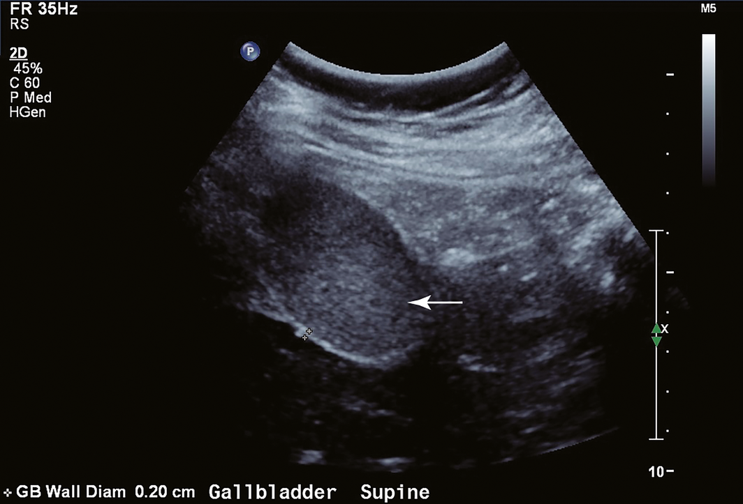
An abdominal ultrasound showed that the gallbladder was filled with thick, immobile and echogenic sludge (Box 3, arrow). There was no evidence of acute cholecystitis or biliary duct dilatation.
Her liver dysfunction was suspected to be related to hyperemesis gravidarum and she was managed with antiemetics, rehydration, correction of electrolyte abnormalities and supplemental nasogastric feeding under the supervision of a dietitian. Follow‐up in hepatology clinic a week later revealed that she was gaining weight and her nausea was controlled. Her blood test results showed bilirubin 26 μmol/L (reference range [RR], < 20 μmol/L), alanine aminotransferase 109 U/L (RR, < 34 U/L), aspartate aminotransferase 40 U/L (RR, < 31 U/L), γ‐glutamyl transferase 31 U/L (RR, < 38 U/L), and alkaline phosphatase 63 U/L (RR, 30–110 U/L). Blood tests repeated one month later had normalised.
Discussion
Pregnancy‐related liver disease tends to be reserved for patients after 20 weeks’ gestation. These disorders can be life‐threating if not identified and managed correctly. They include acute fatty liver of pregnancy, pre‐eclampsia, HELLP (haemolysis, elevated liver enzymes and low platelet count) syndrome, and cholestasis of pregnancy. During the first trimester, few pregnancy‐related liver conditions exist, but one well described is related to hyperemesis gravidarum.
Hyperemesis gravidarum is characterised by nausea, vomiting and typically mild to moderate elevations in serum aminotransferases and bilirubin. It usually manifests in the first trimester and abates by 20 weeks’ gestation. If left untreated, complications of micronutrient deficiency (Wernicke encephalopathy from vitamin B1 deficiency, bleeding from vitamin K deficiency), dehydration (renal injury, electrolyte disturbance) and malnutrition (immunosuppression, muscle wasting) may manifest.
The pathophysiology of jaundice in hyperemesis gravidarum is not well understood. The condition produces a state of ketosis that is known to cause slowing of biliary drainage, which is thought to be in part related to the contraction of the sphincter of Oddi. Pregnancy itself results in delayed gastric emptying, which promotes cholestasis. Histology typically shows evidence of cholestasis and the occasional necrotic hepatocyte.1 More recently, an association has been described between jaundice in hyperemesis gravidarum and the presence of biliary sludge, as evident in our case.2 Formation of biliary sludge has been hypothesised to result from a combination of the above described mechanisms, with the addition of dehydration. Intermittent movement of sludge within the biliary tree causing blockage of bile flow is a possible mechanism contributing to jaundice formation.
Management includes fluids to restore euvolaemia, replacement of electrolytes, supplemental nutrition (enteral and, in severe intractable cases, parenteral) and antiemetics. Medications need to be safe for mother and fetus, and typically ondansetron intravenously followed by glucocorticoid therapy are indicated for intractable cases. Outcomes tend to be favourable for both mother and fetus when patients are managed promptly.3 Recurrence in subsequent pregnancies is common and, as such, patients should be closely monitored to allow early intervention in future pregnancies.
When approaching jaundice in pregnancy, it is important not to overlook exacerbation of existing liver diseases and common causes for liver injury, including viral infections, alcohol, drugs (most commonly antibiotics such as erythromycin, amoxicillin or clavulanic acid), toxins, and biliary pathology. It is important to be systematic and consider pre‐hepatic (haemolytic anaemia), hepatic (intrinsic liver disease) and post‐hepatic (stones, biliary pathology) causes in addition to pregnancy‐specific liver conditions.
Lessons from practice
-
Few pregnancy‐related liver conditions exist in the first trimester but hyperemesis gravidarum is an important one.
-
Elevations in bilirubin and aminotransferases are typically mild to moderate but can also be severe.
-
When approaching jaundice in pregnancy, it is important not to overlook non‐pregnancy causes such as viral infections, alcohol and drug use, and biliary pathology.
-
Outcomes are generally favourable if managed appropriately for both mother and offspring, but recurrence in subsequent pregnancies is likely. ◆
Box 1 – Pertinent blood test results on initial presentation
|
Test |
Result |
Reference range |
|||||||||||||
|
|
|||||||||||||||
|
Haemoglobin, g/L |
146 |
115–155 |
|||||||||||||
|
Platelet count, × 109/L |
285 |
150–400 |
|||||||||||||
|
Total bilirubin, μmol/L |
135 |
< 20 |
|||||||||||||
|
Conjugated bilirubin, μmol/L |
71 |
< 4 |
|||||||||||||
|
Alkaline phosphatase (ALP), U/L |
86 |
30–110 |
|||||||||||||
|
γ‐Glutamyl transferase (GGT), U/L |
64 |
< 38 |
|||||||||||||
|
Alanine aminotransferase (ALT), U/L |
727 |
< 34 |
|||||||||||||
|
Aspartate aminotransferase (AST), U/L |
197 |
< 31 |
|||||||||||||
|
Sodium, mmol/L |
126 |
135–145 |
|||||||||||||
|
Potassium, mmol/L |
2.8 |
3.5–5.2 |
|||||||||||||
|
|
|||||||||||||||
|
|
|||||||||||||||
Box 2 – Laboratory tests to be requested when investigating jaundice in pregnancy
|
Investigation |
Rationale |
||||||||||||||
|
|
|||||||||||||||
|
Non‐pregnancy‐related tests |
|||||||||||||||
|
Pre‐hepatic causes |
|
||||||||||||||
|
Reticulocyte count, haptoglobin, haemoglobin |
Evaluate for haemolytic anaemia |
||||||||||||||
|
Hepatic causes |
|
||||||||||||||
|
CMV IgM and EBV IgM |
Exclude acute cytomegalovirus and Epstein–Barr virus, respectively |
||||||||||||||
|
HAV IgM |
Exclude acute hepatitis A |
||||||||||||||
|
HBsAg, HBc IgM |
Exclude active/chronic hepatitis B |
||||||||||||||
|
HCV antibody |
Exclude chronic hepatitis C |
||||||||||||||
|
ANA |
Often elevated in cases of autoimmune liver disease, SLE |
||||||||||||||
|
Antismooth‐muscle antibody and anti‐LKM antibodies |
Presence raises suspicion of autoimmune hepatitis |
||||||||||||||
|
Antimitochondrial antibody |
If present, suggestive of primary biliary cholangitis |
||||||||||||||
|
Immunoglobulins |
Total IgG often elevated in the setting of autoimmune hepatitis |
||||||||||||||
|
Anti‐tTG IgA |
Exclude coeliac disease, which can manifest as weight loss and rarely cause liver dysfunction |
||||||||||||||
|
TFTs |
Exclude thyrotoxicosis or hypothyroidism, which can rarely cause liver disease |
||||||||||||||
|
Cu/Ceruloplasmin |
Evaluate for evidence of Wilson disease if the patient has cirrhosis or there is a high index of suspicion |
||||||||||||||
|
α‐1‐Antitrypsin |
Deficiency can predispose to liver disease; consider if patient has cirrhosis or family history of cirrhosis |
||||||||||||||
|
Post‐hepatic causes |
|
||||||||||||||
|
Abdomen ultrasound |
Evaluate for stones or biliary pathology |
||||||||||||||
|
Lipase |
Evaluate for evidence of acute pancreatitis or biliary pathology |
||||||||||||||
|
Pregnancy‐related (after 20 weeks’ gestation) tests |
|||||||||||||||
|
Bile acid salts |
Elevated in setting of cholestasis of pregnancy |
||||||||||||||
|
Coagulation studies |
Disseminated intravascular coagulation in the third trimester may be part of acute fatty liver of pregnancy. Assess synthetic function of liver |
||||||||||||||
|
Reticulocyte count, haptoglobin, haemoglobin |
Haemolysis can be a feature of HELLP syndrome |
||||||||||||||
|
|
|||||||||||||||
|
ANA = antinuclear antibody. Anti‐LKM = anti‐liver/kidney microsomal antibody. Anti‐tTg IgA = antitissue transglutaminase IgA antibody. CMV = cytomegalovirus. Cu = copper. EBV = Epstein–Barr virus. HAV = hepatitis A virus. HBc = hepatitis B virus core antibody. HBsAg = hepatitis B surface antigen. HCV = hepatitis C virus. HELLP = haemolysis, elevated liver enzymes and low platelet count. SLE = systemic lupus erythematosus. TFTs = thyroid function tests. ◆ |
|||||||||||||||
Competing interests
No relevant disclosures.
References
- Larrey D, Rueff B, Feldmann, et al. Recurrent jaundice caused by recurrent hyperemesis gravidarum. Gut 1984; 25: 1414–1415.
- Matsubara S, Kuwata T, Kamozawa C, et al. Connection between hyperemesis gravidarum, jaundice or liver dysfunction, and biliary sludge. J Obstet Gynaecol Res 2012; 38: 446–448.
- Nulman I, Rovet J, Barrera M, et at. Long‐term neurodevelopment of children exposed to maternal nausea and vomiting of pregnancy and diclectin. J Pediatr 2009; 2: 45–50.
Linked content
-
MJA Letter: https://www.mja.com.au/journal/2019/211/10/jaundice-and-pregnancy
Provenance: Not commissioned; externally peer reviewed.
Forty Years in the Making: Reporting on the Co-Creation of a National Roadmap for Birthing on Country Services for the Best Start to Life
Res McCalman, Anneka J. Bowman, Roianne West, Kristie Watego, Jyai Allen, Claire Clack, Sue Kildea, Sue Kruske, Melanie Briggs, Cleone Wellington, Rebecca Coddington, Yu Gao, Isabella Garti, Sascha Kowalenko, Sarah Ireland, Catherine Austin, John D. Boffa, Marah Prior, Kelsie Kahl, Bettina Chaseling, Sarah Khaw, Emily Armstrong, Donna Hartz, Yvette Roe
Temporal Trends in Preterm Birth Associated With Hypertensive Disorders of Pregnancy in Victoria, Australia: A Population-Based Interrupted Time-Series Study
Melvin Marzan, Heng Jiang, Daniel Lorber Rolnik, Joanne M. Said, Lisa Hui
Linguistic Manoeuvres: Obstetric Violence Camouflages Harm and Loss of Consent From Birth
Liz Sutton, Harsha Ananthram, Rebecca Matthews
Linguistic Manoeuvres: Obstetric Violence Camouflages Harm and Loss of Consent From Birth
Bashi Kumar-Hazard, Andrew M. Bisits, Sharon L. Settecasse, Jenny Gamble, Hazel Keedle, Hannah G. Dahlen
Linguistic Manoeuvres: Obstetric Violence Camouflages Harm and Loss of Consent From Birth
Wendy Pollock, Alissa Fleming, Ensieh Fooladi, Joy Kloester, Anne Tremayne, Yasmin Zisin, Bethany Carr, Kym Davey, Suzanne Willey, Jenny Gamble
Getting on the Same Page: Why Australia Needs a National Maternity Early Warning System (MEWS) Chart
Briony A. Cutts, Lucy Bowyer, Nisha Khot, Sandra Lowe, Stefan C. Kane