




Medical education
Lessons from practice
Volume 210 - Issue 4
Glaucoma caused by topical corticosteroid application to the eyelids
Authors: Helen HL Chan and John F Salmon
Med J Aust 2019; 210 (4): 142-153. || doi: 10.5694/mja2.50012
Published online: 4 March 2019
Published online: 4 March 2019
A 64-year-old woman was referred to the glaucoma clinic at a tertiary eye hospital with elevated intraocular pressures
Clinical record
A 64‐year‐old woman was referred to the glaucoma clinic at a tertiary eye hospital with elevated intraocular pressure (IOP) noted by her optometrist (right eye, 42 mmHg; left eye, 40 mmHg; reference interval, 10–21 mmHg). She reported being diagnosed with normal tension glaucoma in the left eye at 30 years of age at another hospital, for which she had been prescribed topical pilocarpine 1% three times per day. However, after several years of follow‐up with no progressive change of either the optic disc or visual field, and with IOP consistently in the normal range, treatment had been ceased. She had low myopia; there was no family history of glaucoma. She had no other medical history apart from eczema.
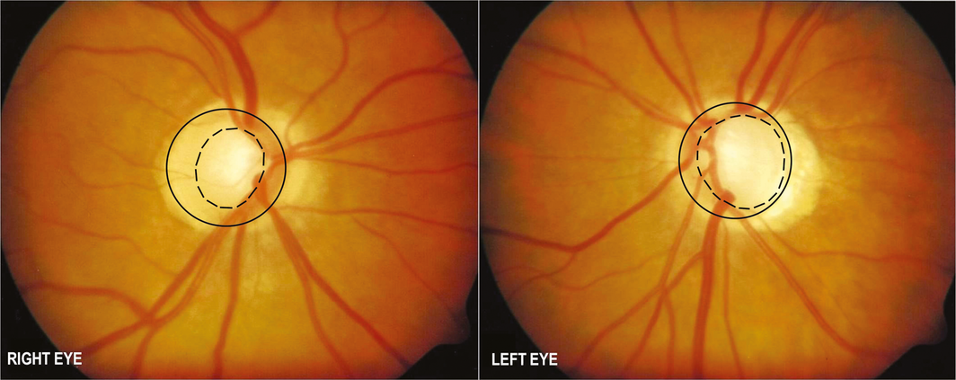
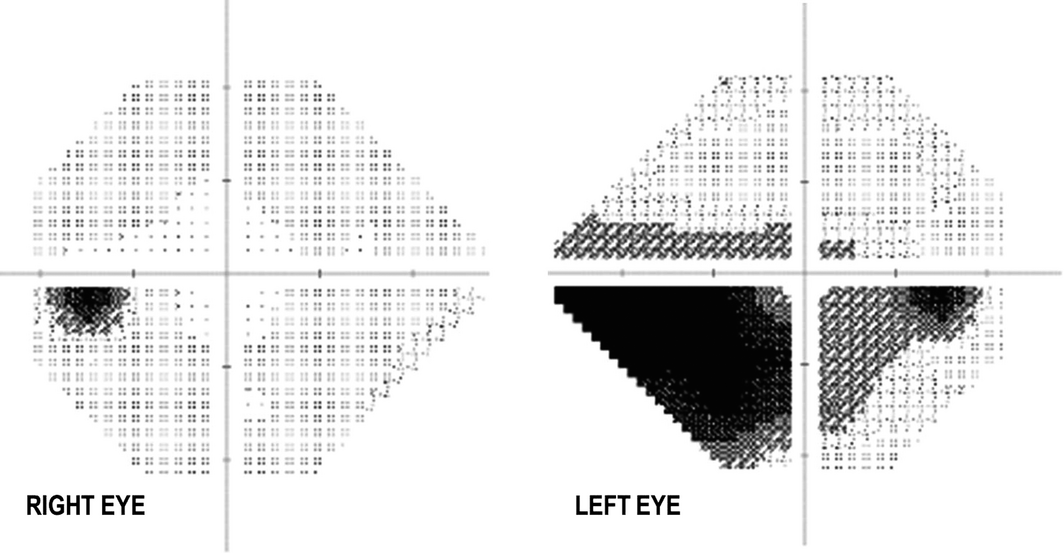
Visual acuity was 6/6 on the right and 6/7.5 on the left. IOP was 31 mmHg on the right and 30 mmHg on the left. Central corneal thickness was 508 microns in each eye. Anterior segments were normal. Gonioscopy revealed open angles. The right optic disc was normal; however, there was glaucomatous cupping of the left optic disc (Box 1). The visual field of the left eye had a corresponding nasal step (Box 2).
She commenced topical therapy in both eyes (latanoprost 0.005% nightly and subsequently timolol 0.25% each morning). However, there was poor response to treatment, with IOP of 29 mmHg in each eye and continued deterioration of the visual field in the left.
Further questioning revealed the daily use of hydrocortisone acetate 1% cream to the eyelids for treatment of eczema for 3–4 months prior to her presentation to the clinic. Two weeks after cessation of the steroid, IOP reduced to 15 mmHg (right) and 18 mmHg (left) on a single agent (timolol 0.5% each morning). Over the next ten years of follow‐up, IOP remained consistently below 18 mmHg in both eyes. The disease has remained stable in the left eye, and early glaucomatous atrophy has developed in the right eye (cup:disc ratio of 0.7 with a superior notch and corresponding inferior arcuate defect) and stable disease in the left eye. One year after her presentation, the patient's brother was also diagnosed with open‐angle glaucoma.
Discussion
Raised intraocular pressure and glaucoma are well known complications of systemic and ocular corticosteroid therapy. However, cutaneous steroid preparations applied to the face and eyelids can also cause a marked elevation in IOP (steroid response).1,2,3 This can lead to glaucomatous optic neuropathy and irreversible visual loss. Elevation in IOP can occur any time after steroid administration, but most commonly within 2–4 weeks.4 The mechanism is obstruction of aqueous outflow, possibly due to alterations in the extracellular matrix or endothelial cells of the trabecular meshwork.4
The degree of steroid response appears to be proportional to the potency, dose and route of administration.5 For example, systemic steroid administration via oral or intravenous routes is less likely to cause IOP elevation than topical intra‐ or peri‐ocular administration (such as eye drops or intravitreal injection); however, it should be stressed that all forms of steroids can induce a significant IOP response. Patients with myopia, connective tissue disease, pre‐existing glaucoma or a family history of glaucoma (one or more first‐degree relatives) are at particular risk.5
In our patient, the early onset glaucomatous atrophy of the left eye most likely developed as a consequence of several years of steroid application to the eyelids in her 20s. The eczema improved, topical steroids were ceased for many years and further IOP elevation was not seen until the recurrence of eczema and resumption of steroid use in her 60s. The patient's brother was also diagnosed with glaucoma, and individuals with a family history of glaucoma are at increased risk of developing a steroid response — the use of steroids may “unmask” individuals with a high baseline risk of IOP elevation.6
The frequency of steroid response following steroid application to the face is unknown.2 It can affect either one or both eyes.4 A consensus statement from the Australasian College of Dermatologists in 2015 reports that despite case observations of steroid‐induced glaucoma from topical steroids, the incidence is rare. Further, it states that “it is currently unclear as to whether there is a threshold of topical corticosteroid use which can induce cataract or glaucoma”.7 To our knowledge, there has been no report of glaucoma after topical steroid application to cutaneous areas other than the face. Once recognised, cessation of steroid use is typically sufficient to lower the IOP within several days to weeks, as in this case.5 However, there are cases in which the IOP remains high and necessitates further medical and surgical therapy.3
Prevention involves using the lowest dose and shortest duration of steroid necessary to control symptoms, and regular screening for patients, particularly those at high risk.2 These medications are effective for common conditions such as eczema and blepharitis, and are therefore frequently used in the community. All patients who are prescribed these should be counselled regarding the risk of IOP elevation and the need for ophthalmic monitoring.1 The monitoring schedule should reflect the patient's individual risk, which is influenced by the dosage and duration of steroid therapy. At a minimum, this should involve IOP and optic nerve evaluation at baseline, during the course of therapy (no later than 2–4 weeks after commencement), and at completion.5
Lessons from practice
-
Topical corticosteroid ointment applied to the face and eyelids can induce a marked elevation in intraocular pressure and subsequent glaucoma.
-
Risk factors for a steroid response include myopia, connective tissue disease and a personal or family history of glaucoma.
-
Patients who are prescribed topical corticosteroids, such as for the treatment of facial eczema, should be counselled regarding the risk of raised intraocular pressure and be monitored at regular intervals by an optometrist or ophthalmologist.
Competing interests
No relevant disclosures.
References
- Sahni D, Darley CR, Hawk JL. Glaucoma induced by periorbital topical steroid use – a rare complication. Clin Exp Dermatol 2004; 29: 617–619.
- Haeck IM, Rouwen TJ, Timmer‐de Mik L, et al. Topical corticosteroids in atopic dermatitis and the risk of glaucoma and cataracts. J Am Acad Dermatol 2011; 64: 275–281.
- Garrott HM, Walland MJ. Glaucoma from topical corticosteroids to the eyelids. Clin Exp Ophthalmol 2004; 32: 224–226.
- Spaeth GL, Rodrigues MM, Weinreb S. Steroid‐induced glaucoma: A. Persistent elevation of intraocular pressure B. Histopathological aspects. Trans Am Ophthalmol Soc 1977; 75: 353–381.
- Spaeth GL, Monteiro de Barros DS, Fudemberg SJ. Visual loss caused by corticosteroid‐induced glaucoma: how to avoid it. Retina 2009; 29: 1057–1061.
- Kersey JP, Broadway DC. Corticosteroid‐induced glaucoma: a review of the literature. Eye 2006; 20: 407.
- Mooney E, Rademaker M, Dailey R, et al. Adverse effects of topical corticosteroids in paediatric eczema: Australasian consensus statement. Aust J Dermatol 2015; 56: 241–51.
Linked content
-
MJA Narrative Review: Current management of glaucoma
-
MJA Perspective: Glaucoma in perspective
-
MJA Podcast: Dr Jed Lusthaus
-
InSight+: Glaucoma: the “sneak thief of sight”
Provenance: Not commissioned; externally peer reviewed.
A Diagnostic Headache
Thomas Glynn, Michelle T. Leech, Stacy K. Goergen, Emily Lin, Nadeem Toodayan, Ralph Junckerstorff
Neisseria meningitidis: an uncommon cause of conjunctivitis
Chloe Story, Tina Marinelli, Simeon Crawford, Amrita Ronnachit
Dismantling barriers to research and clinical care for individuals with a vision impairment
Eden G Robertson, Kate Hetherington, Meredith Prain, Julia Hall, Leighton Boyd AM, Rosemary Boyd OAM, Emily Shepard, Hollie Feller, Sally Karandrews, Fleur O'Hare, Kanae Yamamoto, Matthew P Simunovic, Robyn V Jamieson, Alan Ma, Lauren Ayton AM, Anai Gonzalez‐Cordero
The New South Wales Pharmacy Trial for herpes zoster: on the nose?
Christian P Pappas, Timothy R Holmes, Minas T Coroneo
Skin fragility disorder misdiagnosed as child abuse: a cautionary tale
Heather G Mack, Fred K Chen, John R Grigg
Health economic aspects of inherited retinal diseases: looking for cost‐effective treatments
Benjamin Kamien, Rachael Heath Jeffery, Fred K Chen