




Volume 210 - Issue 11
Deep vein thrombosis: update on diagnosis and management
Authors: Paul C Kruger, John W Eikelboom, James D Douketis and Graeme J Hankey
Med J Aust 2019; 210 (11): 516-524. || doi: 10.5694/mja2.50201
Published online: 17 June 2019
Published online: 17 June 2019
The diagnosis of DVT requires a high index of suspicion because symptoms and signs are often non-specific
Summary
- Diagnosis of deep vein thrombosis (DVT) requires a multifaceted approach that includes clinical assessment, evaluation of pre‐test probability, and objective diagnostic testing.
- Common symptoms and signs of DVT are pain, swelling, erythema and dilated veins in the affected limb.
- The pre‐test probability of DVT can be assessed using a clinical decision rule that stratifies DVT into “unlikely” or “likely”. If DVT is “unlikely”, refer for D‐dimer test. If the D‐dimer level is normal, DVT can be excluded; if the D‐dimer level is increased, refer for compression ultrasound. If DVT is “likely”, refer for compression ultrasound.
- When DVT is confirmed, anticoagulation is indicated to control symptoms, prevent progression and reduce the risk of post‐thrombotic syndrome and pulmonary embolism.
- Anticoagulation may consist of a parenteral anticoagulant overlapped by warfarin or followed by a direct oral anticoagulant (DOAC) (dabigatran or edoxaban), or of a DOAC (apixaban or rivaroxaban) without initial parenteral therapy.
- DOACs are the preferred treatment for DVT because they are at least as effective, safer and more convenient than warfarin. DOACs may require dose reduction or avoidance in patients with renal dysfunction, and should be avoided in pregnancy.
- Recent evidence shows that DVT in patients with cancer may be treated with edoxaban (after discontinuation of 5 days of initial heparin or low molecular weight heparin [LMWH]) or rivaroxaban if patients prefer not to have daily injections of LMWH, but the risk of gastrointestinal bleeding is higher with DOACs than with LMWH in patients with gastrointestinal cancer.
Venous thromboembolism (VTE) most commonly manifests as lower extremity deep vein thrombosis (DVT) and pulmonary embolism and has an annual incidence of 1–2 per 1000 population.1 Mortality is high; death within 30 days occurs in about 6% of patients with DVT, primarily through pulmonary embolism, and in 13% of patients with pulmonary embolism.2 Among treated patients, about 20–50% develop post‐thrombotic syndrome (PTS) after DVT, and 3% develop chronic thromboembolic pulmonary hypertension after pulmonary embolism.3,4 After 3–6 months of anticoagulation, VTE recurs in up to 40% of patients within 10 years. The risk of recurrence is two‐ to threefold higher after unprovoked than provoked VTE.5,6
In the past decade, there have been notable advances in risk prediction, diagnosis and treatment with direct oral anticoagulants (DOACs), thrombolysis and catheters. Nevertheless, there remains uncertainty about the optimal duration of anticoagulation after unprovoked VTE, indications for thrombophilia screening, and the role of catheter‐directed thrombolysis.7
This review summarises contemporary evidence on the diagnosis and management of DVT. We searched PubMed for relevant articles, from 1996 to 2019, using the search terms “venous thromboembolism”, “deep vein thrombosis”, “pulmonary embolism”, “thrombosis”, “anticoagulant” and “anticoagulation”. An accompanying article addresses pulmonary embolism.8 Guidelines from the Thrombosis and Haemostasis Society of Australia and New Zealand (THANZ) underpin evidenced‐based recommendations,9 which have been graded according to the National Health and Medical Research Council levels of evidence (Box 1).10
Diagnosis
The clinical presentation of DVT is often non‐specific. Hence, accurate diagnosis requires sequential integration of clinical features, assessment of pre‐test clinical probability, and confirmatory investigations that include D‐dimer testing and imaging.
Symptoms and signs of deep vein thrombosis
Symptoms and signs of leg or pelvis DVT include leg pain, swelling, erythema and dilated superficial veins. Arm DVT has similar symptoms localised to the arm. Some DVTs are asymptomatic. Differential diagnoses for limb DVT include cellulitis, lymphoedema, chronic venous insufficiency, haematoma and, for leg DVT, ruptured Baker cyst.11
Pre‐test clinical gestalt or impression
The gestalt probability of DVT is an unstructured impression of the likelihood of DVT based on clinical assessment. It can help determine the pre‐test probability of DVT when it is estimated by an experienced physician.12,13 However, validated clinical decision rules are more reliable.14
Pre‐test probability using a clinical decision rule
In primary care and in outpatients with suspected DVT, the Wells rule for DVT helps calculate the pre‐test probability of DVT and guide investigations.9 The Wells rule assigns points for clinical symptoms and risk factors for DVT to produce a total score between −2 and 9 points, which stratifies patients as “unlikely” (≤ 1 point) or “likely” (≥ 2 points) to have DVT.15 A potential weakness is the need for clinicians to subjectively determine whether an alternative (non‐DVT) diagnosis is likely or unlikely, as both groups require investigations.
Among inpatients with suspected DVT, imaging is required because clinical decision rules have not been validated and D‐dimer testing has a high frequency of false positive results.
D‐dimer testing
D‐dimer levels are raised in most patients with DVT (sensitivity, 94–96%) and also in older patients and in patients with malignancy, sepsis, inflammation, chronic kidney failure, recent surgery, trauma, severe burns, and pregnancy (specificity, 42–52%).16,17 Therefore, a negative D‐dimer result helps exclude DVT, particularly when the clinical probability is low, and a positive D‐dimer result requires imaging to confirm DVT.
D‐dimer testing may also be used to predict the risk of recurrent VTE after ceasing anticoagulation, but is not recommended as a screening test for subclinical disease recurrence or to monitor response to anticoagulation.18
Diagnostic imaging
Venous compression ultrasound (CUS) is the first‐line imaging test for suspected DVT.19 There are two acceptable strategies. The first is CUS limited to the proximal leg (thigh and popliteal region) to diagnose proximal DVT, and to repeat one week later to assess for distal DVT extending proximally. The second is CUS of the whole leg which, if negative, avoids the need for a repeat limited CUS, but may diagnose distal DVTs that may not require anticoagulation.20 The risk of missing a DVT in the first 3 months after a negative whole leg CUS is less than 2%.21 Compared with venography, CUS has a sensitivity of 93.8% (95% CI, 92.0–95.3) and specificity of 97.8% (95% CI, 97.0–98.4) for proximal DVT.22
Using an integrated approach to diagnosis of first deep vein thrombosis
The diagnosis of DVT requires an integrated approach because the clinical presentation, clinical decision rule or investigations used in isolation may be insufficient to confirm or exclude DVT (Box 2). Alternative algorithms have been presented by the American Society of Hematology and THANZ.9,14
A low to moderate pre‐test probability by gestalt, or “unlikely” DVT (Wells rule ≤ 1), indicates the need for D‐dimer measurement. D‐dimer has a negative predictive value of 99.1% (95% CI, 96.7–99.9) in populations with a 16% prevalence of DVT.23 Therefore, a normal D‐dimer level in patients with “unlikely” DVT effectively excludes DVT (evidence level A; Box 1). An increased D‐dimer level infers that DVT may be present (along with other alternative diagnoses) and indicates the need for a CUS to confirm or exclude DVT. A CUS that is positive for proximal DVT confirms DVT and the need to initiate anticoagulant therapy, whereas anticoagulant therapy can be withheld with a negative CUS. Ideally, a negative above‐knee CUS should be repeated one week later to exclude the presence of a distal DVT that may have extended proximally. Alternatively, a whole‐leg CUS may be performed and, if negative for DVT, does not need to be repeated (evidence level A).24
A high pre‐test probability by gestalt, or “likely” DVT (Wells rule ≥ 2), indicates CUS, without the need for D‐dimer testing. D‐dimer should not be measured in patients with a high pre‐test probability of DVT because a negative D‐dimer result does not exclude DVT with sufficient certainty in such patients, and an increased D‐dimer level is not specific for DVT.18,23
Investigating for underlying risk factors
Risk factors for DVT (Box 3) determine the risk of recurrent DVT.25
Unprovoked VTE requires assessment for occult cancer, present in up to 10% of patients.26 However, extensive cancer screening with computed tomography of the body or tumour markers is not recommended, unless symptoms of possible cancer are present.27
Testing for hereditary thrombophilia may identify predisposition to the development of VTE, guide testing of family members, and determine the need for long term prophylactic anticoagulation. However, thrombophilia testing is not indicated routinely. Selecting who to test requires consideration because the results will not change management in most patients with VTE, as the most common thrombophilias (factor V Leiden and prothrombin gene mutations) are not strong predictors of recurrent VTE.28 Testing for thrombophilia can be considered in patients with unprovoked VTE who are aged less than 50 years, or who have a strong family history of VTE, or who have recurrent venous or arterial thrombosis.29,30 Thrombophilia testing should not be performed in patients with VTE provoked by surgery or major trauma (evidence level B) because the risk of recurrent VTE is low.
Testing for the antiphospholipid syndrome — an acquired thrombophilia — is indicated when patients develop arterial or venous thrombosis in the setting of thrombocytopenia, haemolytic anaemia, livedo reticularis or cognitive dysfunction in the absence of a stroke.31 Patients with abdominal (portal or hepatic vein) thrombosis may have an underlying myeloproliferative disorder, and Janus kinase 2 (JAK2) V617F mutation testing is recommended in patients without cirrhosis or malignancy.32 When DVT occurs in the setting of pancytopenia or haemolytic anaemia, testing for paroxysmal nocturnal haemoglobinuria should be considered, especially if the DVT is in an unusual location (eg, splanchnic veins or cerebral sinuses).33,34
Investigating patients with deep vein thrombosis for simultaneous pulmonary embolism
About 56% of patients with proximal DVT have pulmonary embolism.35 Chest imaging is not needed routinely because anticoagulation is required regardless, but it may be justified if the patient has significant symptoms of pulmonary embolism, as this may change management (eg, thrombolysis for clinically massive pulmonary embolism) and prognosis (eg, long term risk of chronic thromboembolic pulmonary hypertension).8
Management
Anticoagulation
Anticoagulation is the mainstay of treatment for VTE; it aims to reduce mortality, thrombus extension, recurrence, and the risk of PTS (after DVT) and chronic thromboembolic pulmonary hypertension (after pulmonary embolism). The phases of anticoagulation treatment are “initial” (first week after VTE diagnosis), “long term” (first 3 months after diagnosis) and “extended” (no scheduled stop date) (Box 4).36
Clinically significant bleeding is a contraindication to anticoagulation. Relative contraindications include recent bleeding (eg, gastrointestinal bleeding within 2 weeks, intracranial bleeding within 3 months), recent trauma, known bleeding disorder, severe thrombocytopenia (platelet count < 75 × 109/L), endocarditis and uncontrolled hypertension.
Risk factors for bleeding include age over 65 years, previous bleeding, cancer, metastatic cancer, renal failure, liver failure, thrombocytopenia, previous stroke, diabetes, anaemia, antiplatelet therapy, poor anticoagulation control (for vitamin K antagonists), comorbidity and reduced functional capacity, recent surgery, frequent falls and alcohol misuse. The risk of bleeding can be estimated as low, intermediate or high if none, one, or two or more of these factors are present, respectively.36 Long term concomitant use of non‐steroidal anti‐inflammatory drugs should be avoided if possible, but short (1–2 weeks) courses of treatment can be used, for example, in patients with an inflamed leg or superimposed superficial phlebitis. For patients who require aspirin for cardiovascular prevention, the dose should not exceed 100 mg daily, but, otherwise, it should be avoided during anticoagulant therapy.37
Commencing anticoagulation while waiting for test results is reasonable if the suspicion of DVT is “likely”. Not commencing anticoagulation while waiting for results is reasonable for patients at increased risk of bleeding or if the suspicion of DVT is “unlikely”, provided the results will be available within 24 hours.36
Anticoagulant agents
Options for the initial treatment of VTE include a DOAC (apixaban or rivaroxaban), initial parenteral anticoagulation followed by a DOAC (dabigatran or edoxaban, because initial parenteral therapy was administered in the randomised phase 3 trials of dabigatran and edoxaban) or initial parenteral anticoagulation overlapped by warfarin and continued for at least 5 days and until the international normalised ratio (INR) is more than 2.0 on two occasions 24 hours apart (evidence level A). The DOACs — comprising the direct thrombin inhibitor dabigatran and the factor Xa inhibitors rivaroxaban, apixaban and edoxaban — are as effective as and safer than warfarin (INR, 2.0–3.0) for the treatment of VTE, whereas betrixaban has not been assessed for VTE treatment and is indicated only for VTE prophylaxis in hospitalised medical patients.38,39 Three DOACs — dabigatran, rivaroxaban and apixaban — are approved by the Pharmaceutical Benefits Scheme for the treatment of VTE. Despite the DOACs being classified collectively, each has a unique molecular structure and they should therefore be considered as individual anticoagulants (Box 5).40,41,42,43 Warfarin is associated with a slightly higher bleeding risk than DOACs, with an absolute risk increase for major bleeding of about 1%, and requires routine coagulation monitoring.36,38,44
Anticoagulant selection
The choice of anticoagulant should consider medical issues such as efficacy, safety, renal and hepatic function, and concurrent medications. In addition, practical issues such as availability, familiarity of use, patient preference, and cost should be considered.
DOACs are an ideal first‐line anticoagulant for the treatment of VTE in an uncomplicated patient, have advantages over vitamin K antagonists (Box 6),45 and have few drug–drug interactions (Box 7).46,47,48,49 In patients treated with dabigatran, idarucizumab can reverse anticoagulation in emergency situations (eg, life‐threatening bleeding, urgent surgery).50 In patients treated with rivaroxaban or apixaban, andexanet alfa has been approved in the United States and Europe for reversing the anticoagulant effect but not yet in Australia.51
DOAC treatment is not recommended in pregnant women and is contraindicated in patients with severe renal impairment (creatinine clearance [CrCl] < 30 mL/min for edoxaban, rivaroxaban 15 mg and 20 mg tablets, and dabigatran; or CrCl < 25 mL/min for apixaban; or CrCl < 15 mL/min for rivaroxaban 10 mg tablets), severe hepatic impairment (Child–Pugh score C), mechanical heart valves, and concomitant administration of drug classes that are strong inhibitors of CYP3A4 and P‐glycoprotein.52,53 When treatment with a DOAC is not appropriate, anticoagulation with intravenous unfractionated heparin, low molecular weight heparin (LMWH) or warfarin are alternatives.
Duration of anticoagulation
The duration of the anticoagulation for DVT depends on the risk of recurrence, which is determined by whether the DVT is provoked by a transient or ongoing risk factor, or unprovoked. A DVT may be considered provoked if a transient or permanent or persistent environmental risk factor is present, or unprovoked if no such risk factor is present (Box 3). Risk factors for VTE inform the risk of recurrent VTE. For example, in patients with VTE who are treated with 3–6 months of anticoagulation, stopping anticoagulation is associated with a one‐year risk of recurrence of about 1–3% when the initial VTE occurred in the context of a major transient risk factor, about 10% when the event is unprovoked, and more than 10% when the event occurs in patients with active cancer.25 In patients with thrombophilia, factors such as the factor V Leiden and prothrombin gene mutations are weak determinants of disease recurrence, whereas less common factors such as inherited deficiencies of protein C, protein S and antithrombin and the antiphospholipid antibody syndrome are stronger predictors of recurrent VTE, for which the THANZ guidelines suggest extended anticoagulant therapy.29
Six weeks of anticoagulation are recommended for treatment of distal DVT caused by a major transient risk factor that is no longer present (evidence level B). Three months of anticoagulation are recommended for the treatment of proximal DVT provoked by surgery or trauma (evidence level A), proximal DVT provoked by a non‐surgical transient risk factor, unprovoked isolated distal DVT (evidence level B), and for unprovoked DVT when the bleeding risk is high. A minimum of 3 months of anticoagulation, followed by re‐evaluation of the risk–benefit ratio, is indicated for unprovoked proximal DVT (evidence level A). At least 6 months of anticoagulation are recommended for DVT that is provoked by active cancer (evidence level A). Extended anticoagulation is recommended for a second or unprovoked DVT, if risk factors such as active cancer persist, or for unprovoked proximal DVT when the bleeding risk is low.44 Patients receiving extended anticoagulation should be followed up at least once per year to re‐evaluate their individual risk of thrombosis and bleeding, monitor for adverse effects from the anticoagulant, and detect changes that may affect the half‐life of the anticoagulant (eg, renal impairment). DOACs are preferred over warfarin for long term and extended anticoagulation therapy, provided there are no contraindications (evidence level A), but warfarin is a reasonable alternative when INR monitoring is feasible and acquisition costs of DOACs are an issue. After long term treatment of VTE, extended treatment with low dose apixaban (2.5 mg twice daily) or low dose rivaroxaban (10 mg once daily) is effective at reducing the risk of recurrent VTE compared with placebo or aspirin, respectively, without increasing the rate of major bleeding.54,55
Inferior vena cava filter
Placement of an inferior vena cava (IVC) filter should be considered in specific clinical circumstances. Among 2055 patients with acute proximal DVT and a contraindication to anticoagulation, IVC filter insertion reduced the risk of subsequent pulmonary embolism by 50% compared with no filter.56 However, IVC filters are associated with a 70% increased risk of subsequent DVT when compared with no IVC filter insertion, and carry unique risks such as filter thrombosis (about 2% of cases), migration, and penetration of the wall of the IVC. In a cohort study of 126 030 patients with VTE and a contraindication to anticoagulation, 30‐day mortality was significantly higher in patients who had an IVC filter inserted compared with no IVC filter (hazard ratio [HR], 1.18; 95% CI, 1.13–1.22; P < 0.001).57 IVC filters should only be considered in patients with an absolute contraindication to anticoagulation or selected patients who develop recurrent pulmonary embolism despite anticoagulation and have significant residual DVT (evidence level C).44 Massive pulmonary embolism is not an indication for an IVC filter.56,58 The IVC filter should be removed preferably within 3 weeks of placement and when it is safe to resume anticoagulation.
Thrombolysis
Catheter‐directed thrombolysis involves the percutaneous insertion of a catheter and infusion of a thrombolytic — typically recombinant tissue plasminogen activator (tPA) — directly to the DVT. A randomised trial of anticoagulation plus tPA versus anticoagulation alone in patients with acute proximal leg DVT showed no reduction in the risk of PTS and increased bleeding with dual therapy.59 Patients with recent (< 1 week) extensive, typically iliofemoral, DVT or with phlegmasia cerulea dolens and at low bleeding risk were poorly represented in this trial and may yet benefit from catheter‐directed thrombolysis (evidence level C).60 In contrast to leg DVT, there have been no randomised trials of thrombolysis for treatment of arm DVT.61
Surgical thrombus removal
Surgical removal of DVT does not have a proven role. Some suggest that venous thrombectomy should only be considered in patients with impending venous gangrene despite optimal anticoagulation and all of the following: catheter‐directed thrombolysis not available, iliofemoral DVT, symptoms for less than 7 days, good functional status, life expectancy greater than one year, and availability of appropriate resources and expertise.36
Compression stockings
Current evidence suggests a beneficial effect of early application of elastic compression stockings on PTS and residual vein occlusion (evidence level C). In the IDEAL DVT study, compression stockings worn for at least a minimum of 6 months until the Villalta score decreased to 4 or less on two consecutive readings, compared with 2 years, were non‐inferior for preventing PTS in patients with acute proximal DVT (29% v 28%; odds ratio [OR], 1.06; 95% CI, 0.78–1.44).62 In a pre‐specified substudy of the IDEAL DVT study, acute compression of the leg within 24 hours of DVT diagnosis, compared with no compression, was associated with a decrease in residual vein obstruction (46% v 66%; OR, 0.46; 95% CI, 0.27–0.80) and PTS (46% v 54%; OR, 0.65; 95% CI, 0.46–0.92).63
Secondary prevention of venous thromboembolism
Aspirin 100 mg daily is reasonable to decrease the risk of recurrent VTE in patients who have completed anticoagulation treatment for a first unprovoked DVT or pulmonary embolism, and in whom ongoing anticoagulation is not appropriate or cannot be accessed.64 Since the introduction of DOACs, new secondary prevention strategies have been tested. In patients with VTE who had completed 6–12 months of anticoagulant therapy, and in whom there was clinical equipoise regarding the continuation or cessation of anticoagulant therapy, extended anticoagulation with low dose apixaban 2.5 mg twice daily for a further 12 months was more effective than placebo in reducing recurrent VTE or death from any cause (relative risk [RR], 0.33; 95% CI, 0.22–0.48) without increasing major bleeding (RR, 0.49; 95% CI, 0.09–2.64).54 In another similar population, extended anticoagulation with rivaroxaban was more effective than aspirin in reducing recurrent VTE (HR, 0.34; 95% CI, 0.20–0.59, for 20 mg daily dose; HR, 0.26; 95% CI, 0.14–0.47, for 10 mg daily dose v aspirin), without a significant increase in bleeding rates (major bleeding 0.5% with rivaroxaban 20 mg, 0.4% with rivaroxaban 10 mg, and 0.3% with aspirin 100 mg daily).55
Special circumstances affecting the diagnosis and management of deep vein thrombosis
Pregnancy
Pregnant women may not be assessed accurately using the Wells rule for DVT because the rule was derived from a non‐pregnant population. The LEFt rule is an alternative clinical prediction rule that assigns one point each for symptoms in the left leg, calf circumference difference of 2 cm or more, and first trimester of pregnancy.65 A LEFt score of zero points is accurate to rule out DVT in pregnant women.66 D‐dimer level is less likely to rule out DVT in pregnancy because it steadily increases until delivery; therefore, reports conflict over whether to perform this test for suspected VTE in pregnancy.18,66 When DVT is confirmed, the most appropriate anticoagulation is LMWH (evidence level A). Warfarin use during pregnancy is associated with an increased risk for fetal embryopathy, especially when used during weeks 6–12 of gestation.67 DOACs have been used in pregnancy, but experience is limited.68 As DOACs are small molecules that cross the placenta, there is potential for adverse effects on the fetus.
Cancer
Active cancer may be associated with an increased D‐dimer level even if DVT is not present, which reduces the negative predictive value for DVT.18 Previously, LMWH has been recommended for treatment of DVT for patients with cancer because, compared with warfarin, the rate of recurrent VTE is lower and the rate of bleeding is similar.69 The optimal duration of anticoagulation for cancer‐associated VTE has not been formally assessed beyond 6 months. Continuing anticoagulation for longer than 6 months is indicated if the cancer is still active or treatment with chemotherapy, radiotherapy or hormonal therapy is ongoing.27,70 Two DOACs have recently been compared with LMWH for cancer‐related VTE. Edoxaban (after 5 days of initial LMWH), compared with LMWH (dalteparin), was non‐inferior for the composite outcome of recurrent VTE or major bleeding. The rate of recurrent VTE was not different (7.9% v 11.3%; HR, 0.71; 95% CI, 0.48–1.06), but the risk of major bleeding was higher with edoxaban (6.9% v 4.0%; HR, 1.77; 95% CI, 1.03–3.04) and there was no difference in mortality.71 Rivaroxaban (no initial LMWH), compared with dalteparin, decreased the rate of recurrent VTE (HR, 0.43; 95% CI, 0.19–0.99) and increased the risk of clinically relevant non‐major bleeding (HR, 3.76; 95% CI, 1.63–8.69), but the risk of major bleeding was not significantly different (HR, 1.83; 95% CI, 0.68–4.96).72 In both trials, the increased rate of bleeding with DOACs was mainly due to the higher rate of upper gastrointestinal bleeding in patients enrolled with gastrointestinal cancer. These data support rivaroxaban and edoxaban as effective oral treatment options for patients with VTE and cancer (evidence level B).
Distal deep vein thrombosis
Isolated distal (calf) DVTs have a lower risk of extension than proximal DVTs and do not always require anticoagulation. About 15% of isolated distal DVTs will extend to the proximal veins if not treated, usually in less than 2 weeks.73 Treatment with anticoagulation for 3 months may be considered for patients with distal DVT who have a significant symptom burden, or if risk factors for extension are present (eg, thrombosis > 5 cm in length, > 7 mm maximum diameter, involving multiple veins), the DVT is unprovoked, there is a history of active cancer or previous VTE, or the patient is currently admitted to hospital. If the patient is asymptomatic and does not initially receive anticoagulation, it is suggested to repeat the CUS after 1–2 weeks to detect possible proximal extension.73
Recurrent deep vein thrombosis
Recurrent DVT is more difficult to diagnose than first DVT because the features of the initial episode of DVT may persist on CUS.74 Diagnosis of recurrent DVT may be assisted by comparing the CUS with a previous one at the end of treatment of the first DVT. Hence, repeating the CUS at the end of anticoagulation, although not performed routinely,24 can be valuable in patients with high risk of recurrence (eg, unprovoked proximal DVT). When VTE recurs during anticoagulation treatment, the patient should be evaluated for suboptimal anticoagulation therapy (eg, incorrect dose, poor adherence) and the emergence of conditions that increase the risk of recurrent VTE (eg, cancer).75
Antiphospholipid syndrome
Patients with antiphospholipid syndrome should be anticoagulated with warfarin.31 In high risk patients with antiphospholipid syndrome, rivaroxaban treatment was associated with no benefit and increased risk compared with warfarin (evidence level C).76 Further studies are needed to assess the safety of DOACs in selected, lower risk patients with antiphospholipid syndrome.
Superficial vein thrombosis
Superficial vein thrombosis manifests as thrombosis of a superficial venous system and is associated with a low incidence of proximal DVT and pulmonary embolism. Anticoagulation with a prophylactic dose of LMWH or fondaparinux for 45 days is recommended in patients with leg superficial vein thrombosis greater than 5 cm in length, severe symptoms, involvement of the greater saphenous vein, history of VTE or superficial vein thrombosis, active cancer, and recent surgery (evidence level C). Patients with superficial vein thrombosis above the knee should have a CUS to investigate for DVT. Non‐steroidal anti‐inflammatories can alleviate pain in patients who are not anticoagulated.36
Conclusion
The diagnosis of DVT requires a high index of suspicion because symptoms and signs are often non‐specific. Anticoagulation continues to be the cornerstone of therapy. The optimal anticoagulant and duration of therapy are determined by the clinical assessment.
Box 1 – The National Health and Medical Research Council levels of evidence10
|
Component |
Evidence base |
Definition |
|||||||||||||
|
|
|||||||||||||||
|
A |
Excellent |
One or more level I studies with a low risk of bias or several level II studies with a low risk of bias |
|||||||||||||
|
B |
Good |
One or two level II studies with a low risk of bias or a systematic review or several level III studies with a low risk of bias |
|||||||||||||
|
C |
Satisfactory |
One or two level III studies with a low risk of bias, or level I or II studies with a moderate risk of bias |
|||||||||||||
|
D |
Poor |
Level IV studies, or level I to III studies or systematic reviews with a high risk of bias |
|||||||||||||
|
|
|||||||||||||||
|
|
|||||||||||||||
Box 2 – Diagnosing first deep vein thrombosis (DVT) presenting in an outpatient or in primary care
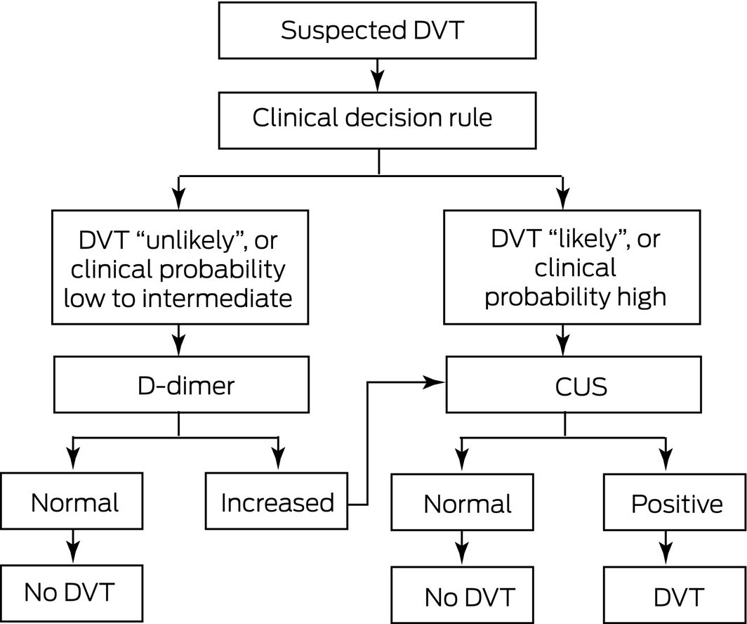
CUS = compression ultrasound. ◆
Box 3 – Risk factors for venous thromboembolism*25
|
VTE risk factor category |
Examples |
||||||||||||||
|
|
|||||||||||||||
|
Transient |
|
||||||||||||||
|
Permanent/persistent |
|
||||||||||||||
|
Unprovoked |
|
||||||||||||||
|
Non‐environmental |
|
||||||||||||||
|
|
|||||||||||||||
|
* Environmental (or acquired) risk factors for venous thromboembolism (VTE) may be transient or persistent. A transient risk factor is one that resolves after it has provoked the VTE. Resolution of the transient risk factor should be confirmed before stopping anticoagulation therapy. A permanent/persistent risk factor is one that is still present after it provokes the VTE. A VTE that occurs without transient or permanent/persistent risk factors is considered unprovoked. Non‐environmental (or intrinsic) risk factors do not influence whether a VTE is considered provoked or unprovoked but may influence the risk of recurrence. ◆ |
|||||||||||||||
Box 4 – Anticoagulants for venous thromboembolism (VTE)36
|
Agent and VTE treatment dose |
Phase |
||||||||||||||
|
Initial |
Long term |
Extended |
|||||||||||||
|
|
|||||||||||||||
|
Unfractionated heparin 80 IU/kg intravenous bolus, then 18 IU/kg per hour intravenous infusion, target aPTT is hospital‐specific |
● |
|
|
||||||||||||
|
Enoxaparin 1.5 mg/kg subcutaneous daily, or 1.0 mg/kg subcutaneous twice daily |
● |
●(cancer) |
●(cancer) |
||||||||||||
|
Dalteparin 100 IU/kg subcutaneous twice daily, or for patients with cancer 200 IU/kg subcutaneous daily (maximum 18 000 IU/day) for 30 days, 150 IU/kg thereafter |
● |
●(cancer) |
●(cancer) |
||||||||||||
|
Nadroparin 86 anti‐Xa IU/kg body weight subcutaneous twice daily |
● |
|
|
||||||||||||
|
Apixaban 10 mg oral twice daily for 7 days, then 5 mg twice daily. For extended treatment, decrease to 2.5 mg twice daily |
● |
● |
● |
||||||||||||
|
Rivaroxaban 15 mg oral twice daily for 21 days, then 20 mg daily. For extended treatment, decrease to 10 mg daily |
● |
● |
● |
||||||||||||
|
Dabigatran* 150 mg oral twice daily. Decrease to 110 mg twice daily if age > 75 years or CrCl 30–49 mL/min |
|
● |
● |
||||||||||||
|
Warfarin* once daily, oral administration; target INR 2.0–3.0 |
|
● |
● |
||||||||||||
|
Aspirin† 100 mg daily (after anticoagulation ceased) |
|
|
● |
||||||||||||
|
|
|||||||||||||||
|
aPTT = activated partial thromboplastin time; CrCl = creatinine clearance; INR = international normalised ratio. * Initial parenteral anticoagulation (unfractionated heparin or low molecular weight heparin) is required for dabigatran for a minimum of 5 days, and for warfarin until the INR is 2.0–3.0. † For patients with first unprovoked VTE who cannot access or tolerate ongoing anticoagulation but require reduction of thrombosis risk. ◆ |
|||||||||||||||
Box 5 – Efficacy of direct oral anticoagulants versus warfarin (international normalised ratio, 2.0–3.0) for symptomatic venous thromboembolism (VTE)
|
|
RE‐COVER40 |
EINSTEIN‐DVT41 |
AMPLIFY42 |
Hokusai‐VTE43 |
|||||||||||
|
|
|||||||||||||||
|
Treatment |
Dabigatran* (n = 1274) v warfarin* (n = 1265) |
Rivaroxaban* (n = 1731) v warfarin* (n = 1718) |
Apixaban (n = 2691) v warfarin* (n = 2704) |
Edoxaban* (n = 4118) v warfarin* (n = 4122) |
|||||||||||
|
Index event |
|
|
|
|
|||||||||||
|
Outcomes |
HR (95% CI) |
HR (95% CI) |
RR (95% CI) |
HR (95% CI) |
|||||||||||
|
Primary efficacy† |
1.10 (0.65–1.84) |
|
0.84 (0.60–1.18)‡ |
0.89 (0.70–1.13)‡ |
|||||||||||
|
Recurrent VTE |
|
0.68 (0.44–1.04)‡ |
|
|
|||||||||||
|
Major bleeding |
0.82 (0.45–1.48) |
0.65 (0.33–1.30) |
0.31 (0.17–0.55)‡ |
0.84 (0.59–1.21) |
|||||||||||
|
All deaths |
0.98 (0.53–1.79) |
0.67 (0.44–1.02) |
0.79 (0.53–1.19) |
|
|||||||||||
|
|
|||||||||||||||
|
AMPLIFY = Apixaban for the Initial Management of Pulmonary Embolism and Deep Vein Thrombosis as First‐line Therapy; DVT = deep vein thrombosis; EINSTEIN‐DVT = Oral Direct Factor Xa Inhibitor Rivaroxaban in Patients with Acute Symptomatic Deep Vein Thrombosis without Symptomatic Pulmonary Embolism; Hokusai‐VTE = Edoxaban versus Warfarin for the Treatment of Symptomatic Venous Thromboembolism; HR = hazard ratio; RE‐COVER = Efficacy and Safety of Dabigatran Compared with Warfarin for 6‐month Treatment of Acute Symptomatic Venous Thromboembolism; RR = relative risk. * Initial parenteral anticoagulation (heparin or low molecular weight heparin) was administered. † Recurrent symptomatic VTE or VTE‐related death. ‡ P < 0.05. ◆ |
|||||||||||||||
Box 6 – Key factors that influence the choice of anticoagulant for venous thromboembolism (VTE)*45
|
Anticoagulant |
Indications |
Routine monitoring |
Daily injections |
Adverse effects |
Contraindications† |
Reversal agent |
Cost |
||||||||
|
|
|||||||||||||||
|
Dabigatran |
|
No |
No |
Bleeding,‡ dyspepsia |
CrCl < 30 mL/min, severe hepatic impairment, pregnancy, breastfeeding |
Idarucizumab |
$$$ |
||||||||
|
Rivaroxaban |
|
No |
No |
Bleeding‡ |
CrCl < 30 mL/min, severe hepatic impairment, pregnancy, breastfeeding |
|
$$$ |
||||||||
|
Apixaban |
|
No |
No |
Bleeding‡ |
CrCl < 25 mL/min, severe hepatic impairment, pregnancy, breastfeeding |
|
$$$ |
||||||||
|
Enoxaparin |
|
No |
Yes |
Bleeding |
Heparin‐induced thrombocytopenia within previous 100 days |
|
$$$ |
||||||||
|
Dalteparin |
|
No |
Yes |
Bleeding |
Heparin‐induced thrombocytopenia |
|
$$$ |
||||||||
|
Warfarin |
|
Yes |
No |
Bleeding, intracranial haemorrhage, not safe in pregnancy |
Intracranial haemorrhage, skin necrosis, pregnancy, breastfeeding |
Vitamin K, prothrombin complex concentrate |
$ |
||||||||
|
Unfractionated heparin |
|
Yes |
Yes |
|
Heparin‐induced thrombocytopenia |
Protamine |
$ |
||||||||
|
|
|||||||||||||||
|
CrCl = creatinine clearance; DVT = deep vein thrombosis; PE = pulmonary embolism. * Patients with acute VTE should be evaluated for treatment with a direct oral anticoagulant (DOAC). In patients who are eligible for treatment with a DOAC, there is no evidence to recommend one agent over another because the DOACs have not been compared directly. The choice of DOAC is guided by considering renal function, whether initial parenteral anticoagulation (with dabigatran) is cumbersome, and whether once per day or twice per day dosing is preferred. † Active bleeding and known hypersensitivity are contraindications for all anticoagulants. ‡ Including upper gastrointestinal tract bleeding. ◆ |
|||||||||||||||
Box 7 – Direct oral anticoagulant (DOAC) interaction with other medicines
|
DOAC |
Contraindicated |
Caution |
|||||||||||||
|
|
|||||||||||||||
|
Rivaroxaban46 |
|
|
|||||||||||||
|
Apixaban47 |
|
|
|||||||||||||
|
Dabigatran48 |
|
|
|||||||||||||
|
Edoxaban49 |
|
|
|||||||||||||
|
|
|||||||||||||||
|
HIV = human immunodeficiency virus. ◆ |
|||||||||||||||
Competing interests
No relevant disclosures.
References
- Heit JA, Spencer FA, White RH. The epidemiology of venous thromboembolism. J Thromb Thrombolysis 2016; 41: 3–14.
- Spencer FA, Gore JM, Lessard D, et al. Patient outcomes after deep vein thrombosis and pulmonary embolism: the Worcester Venous Thromboembolism Study. Arch Intern Med 2008; 168: 425–430.
- Galanaud JP, Monreal M, Kahn SR. Epidemiology of the post‐thrombotic syndrome. Thromb Res 2018; 164: 100–109.
- Ende‐Verhaar YM, Cannegieter SC, Vonk Noordegraaf A, et al. Incidence of chronic thromboembolic pulmonary hypertension after acute pulmonary embolism: a contemporary view of the published literature. Eur Respir J 2017; 49. pii: 1601792.
- Prandoni P, Noventa F, Ghirarduzzi A, et al. The risk of recurrent venous thromboembolism after discontinuing anticoagulation in patients with acute proximal deep vein thrombosis or pulmonary embolism. A prospective cohort study in 1,626 patients. Haematologica 2007; 92: 199–205.
- Iorio A, Kearon C, Filippucci E, et al. Risk of recurrence after a first episode of symptomatic venous thromboembolism provoked by a transient risk factor: a systematic review. Arch Intern Med 2010; 170: 1710–1716.
- Wallace R, Anderson MA, See K, et al. Venous thromboembolism management practices and knowledge of guidelines: a survey of Australian haematologists and respiratory physicians. Intern Med J 2017; 47: 436–446.
- Kruger PC, Eikelboom JW, Douketis JD, Hankey GJ. Pulmonary embolism: update on diagnosis and management. Med J Aust 2019. In press.
- Tran HA, Gibbs H, Merriman E, et al. New guidelines from the Thrombosis and Haemostasis Society of Australia and New Zealand for the diagnosis and management of venous thromboembolism. Med J Aust 2019; 210: 227–235. https://www.mja.com.au/journal/2019/210/5/new-guidelines-thrombosis-and-haemostasis-society-australia-and-new-zealand
- National Health and Medical Research Council. A guide to the development, implementation and evaluation of clinical practice guidelines. Canberra: NHMRC, 1999. https://nhmrc.gov.au/about-us/publications/guide-development-evaluation-and-implementation-clinical-practice-guidelines (viewed Feb 2019).
- Divittorio R, Bluth EI Sullivan MA. Deep vein thrombosis: diagnosis of a comon clinical problem. Ochsner J 2002; 4: 14–17.
- Sanders S, Doust J, Glasziou P. A systematic review of studies comparing diagnostic clinical prediction rules with clinical judgment. PLoS One 2015; 10: e0128233.
- Geersing GJ, Janssen KJ, Oudega R, et al. Diagnostic classification in patients with suspected deep venous thrombosis: physicians’ judgement or a decision rule? Br J Gen Pract 2010; 60: 742–748.
- Lim W, Le Gal G, Bates SM, et al. American Society of Hematology 2018 guidelines for management of venous thromboembolism: diagnosis of venous thromboembolism. Blood Adv 2018; 2: 3226–3256.
- Geersing GJ, Zuithoff NP, Kearon C, et al. Exclusion of deep vein thrombosis using the Wells rule in clinically important subgroups: individual patient data meta‐analysis. BMJ 2014; 348: g1340.
- Di Nisio M, Squizzato A, Rutjes AW, et al. Diagnostic accuracy of D‐dimer test for exclusion of venous thromboembolism: a systematic review. J Thromb Haemost 2007; 5: 296–304.
- Harper PL, Theakston E, Ahmed J, Ockelford P. D‐dimer concentration increases with age reducing the clinical value of the D‐dimer assay in the elderly. Intern Med J 2007; 37: 607–613.
- Adam SS, Key NS, Greenberg CS. D‐dimer antigen: current concepts and future prospects. Blood 2009; 113: 2878–2887.
- Karande GY, Hedgire SS, Sanchez Y, et al. Advanced imaging in acute and chronic deep vein thrombosis. Cardiovasc Diagn Ther 2016; 6: 493–507.
- Bernardi E, Camporese G, Buller HR, et al. Serial 2‐point ultrasonography plus D‐dimer vs whole‐leg color‐coded Doppler ultrasonography for diagnosing suspected symptomatic deep vein thrombosis: a randomized controlled trial. JAMA 2008; 300: 1653–1659.
- Johnson SA, Stevens SM, Woller SC, et al. Risk of deep vein thrombosis following a single negative whole‐leg compression ultrasound: a systematic review and meta‐analysis. JAMA 2010; 303: 438–445.
- Goodacre S, Sampson F, Thomas S, et al. Systematic review and meta‐analysis of the diagnostic accuracy of ultrasonography for deep vein thrombosis. BMC Med Imaging 2005; 5: 6.
- Wells PS, Anderson DR, Rodger M, et al. Evaluation of D‐dimer in the diagnosis of suspected deep‐vein thrombosis. N Engl J Med 2003; 349: 1227–1235.
- Wells PS, Ihaddadene R, Reilly A, Forgie MA. Diagnosis of venous thromboembolism: 20 years of progress. Ann Intern Med 2018; 168: 131–140.
- Kearon C, Ageno W, Cannegieter SC, et al. Categorization of patients as having provoked or unprovoked venous thromboembolism: guidance from the SSC of ISTH. J Thromb Haemost 2016; 14: 1480–1483.
- Gheshmy A, Carrier M. Venous thromboembolism and occult cancer: impact on clinical practice. Thromb Res 2016; 140 (Suppl): S8–S11.
- Khorana AA, Carrier M, Garcia DA, Lee AY. Guidance for the prevention and treatment of cancer‐associated venous thromboembolism. J Thromb Thrombolysis 2016; 41: 81–91.
- Douketis J, Tosetto A, Marcucci M, et al. Patient‐level meta‐analysis: effect of measurement timing, threshold, and patient age on ability of D‐dimer testing to assess recurrence risk after unprovoked venous thromboembolism. Ann Intern Med 2010; 153: 523–531.
- Stevens SM, Woller SC, Bauer KA, et al. Guidance for the evaluation and treatment of hereditary and acquired thrombophilia. J Thromb Thrombolysis 2016; 41: 154–164.
- Connors JM. Thrombophilia testing and venous thrombosis. N Engl J Med 2017; 377: 1177–1187.
- Garcia D, Erkan D. Diagnosis and management of the antiphospholipid syndrome. N Engl J Med 2018; 378: 2010–2021.
- Foucar CE, Stein BL. JAK2 V617F mutation testing in patients presenting with hepatic and portal vein thrombosis. JAMA 2017; 317: 2228–2229.
- Donadini MP, Ageno W. Unusual site thrombosis. Semin Hematol 2011; 48: 264–270.
- Hill A, Kelly RJ, Hillmen P. Thrombosis in paroxysmal nocturnal hemoglobinuria. Blood 2013; 121: 4985–4996; quiz 5105.
- Hirsh J, Hoak J. Management of deep vein thrombosis and pulmonary embolism. A statement for healthcare professionals. Council on Thrombosis (in consultation with the Council on Cardiovascular Radiology), American Heart Association. Circulation 1996; 93: 2212–2245.
- Kearon C, Akl EA, Comerota AJ, et al. Antithrombotic therapy for VTE disease: antithrombotic therapy and prevention of thrombosis, 9th ed, American College of Chest Physicians evidence‐based clinical practice guidelines. Chest. 2012; 141: e419S–e496S.
- Weitz JI, Jaffer IH, Fredenburgh JC. Recent advances in the treatment of venous thromboembolism in the era of the direct oral anticoagulants. F1000Res 2017; 6: 985.
- van Es N, Coppens M, Schulman S, et al. Direct oral anticoagulants compared with vitamin K antagonists for acute venous thromboembolism: evidence from phase 3 trials. Blood 2014; 124: 1968–1975.
- Cohen AT. Extended thromboprophylaxis with betrixaban: a new standard for acute medically ill patients. Eur Heart J Suppl 2018; 20 (Suppl): E1–E2.
- Schulman S, Kearon C, Kakkar AK, et al. Dabigatran versus warfarin in the treatment of acute venous thromboembolism. N Engl J Med 2009; 361: 2342–2352.
- Bauersachs R, Berkowitz SD, Brenner B, et al. Oral rivaroxaban for symptomatic venous thromboembolism. N Engl J Med 2010; 363: 2499–2510.
- Agnelli G, Buller HR, Cohen A, et al. Oral apixaban for the treatment of acute venous thromboembolism. N Engl J Med 2013; 369: 799–808.
- Buller HR, Decousus H, Grosso MA, et al. Edoxaban versus warfarin for the treatment of symptomatic venous thromboembolism. N Engl J Med 2013; 369: 1406–1415.
- Kearon C, Akl EA, Ornelas J, et al. Antithrombotic Therapy for VTE Disease: CHEST Guideline and Expert Panel Report. Chest 2016; 149: 315–352.
- Eikelboom JW, Weitz JI. New anticoagulants. Circulation 2010; 121: 1523–1532.
- Bayer Australia. Xarelto® (rivaroxaban) product information. Sydney, NSW: Bayer, 2017. https://www.bayer.com.au/products/product-details.php?id=559 (viewed Feb 2019).
- Pfizer Australia. Eliquis® (apixaban) product information. Sydney, NSW: Pfizer. http://www.medicines.org.au/files/bqpeliqu.pdf (viewed Feb 2019).
- Boehringer Ingelheim. Pradaxa® (dabigatran) product information. Sydney, NSW: Boehringer Ingelheim, 2017. https://www.ebs.tga.gov.au/ebs/picmi/picmirepository.nsf/pdf?OpenAgent&id=CP-2009-PI-01177-3&d=201902071016933 (viewed Feb 2019).
- Servier Canada. Lixiana® (edoxaban) product information. Laval, Quebec: Servier, 2017. https://www.servier.ca/sites/default/files/webform/products/PM_Lixiana_%2026%20JUL%202017%20EN.pdf?ts=1535161192 (viewed Feb 2019).
- Levy JH, Ageno W, Chan NC, et al. When and how to use antidotes for the reversal of direct oral anticoagulants: guidance from the SSC of the ISTH. J Thromb Haemost 2016; 14: 623–627.
- Heo YA. Andexanet alfa: first global approval. Drugs 2018; 78: 1049–1055.
- Turpie AGG, Purdham D, Ciaccia A. Nonvitamin K antagonist oral anticoagulant use in patients with renal impairment. Ther Adv Cardiovasc Dis 2017; 11: 243–256.
- Burnett AE, Mahan CE, Vazquez SR, et al. Guidance for the practical management of the direct oral anticoagulants (DOACs) in VTE treatment. J Thromb Thrombolysis 2016; 41: 206–232.
- Agnelli G, Buller HR, Cohen A, et al. Apixaban for extended treatment of venous thromboembolism. N Engl J Med 2013; 368: 699–708.
- Weitz JI, Lensing AWA, Prins MH, et al. Rivaroxaban or Aspirin for Extended Treatment of Venous Thromboembolism. N Engl J Med 2017; 376: 1211–1222.
- Bikdeli B, Chatterjee S, Desai NR, et al. Inferior Vena cava filters to prevent pulmonary embolism: systematic review and meta‐analysis. J Am Coll Cardiol 2017; 70: 1587–1597.
- Turner TE, Saeed MJ, Novak E, Brown DL. Association of inferior vena cava filter placement for venous thromboembolic disease and a contraindication to anticoagulation with 30‐day mortality. JAMA Network Open 2018; 1: e180452.
- Caplin DM, Nikolic B, Kalva SP, et al. Quality improvement guidelines for the performance of inferior vena cava filter placement for the prevention of pulmonary embolism. J Vasc Interv Radiol 2011; 22: 1499–1506.
- Vedantham S, Goldhaber SZ, Julian JA, et al. Pharmacomechanical catheter‐directed thrombolysis for deep‐vein thrombosis. N Engl J Med 2017; 377: 2240–2252.
- Vedantham S, Piazza G, Sista AK, Goldenberg NA. Guidance for the use of thrombolytic therapy for the treatment of venous thromboembolism. J Thromb Thrombolysis 2016; 41: 68–80.
- Feinberg J, Nielsen EE, Jakobsen JC. Thrombolysis for acute upper extremity deep vein thrombosis. Cochrane Database Syst Rev 2017; (12): CD012175.
- Cate‐Hoek T, Amin EE, Bourman AC, et al. Individualised versus standard duration of elastic compression therapy for prevention of post‐thrombotic syndrome (IDEAL DVT): a multicentre, randomised, single‐blind, allocation‐concealed, non‐inferiority trial. Lancet Haematol 2018; 5: e25–e33.
- Amin EE, Bistervels IM, Meijer K, et al. Reduced incidence of vein occlusion and postthrombotic syndrome after immediate compression for deep vein thrombosis. Blood 2018; 132: 2298–2304.
- Simes J, Becattini C, Agnelli G, et al. Aspirin for the prevention of recurrent venous thromboembolism: the INSPIRE collaboration. Circulation 2014; 130: 1062–1071.
- Chan WS, Lee A, Spencer FA, et al. Predicting deep venous thrombosis in pregnancy: out in “LEFt” field? Ann Intern Med 2009; 151: 85–92.
- Righini M, Jobic C, Boehlen F, et al. Predicting deep venous thrombosis in pregnancy: external validation of the LEFt clinical prediction rule. Haematologica 2013; 98: 545–548.
- Wainwright H, Beighton P. Warfarin embryopathy: fetal manifestations. Virchows Arch 2010; 457: 735–739.
- Beyer‐Westendorf J, Michalski F, Tittl L, et al. Pregnancy outcome in patients exposed to direct oral anticoagulants — and the challenge of event reporting. Thromb Haemost 2016; 116: 651–658.
- Lyman GH, Bohlke K, Khorana AA, et al. Venous thromboembolism prophylaxis and treatment in patients with cancer: American Society of Clinical Oncology clinical practice guideline update 2014. J Clin Oncol 2015; 33: 654–656.
- Noble SI, Shelley MD, Coles B, et al. Management of venous thromboembolism in patients with advanced cancer: a systematic review and meta‐analysis. Lancet Oncol 2008; 9: 577–584.
- Raskob GE, van Es N, Verhamme P, et al. Edoxaban for the treatment of cancer‐associated venous thromboembolism. N Engl J Med 2018; 378: 615–624.
- Young AM, Marshall A, Thirlwall J, et al. Comparison of an oral factor Xa inhibitor with low molecular weight heparin in patients with cancer with venous thromboembolism: results of a randomized trial (SELECT‐D). J Clin Oncol 2018; 36: 2017–2023.
- Palareti G. How I treat isolated distal deep vein thrombosis (IDDVT). Blood 2014; 123: 1802–1809.
- Piovella F, Crippa L, Barone M, et al. Normalization rates of compression ultrasonography in patients with a first episode of deep vein thrombosis of the lower limbs: association with recurrence and new thrombosis. Haematologica 2002; 87: 515–522.
- Schulman S. How I treat recurrent venous thromboembolism in patients receiving anticoagulant therapy. Blood 2017; 129: 3285–3293.
- Pengo V, Denas G, Zoppellaro G, et al. Rivaroxaban vs warfarin in high‐risk patients with antiphospholipid syndrome. Blood 2018; 132: 1365–1371.
Linked content
-
MJA Podcast: Dr Paul Kruger
-
InSight+: Deep vein thrombosis: maintain the suspicion
Provenance: Commissioned; externally peer reviewed.
Splenic injury in severe cases of the zoonoses Q fever and rickettsial infection: diagnostic challenges
Ashleigh Drury, Philippa Harrison, Aiveen Bannan
Health and economic benefits of improving pre‐hospital identification of stroke in Australian women: a modelling study
Thomas Gadsden, Lei Si, Emily R Atkins, Cheryl Carcel, Xia Wang, Stephen Jan, Mark Woodward, Laura E Downey
Updating the diagnosis and management of elevated serum ferritin levels in the era of routine ferritin testing of blood donors by Australian Red Cross Lifeblood
Gary D Zhang, James Chen, Daniel M Johnstone, Martin B Delatycki, Katie Allen, John K Olynyk
Early cardiovascular collapse after envenoming by snakes in Australia, 2005–2020: an observational study (ASP‐31)
Geoffrey K Isbister, Katherine Z Isoardi, Angela L Chiew, Shane Jenkins, Nicholas A Buckley
Updated recommendations for warfarin reversal in the setting of four‐factor prothrombin complex concentrate
Danielle Robinson, James McFadyen, Eileen Merriman, Chee Wee Tan, Ross Baker, Huyen Tran
Updating the diagnosis and management of iron deficiency in the era of routine ferritin testing of blood donors by Australian Red Cross Lifeblood
Gary D Zhang, Daniel Johnstone, Michael F Leahy, John K Olynyk